Skin Cancer


Cancer of the skin is the most common human malignancy, and its incidence is rising worldwide. There are approximately 900,000 to 1,200,000 new cases of skin cancer annually in the United States, with melanoma representing about 59,940 cases and about 95% of deaths due to skin cancer ( ; Jemal et al., 2007). Although the majority of deaths due to cutaneous cancer are caused by malignant melanoma, nonmelanoma skin cancer is responsible for significant morbidity. Approximately 80% of nonmelanoma skin cancers are basal cell carcinomas, and 20% are squamous cell carcinomas (SCCs) ( ). SCC is the second most common cancer among whites. Unlike basal cell carcinomas, cutaneous SCCs are associated with a substantial risk of metastasis ( ). The major factors involved in the development of skin cancer today seem to be a combination of environmental ultraviolet (UV) light exposure and the ability to tan as controlled by genetic differences in skin color. These observations explain the high incidence of skin cancer in fair-skinned individuals and in those living in lower latitudes and higher altitudes. Dark skin is highly protective against the development of skin cancer. Exposure to ionizing radiation, either as part of therapy for a variety of benign disorders or as an occupational risk (e.g., dentists, radiologists), has also been implicated in the development of SCC. Arsenic, which is used in insecticides, has continued to be a significant cause of skin cancer in farmers and industrial workers. Finally, each of these causes may be enhanced by genetic defects in the body’s ability to repair DNA and by immunosuppression. Two disorders, xeroderma pigmentosum and basal cell nevus syndrome, are important, genetically transmitted conditions characterized by a much higher than average incidence of skin cancer. Human papillomavirus (HPV), especially in certain sites and in the setting of immunosuppression, has been shown to be implicated in the pathogenesis of SCCs.


Because skin cancer occurs on the body surface, careful inspection is the first step toward early diagnosis. Although each tumor described below demonstrates certain typical features that aid in the diagnosis, any lesion that shows biologic activity—as indicated by change in size, shape, or color—should be considered suspicious. Bleeding and ulceration are generally characteristics of more advanced lesions.


Benign Skin Tumors


Different forms of benign tumors may arise from the skin, reflecting the heterogeneity of resident cell types. It is important to identify these tumors so as to distinguish them from malignancies. Furthermore, it should be noted that many of these benign neoplasms are capable of causing functional disturbances, as well as cosmetic problems.


One of the most common benign tumors of the skin is seborrheic keratosis, which usually affect patients older than 30 years of age ( Figs. 13.1 and 13.2 ). Most people will develop at least one such lesion in their lifetime. Appendage tumors of the skin differentiate toward adnexal structures, including eccrine and apocrine sweat glands, hair follicles, and sebaceous glands. They may be solitary or multiple. Histopathologic examination is necessary for a correct diagnosis and classification of adnexal neoplasms ( Fig. 13.3 ). Other common benign tumors include those of vascular origin (hemangioma and variants), adipose tissue origin (lipoma and variants), fibrohistiocytic tumors (i.e., dermatofibroma), smooth muscle tumors, and neural tumors. Florid reparative processes such as hypertrophic scars and keloids may mimic true tumors ( Fig. 13.4 ). Mastocytosis (mast cell disease) can be classified in cutaneous and systemic variants ( ). Cutaneous mastocytosis includes mastocytoma ( Figs. 13.5 and 13.6 ), urticaria pigmentosa ( Fig. 13.7 ), and diffuse cutaneous mastocytosis. Cutaneous mastocytosis is frequently a benign condition and especially in children, tends to resolve spontaneously.


FIGURE 13.1


SEBORRHEIC KERATOSIS. A large, dark brown tumor appears to have been “stuck on” the upper portion of the antihelix. Fine cystic inclusions of keratin (“horn cysts”) appear as black pits.


FIGURE 13.2


SEBORRHEIC KERATOSIS. A brown, circumscribed lesion is located on the upper portion of the antihelix.


FIGURE 13.3


CYLINDROMA. The scalp is a common site for this benign apocrine gland tumor. The nodule has a smooth surface, and telangiectasa may be present.


FIGURE 13.4


KELOIDS. These tumors represent an abnormal reparative reaction to skin injury. Their frequent extension beyond the original injury distinguishes them from hypertrophic scars, which are confined to the wound margins. Histologically, they are characterized by proliferation of fibroblasts and collagen.


FIGURE 13.5


MASTOCYTOMA. (A) Isolated lesions, representing dermal accumulations of mast cells, may be seen in neonates and infants. They are usually skin-colored, slightly indurated, infiltrated plaques 1–2 cm in size. (B) The clue to the diagnosis is a wheal-and-flare reaction following slight pressure on the lesions (Darier sign). This response results from the effects on the local vasculature of histamine released from infiltrating mast cells.


FIGURE 13.6


MASTOCYTOMA. Histologically, this lesion is characterized by the presence of numerous monotonous mast cells filling the dermis (A) . Scattered eosinophils are often present. (B) Positivity of mast cells with chloroacetate esterase stain is shown.


FIGURE 13.7


URTICARIA PIGMENTOSA IN A 48-YEAR-OLD WOMAN. There is a generalized hyperpigmented macular skin eruption. A pronounced urticarial reaction (Darier sign) occurred after the skin was stroked with a pointed object. Urticaria pigmentosa in adults is less frequently associated with spontaneous regression and more commonly associated with systemic involvement (particularly bone marrow).


Among benign melanocytic neoplasias (nevi), Spitz nevus (spindle and epithelioid nevus) deserves special mention because of its unique characteristics ( Figs. 13.8 and 13.9 ) ( ) and the difficulty that it poses to clinicians and pathologists alike when it presents with atypical features. The original name of juvenile melanoma is confusing and should be avoided.


FIGURE 13.8


SPITZ NEVUS. (A) This picture illustrates a classical clinical presentation of a well-circumscribed and symmetrical, red to brown papule on the cheek of a young child. (B) Histologically, Spitz nevus is characterized by epithelioid and spindle cells with abundant eosinophilic cytoplasm, large vesicular nuclei, and evident nucleoli.


FIGURE 13.9


SPITZ NEVUS AND MALIGNANT MELANOMA. Comparative diagram illustrating the most distinctive features. (A) Spitz nevus tends to be symmetrical and better circumscribed, without prominent pagetoid spread. Dermal melanocytes in Spitz nevus tend to mature (become smaller) in deeper areas of the lesion. Mitosis may be seen in Spitz nevus; however, they are not atypical and they are not seen at the base of the dermal component. (B) Malignant melanoma tends to be asymmetrical with prominent pagetoid spread and lack of maturation. Distinction between the two entities, however, is not always so straightforward, and a gray area of borderline and difficult lesions exists. Spitz nevi are benign lesions more frequently encountered in children. Special caution is recommended with Spitz-like lesions in older adults, since with increasing patient age the likelihood of a Spitz-like lesion representing a melanoma also increases.

(Diagram adapted from Smith NP: The pigmented spindle cell tumor of Reed: an underdiagnosed lesion. Semin Diagn Pathol 4:75–87; 1987.)


Premalignant Skin Tumors


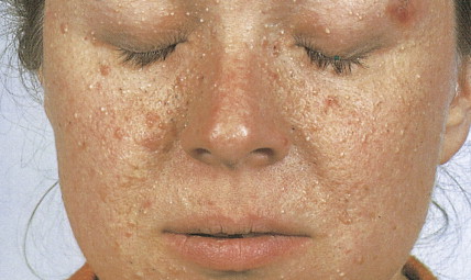
Actinic keratoses, also called solar keratoses, appear as thin, scaly, red lesions with epidermal hyperplasia and keratinocytic atypia ( Figs. 13.10 through 13.15 ). Although they are allegedly precursors of SCC, most do not proceed to frank malignancy, and conservative treatment is indicated. Because lesions tend to be multifocal and numerous, nonscarring methods of destruction, such as cryosurgery, electrodessication, or topical 5-fluorouracil cream, imiquimod 5% cream, and diclofenac 3% gel, are usually effective alternative therapies. Lesions that persist after treatment should be biopsied to rule out a malignant component. Topical sunscreens and other protective measures against sun exposure seem to be effective in preventing the development of new lesions.


FIGURE 13.10


SOLAR KERATOSES. Chronic skin changes on the chest of a 76-year-old man with long-standing sun exposure. There are numerous thin, scaly, erythematous lesions and hyperpigmented areas. Regular use of sunscreens would help to prevent development of these skin changes.


FIGURE 13.11


SOLAR KERATOSIS. This tumor appears as either a well-defined, raised red papule or, as shown here, a raised red plaque with an adherent scale.


FIGURE 13.12


SOLAR KERATOSIS. This patient’s scaly plaque (A) was treated successfully with 5-fluorouracil cream (B) .

(Courtesy of St. Mary’s Hospital, London, UK.)


FIGURE 13.13


SOLAR KERATOSIS. (A) Ultraviolet light exposure has produced a dry, elevated, white, scaly lesion on the posterior helical rim. (B) A solar keratosis arising from the antihelix has produced a small keratin horn.


FIGURE 13.14


Cutaneous horn refers to a reaction pattern characterized by a conical hyperkeratotic protuberans that can be associated with several different lesions. In most cases the underlying lesion will be an actinic keratosis. Other lesions that may be associated with a cutaneous horn pattern include verrucae, seborrheic keratosis, and squamous cell carcinoma.


FIGURE 13.15


SOLAR KERATOSIS. There are alternating areas of ortho- and parakeratosis. Underneath the parakeratosis, the epidermis shows keratinocytic atypia. The epidermis underneath the areas of orthokeratosis does not show significant pathologic change.


Arsenical keratoses are small, hard, punctate tumors that usually occur on the hands and feet ( Fig. 13.16 ). Increase in depth or diameter and ulceration of the lesions usually indicate progression to SCC.


FIGURE 13.16


ARSENICAL KERATOSIS. Exposure to arsenic, often as a pesticide, produces keratoses on the palms and fingers.


Xeroderma pigmentosum is an autosomal-recessive disorder characterized by the inability to repair UV light–induced DNA damage and, consequently, a pronounced cutaneous hypersensitivity to the effects of the sun’s rays. The disease, which starts in childhood, primarily affects the exposed parts of the body; lesions on the trunk may occur late in the course of the disease ( Figs. 13.17 and 13.18 ). Dryness, desquamation, and freckling are followed first by atrophic and telangiectatic spots, then by verrucous keratotic lesions. The most frequent malignancy is basal cell carcinoma, followed by SCC. Rarely, melanomas or sarcoma may also develop.


FIGURE 13.17


XERODERMA PIGMENTOSUM. Extreme photosensitivity is the feature of this condition. Persistent erythema occurs after seemingly innocent solar exposure. Multiple premalignant keratoses are evident.


FIGURE 13.18


XERODERMA PIGMENTOSUM. (A) After solar exposure, permanent freckling of exposed skin quickly ensues. Malignant change occurs early in life. This patient had her first SCC at the age of 2 years. (B) This keratoacanthoma developed when she was 12 years old. The lesion resolved spontaneously.


Patients with epidermodysplasia verruciformis, a generalized virally induced (HPV 5–associated) dermatosis, are also prone to develop SCCs.


Bowen disease is a term used to describe a characteristic clinical lesion that presents as a well-demarcated, scaly, red plaque and that histologically corresponds to SCC in situ ( Figs. 13.19 through 13.22 ). Usually Bowen disease affects skin unexposed to sunlight. It should be emphasized that the diagnosis of Bowen disease is a clinicopathologic one. Lesions showing similar microscopic changes may not show the classical clinical features of Bowen disease. Some studies have shown an apparent increase in the incidence of visceral cancer in patients with Bowen disease, but others have failed to document such an association. Lesions of Bowen disease seem to be capable of developing into invasive SCC. Removal by simple excision is usually an adequate treatment.


FIGURE 13.19


Bowen disease is characterized by well-defined erythematous scaly plaques histologically representing SCC in situ. In this patient, however, the right side of the lesion has already undergone transformation to invasive SCC.


FIGURE 13.20


BOWEN DISEASE. Involvement of the hands is quite common and can present diagnostic and therapeutic problems.


FIGURE 13.21


BOWEN DISEASE. The surface of the lesion, seen here in its characteristic presentation as a well-defined, slightly raised, red patch with an adherent scale, may become eroded.


FIGURE 13.22


BOWEN DISEASE. There is full-thickness keratinocytic atypia with marked nuclear pleomorphism and numerous mitotic figures. Bowen disease is synonymous with SCC in situ.


Common Skin Cancers


BASAL CELL CARCINOMA


Basal cell carcinoma is the most common malignancy in white people, with an incidence that is increasing worldwide by up to 10% annually. Exposure to UV radiation is the main pathogenetic factor. The most classical clinical presentation is that of a pearly-gray papule or nodule with prominent telangiectasia ( Figs. 13.23 through 13.25 ). Histologically the lesions consist of small, undifferentiated basal cells with minimal nuclear atypia ( Fig. 13.26 ). Several clinical and pathologic variants exist, including nodular, micronodular, superficial, and infiltrative/morpheaform types ( Figs. 13.27 and 13.28 ). The majority of tumors are located on the face, neck, and dorsum of the hands. Superficial variants tend to be located on the trunk. Some basal cell carcinomas may be pigmented and can be clinically confused with melanocytic lesions, especially melanoma ( Figs. 13.29 and 13.30 ). Basal cell carcinomas can be locally destructive, but only exceptional reports of cases with metastatic behavior exist in the literature ( Figs. 13.31 through 13.34 ). The treatment of choice is surgery, but for superficial variants that tend to be broad and multifocal, cryotherapy, photodynamic therapy, and more recently topical treatment with immune response modifiers such as imiquimod have shown to be effective. Infiltrative variants with irregular borders and sometimes perineural invasion are more prone to recur, probably due to insufficient surgery. Recurrent tumors incur serious complications. Treating such lesions in a combined clinic, comprising a dermatologist, a plastic surgeon, and an oncologist, is recommended.


FIGURE 13.23


BASAL CELL CARCINOMA. This early lesion is beginning to show the translucent, pearly appearance typical of these nodules as they begin to undergo central ulceration.


FIGURE 13.24


BASAL CELL CARCINOMA. A typical lesion, with a rolled edge, appears on this patient’s chin. Small vessels sweep over the edge, and the center is ulcerated.


FIGURE 13.25


BASAL CELL CARCINOMA. This is the most common malignant tumor of the eyelid and usually occurs either on the lower eyelid or at the medial canthus. This is an example of a relatively benign type of basal cell carcinoma with a classic pearly margin laced with blood vessels and a shallow ulcerated base at the center.


FIGURE 13.26


BASAL CELL CARCINOMA. (A) Low-power photomicrograph shows dermal aggregates of small basaloid cells. (B) Higher-power view illustrating the peripheral palisading of basaloid cells.


FIGURE 13.27


SUPERFICIAL BASAL CELL CARCINOMA. This variant of basal cell carcinoma presents as a plaque with a rolled, pearly margin. It is a less aggressive version of the ulcerative type of basal cell tumor.


FIGURE 13.28


MORPHEAFORM OR INFILTRATIVE BASAL CELL CARCINOMA. This is a more locally aggressive variant characterized by infiltrative, less clearly defined borders and sometimes perineural invasion. A wider surgical excision is required to prevent recurrence.


FIGURE 13.29


PIGMENTED BASAL CELL CARCINOMA. This subtype of basal cell tumor is similar in presentation to the nodular-ulcerative type except that the margin of the ulcer is rolled and pigmented. The clinical significance of this variant is that it may be mistaken for malignant melanoma. This lesion occurred on the leg, an unusual site.


FIGURE 13.30


PIGMENTED BASAL CELL CARCINOMA. Histologically, this lesion shows features of a nodular basal cell carcinoma. The neoplastic cells, however, contain melanin in their cytoplasm. Melanin is also seen within melanophages in the dermis.


FIGURE 13.31


BASAL CELL CARCINOMA. These tumors spread by direct extension and may be highly invasive, although they rarely metastasize. In this example an extensive basal cell carcinoma has spread to involve surrounding structures.


FIGURE 13.32


BASAL CELL CARCINOMA. If neglected the tumor grows inexorably, causing marked destruction of normal structures.

(Courtesy of Dr. D.E. Sharvill, Canterbury, UK.)


FIGURE 13.33


RADIATION-INDUCED BASAL CELL CARCINOMA. A tumor developed on this patient’s scalp 60 years after she underwent irradiation for tinea capitis as a child. She had chronic alopecia following the overirradiation. The lesion was successfully excised.

(Courtesy of J.P. Bennett.)


FIGURE 13.34


RECURRENT BASAL CELL CARCINOMA. This tumor has recurred on the area of a skin graft used to repair the site of a previously excised lesion. This is more frequently seen with infiltrative/morpheaform variants, advanced/large lesions, and multifocal tumors.


The basal cell nevus syndrome (Gorlin syndrome) refers to an autosomal-dominant disorder characterized by multiple basal cell carcinomas, associated with jaw cysts and skeletal anomalies ( Fig. 13.35 ). Patients have peculiar cutaneous pits in the palms and soles; the pits have histologic features of miniature basal cell carcinomas ( Fig. 13.36 ).


FIGURE 13.35


BASAL CELL–NEVUS SYNDROME. In this autosomal-dominant disorder, multiple basal cell carcinomas develop from childhood onward, as shown in this patient.

(Courtesy of Dr. A.C. Pembroke.)


FIGURE 13.36


BASAL CELL–NEVUS SYNDROME. Tiny pits on the palms, as shown in this low-power magnified view, are characteristic features of the condition.

(Courtesy of Dr. Eugene van Scott, Skin and Cancer Hospital, Philadelphia, PA.)


SQUAMOUS CELL CARCINOMA


SCC is the second most frequent cutaneous malignancy (next to basal cell carcinoma) ( ). Tumors tend to occur in sun-exposed areas such as the face, neck, arms, and hands. The pathogenesis is multifactorial, with chronic actinic damage, particularly, in light-skinned patients, being the most important factor ( Figs. 13.37 through 13.40 ). One of the postulated pathogenetic mechanisms is the induction of TP53 mutations by UV light (Burnworth et al., 2006). Tumors affecting external genitalia and perianal and periungual regions seem to be HPV-associated in an important percentage of cases. The overall recurrence of SCC seems to be between 3.7% and 10%, and the metastatic rate 5.2% ( Fig. 13.41 ). However, when tumors are located in special sites such as the lip, ear, and anogenital areas, they show a higher rate of recurrence and metastasis. Tumors arising in the setting of inflammatory, scarring, and degenerative processes are also associated with a worse prognosis than those developing in sun-damaged skin ( Figs. 13.42 through 13.44 ). SCCs are usually classified based on a three-grade system as well-differentiated, moderately differentiated, and poorly differentiated tumors. Poorly differentiated tumors tend to have a more aggressive behavior. In addition to the usual classic type, several SCC variants that may show variable clinical behavior have been described ( ). Some of these variants include verrucous carcinoma ( Figs. 13.45 and 13.46 ), carcinoma cuniculatum ( Fig. 13.47 ) ( ), warty carcinoma ( Fig. 13.48 ), basaloid carcinoma ( Fig. 13.49 ), sarcomatoid carcinoma ( Fig. 13.50A, B ), adenoid (acantholytic) carcinoma, angiosarcomatoid (pseudovascular) carcinoma, clear cell carcinoma, mucoepidermoid (adenosquamous cell carcinoma), and lymphoepithelioma-like carcinoma. Independently of the histologic variant and histologic grade and has been shown that depth of invasion and tumor thickness are the most helpful pathologic prognostic parameters ( ). SCCs less than 2 mm thick, which represent a high percentage of tumors, almost never metastasize. The risk of metastasis increases with tumors infiltrating the subcutaneous tissue. The risk of metastasis for undifferentiated carcinomas greater than 6 mm thick that have infiltrated the musculature, the perichondrium, or the periosteum, however, is quite high. Tumors between 2 and 6 mm thick with moderate differentiation and a depth of invasion that does not extend beyond the subcutis can be classified as low-risk carcinomas. In addition to high histologic grade, infiltration of subcutaneous tissue and deeper structures, and perineural and lymphovascular invasion are also indicators of poor prognosis.


FIGURE 13.37


SQUAMOUS CELL CARCINOMA. The back of the hand is a common site for SCC. This ulcerated lesion consists of a purulent base surrounded by a firm, everted, and irregular margin. Note the surrounding atrophic, sun-damaged skin.


FIGURE 13.38


SQUAMOUS CELL CARCINOMA. This lesion, which arose within a preceding solar keratosis, presents as a firm, indurated nodule. The ear is a common site.


FIGURE 13.39


SQUAMOUS CELL CARCINOMA. The lesion may also present as an ulcer having a raised, firm, indurated margin.


FIGURE 13.40


INVASIVE SQUAMOUS CELL CARCINOMA. Histologic features. This is a classical example of an invasive SCC of the usual type arising in sun-damaged skin showing actinic keratosis. Emanating from a dysplastic epidermis and infiltrating the reticular dermis, there are aggregates of neoplastic keratinocytes. The subcutaneous tissue is not affected, and this is important because the invasion of the subcutaneous tissue would have been an adverse prognostic factor.


FIGURE 13.41


SQUAMOUS CELL CARCINOMA. Spread of metastases to the right anterior shoulder from a primary SCC of the hand. There is extensive involvement of the axilla causing lymphatic obstruction and arm edema. Note ulceration of the tumor. Skin hyperemia is secondary to radiation therapy. In some patients metastases can develop in the lung, bone, liver, and other sites.


FIGURE 13.42


SQUAMOUS CELL CARCINOMA. Arising in an area of chronic ulceration, this large lesion, showing a purulent base and indurated margin, had been misdiagnosed as a benign varicose ulcer.


FIGURE 13.43


TREATMENT-INDUCED SQUAMOUS CELL CARCINOMA. This early-stage lesion of the scrotum developed after a decade of nitrogen mustard therapy for mycosis fungoides. Topical nitrogen mustard and psoralen + UV A therapy for this disorder are known cutaneous carcinogens.

(Courtesy of Dr. Eugene van Scott, Skin and Cancer Hospital, Philadelphia, PA.)


FIGURE 13.44


SQUAMOUS CELL CARCINOMA IN XERODERMA PIGMENTOSUM. Development of a squamous cell cancer in the rare syndrome of xeroderma pigmentosum is especially common. The lesion may arise de novo or from a preexisting senile keratosis or keratoacanthoma and is relatively more common in sun-exposed areas. Characteristically, this lesion has an everted edge and is irregular in shape.


FIGURE 13.45


VERRUCOUS CARCINOMA. Clinical picture illustrating two separate verrucous carcinomas of the foreskin arising in a background of long-standing lichen sclerosis.


FIGURE 13.46


VERRUCOUS CARCINOMA. Histologically, verrucous carcinoma is characterized by papillary surface and bulbous deep borders. It is an extremely well differentiated tumor without koilocytosis. Between the papillae there is a characteristic piling up of keratin. Pure verrucous carcinomas (not associated with higher-grade areas or infiltrative borders) have an excellent prognosis. Hybrid or mixed verrucous carcinomas are associated with a worse prognosis.


FIGURE 13.47


CARCINOMA CUNICULATUM. Unusual variant of verrucous carcinoma showing deep tumoral invaginations simulating rabbit’s burrows. The picture shows a cut section of a partial penectomy specimen with carcinoma cuniculatum.


FIGURE 13.48


Warty carcinoma is an HPV-related tumor usually affecting genital/perianal areas. The lesion has some features of condyloma but architectural (infiltrative borders) and cytologic (nuclear pleomorphism) features of carcinoma. There is marked koilocytosis throughout the neoplasm. This tumor should be distinguished from verrucous carcinoma. Warty carcinoma is associated with local-regional metastasis in approximately a fourth of the cases.


FIGURE 13.49


BASALOID CARCINOMA. Aggressive HPV-related variant of SCC usually affecting genital/perianal areas. The lesion is characterized by aggregates of poorly differentiated basaloid cells with prominent apoptosis and numerous mitoses. Central areas of comedonecrosis and vascular and perineural invasion are common findings in this lesion. It should be distinguished from basal cell carcinomas; the latter are highly unusual in mucosal genital areas.


FIGURE 13.50


SARCOMATOID SQUAMOUS CELL CARCINOMA. Not infrequently, squamous cell carcinomas are predominantly composed of spindle cells (A) . Immunohistochemical studies are necessary to confirm the epithelial nature of these lesions and differentiate them from spindle cell melanomas, atypical fibroxanthomas, and sarcomas. (B) Expression of cytokeratin 34Beta12 by the tumor cells in this example of sarcomatoid carcinoma.


Transplant recipient patients who undergo long-term immunosuppression and patients with other forms of immune suppression are at increased risk for different neoplasms, and in the skin, especially SCC. These tumors tend to affect younger patients and are more frequently located in sun-exposed areas. The lesions tend to be clinically problematic, since they tend to be multiple and arise in a background of dysplastic epidermis ( Fig. 13.51 ). It appears that a good percentage of tumors in this setting may be HPV-related, and in fact it is not unusual to find features suggestive of a viral wart associated with frankly carcinomatous areas ( Fig. 13.52 ). It seems that tumors associated with immunosuppression tend to have a more aggressive behavior with a significant incidence of metastasis and even mortality ( Fig. 13.53 ) ( ).


FIGURE 13.51


Arm of a man who had received a kidney transplant more then 30 years earlier. There is a background of actinic keratosis and several invasive SCCs. In transplant recipient patients the risk for developing cutaneous squamous cell carcinoma is related to the duration and level of immunosuppression.

(Courtesy of Dr. Danielle Miller, Department of Dermatology, Brigham and Women’s Hospital, Boston, MA.)


FIGURE 13.52


PATIENT WITH HEART TRANSPLANT. There is an SCC with “warty features” of the left groin.

(Courtesy of Dr. Danielle Miller, Department of Dermatology, Brigham and Women’s Hospital, Boston, MA.)


FIGURE 13.53


Elderly patient who had a history of significant sun exposure throughout his life with an SCC of the scalp associated with in-transit metastasis.

(Courtesy of Dr. Danielle Miller, Department of Dermatology, Brigham and Women’s Hospital, Boston, MA.)


KERATOACANTHOMA


Keratoacanthoma is a rapidly growing tumor usually affecting sun-exposed skin of elderly patients. The classical presentation is that of a solitary discrete, round to oval, flesh-colored umbilicated nodule with a central keratin-filled crater ( Figs. 13.54 and 13.55 ). Lesions have a rapid clinical evolution and usually regress within 4–6 months ( Fig. 13.55 ). There has been a lot of controversy around this entity concerning whether it represents a benign or a malignant tumor, and multiple clinical and histologic criteria have been proposed to differentiate it from SCC ( Fig. 13.56 ). In unusual cases, however, lesions in the histologic spectrum of typical keratoacanthomas have been shown to follow an aggressive clinical course. Modern immunohistochemical and molecular techniques have not proved to be more useful in this distinction. With all of this controversy, and since there are no clinical or histologic criteria to classify a potential keratoacanthoma as a benign tumor that might spontaneously regress or as a neoplasm with metastatic potential, it seems most appropriate to consider keratoacanthoma as an extremely well-differentiated variant of SCC and to treat it as such ( ).


Aug 13, 2019 | Posted by in ONCOLOGY | Comments Off on Skin Cancer

Full access? Get Clinical Tree

Get Clinical Tree app for offline access