Age Changes
Physiology
Age-related changes affect virtually all aspects of the respiratory system. Structural and functional changes occur, decreasing efficiency of gas transfer. However, because the lungs have huge reserve capacity, significant clinical issues only arise when an elderly person becomes sick unless lung function has been progressively damaged by smoking or air pollution. Older people may have difficulty performing many lung function tests, but spirometry is usually possible.
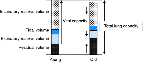
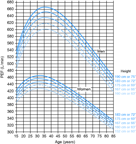
- Reduced lung elasticity and chest wall compliance lead to air trapping, a rise in residual volume and a fall in the forced vital capacity (FVC), forced expiratory volume in 1 s (FEV1) and peak expiratory flow as shown in Figures 10.1 and 10.2. Some normal old people will have an FEV1/FVC ratio of < 0.7. Do not treat (beyond smoking cessation advice) unless they are symptomatic.
- An increase in airway size and loss of alveolar surface decrease the lung volume available for gas exchange and increase dead space, reducing efficiency of gas exchange. Premature closure of small airways results in ventilation–perfusion mismatching, contributing to an increase in the alveolar-arterial oxygen gradient. Arterial oxygen tension (PaO2) falls from 12.7 kPa at age 30 to 10 kPa at age 60.
- Oxygen delivery to tissues (VO2 max) decreases due to age-related decreases in cardiac output and body muscle mass, as well as ventilation–perfusion mismatching and decreased alveolar volume.
- Mucociliary protection of the lower airway is impaired.
Examination
Check the rate and pattern of respiration. Tachypnoea suggests a cardiorespiratory problem, which can be very useful if the patient cannot give much history. Cheyne–Stokes respiration, in which the breathing becomes progressively shallower, sometimes culminating in an apnoeic episode before becoming progressively deeper again in a cyclical pattern, is more common in the elderly. It is often seen in stroke but may occur in apparently normal individuals. Note the chest shape. Significant kyphosis is usually now due to osteoporosis (previously TB). Look for thyroid and thoracotomy scars. The normal trachea may deviate slightly to the right around an unfolded aorta. Many older patients have basal crackles that clear on coughing and have no significance. Conversely, if the patient has poor air entry, an area of consolidation may appear silent. A silent chest is a danger sign in airway obstruction.
After a fall, check for bruising of the chest wall and ‘spring’ the ribcage for fractures (pneumonia usually follows). Remember other systems associated with chest problems, e.g. aspiration in PD, and check for heart failure. Oxygen saturation measured by a fingertip probe is a useful bedside test. The admission CXR in an ill old patient is often difficult to interpret; usually taken AP or even supine (check for scapular lines), often with rotation (check relation of heads of clavicles to spinous processes), sometimes with the head in the chest and with poor inspiration. A subtle diagnosis may require a repeat film when the patient is improving.
Upper Respiratory Tract Infection
Rhinoviruses (species of the genus Enterovirus from the Picorna virus family) and corona viruses cause coryza, the common cold. This is a mild systemic upset with nasal symptoms, but older people, particularly smokers and those with pre-existing chronic illness, may develop lower respiratory complications.
Influenza is usually debilitating in the elderly, particularly in the presence of chronic heart, chest or renal disease, or diabetes and may be complicated by pneumonia, especially due to Staphylococcus aureus. Everyone aged over 65 years should be offered immunization in October/November. The influenza viruses are constantly altering their antigenic structure, so every year WHO recommends which strains should be included in the flu vaccine. The vaccine may cause a mild local reaction and hypersensitivity to egg products is a contraindication. Immunity takes 2–4 weeks to develop and lasts 6–8 months. There is evidence that immunization of staff is the best way to protect residents of institutions, and some hospitals offer free vaccination to their staff. The prevalence of influenza A/H1N1 (pandemic swine flu) increased in 2010 but unlike seasonal flu is more common and severe in the young and pregnant. Oseltamivir (a neuraminidase inhibitor) is given if H1N1 is suspected.
Acute Breathlessness
The acutely dyspnoeic elderly patient is a very common medical emergency.
Common Causes of Acute Respiratory Distress
- Left ventricular failure.
- Pneumonia.
- Exacerbation of COPD.
- Exacerbation of asthma.
- Pulmonary embolism.
- Pneumothorax.
- Inhaled foreign body (stridor, choking).
Airway Obstruction
Most cases are due to COPD but a number of patients have long-standing asthma and late-onset asthma may be increasing. COPD includes chronic bronchitis, defined clinically as sputum production on most days for 3 months of two successive years, and emphysema, defined histologically as air space dilatation due to destruction of alveolar walls. There may be a reversible component especially in asthmatics who have smoked. COPD causes progressive breathlessness and reduction in exercise capacity. The presence of airflow obstruction should be confirmed by post-bronchodilator spirometry. The death rate for COPD has increased in recent years whilst rates of other causes of mortality (heart, neoplasm and cerebrovascular diseases) are falling. About 15% of those dying from COPD have never smoked and air pollution is thought to be a significant factor.
Features of Severe Attack of Airways Obstruction Requiring Admission
- Unable to cope at home.
- Already receiving long-term oxygen therapy (LTOT).
- Cannot complete sentences.
- Respiratory rate > 25/min.
- Pulse rate > 110 b.p.m. (unreliable in AF).
- Peak flow < 50% of patient’s normal or predicted.
- SaO2 < 90%.
- PO2 < 7 kPa, PCO2 > 6.7 kPa on air.
Management of Acute Exacerbation of Airway Obstruction
- Non-invasive ventilation (NIV) has replaced doxapram and is a good option for patients who are poor candidates for intubation because extubation may be difficult.
- Intensive care for ventilation if above measures ineffective. Find out about the patient’s functional status prior to this illness before contacting ITU to discuss admission. Patients with COPD may do better than anticipated in this situation.
Post-acute/chronic phase
Patients with mild COPD need a SAMA or SABA.
If the patient remains breathless or has exacerbations and FEV1 ≥ 50% predicted, offer a LAMA or a LABA. If FEV1 < 50% predicted, offer a LAMA or LABA/ICS.
Tiotropium and the combination inhalers are expensive but persistent breathlessness or frequent exacerbations warrant a LAMA and a LABA/ICS regardless of FEV1. This is common in frail older people with recurrent admissions with COPD. ICS have been shown to increase pneumonia but the benefit in terms of reducing exacerbations is worth it. Recurrent exacerbations are associated with increasing morbidity and a strong predictor of mortality (see also 3 below).
Different types of inhaler are available requiring different levels of manual dexterity, cognition and inspiratory flow. Ensure inhaler and spacer device are understood by the patient. Prescribe SABA as ‘rescue medication’.
Stay updated, free articles. Join our Telegram channel

Full access? Get Clinical Tree


