Rare Pediatric Tumors


Rare Pediatric Tumors


Carlos Rodriguez-Galindo

Wayne L. Furman

Alberto S. Pappo


INTRODUCTION

The Office of Rare Diseases of the National Institutes of Health defines rare diseases as those diseases having a prevalence of less than 200,000 affected individuals in the United States and according to this definition all childhood cancers are rare.1,2 Many cancers considered rare in pediatrics are adult cancers of early onset, such as melanoma, nasopharyngeal carcinoma, or gastrointestinal cancers; however, a significant number of cases correspond to truly rare embryonal malignancies with almost exclusive pediatric occurrence, such as pleuropulmonary blastoma or pancreatoblastoma.

Registries dedicated to describing the epidemiology, biology, and clinical characteristics of specific pediatric rare cancers have been developed and are great tools to advance our knowledge on these rare malignancies. Advances in the study and treatment of rare pediatric cancers are limited by the small numbers and the lack of expertise; for this reason, cooperative group initiatives, such as those led by the Children’s Oncology Group,3 the Italian TREP (Tumori Rari in Età Pediatrica) project,2 or the European ExPERT group,4 among others, are key to developing consensus guidelines for therapy and prospective studies that integrate biologic and therapeutic aims.

This chapter summarizes the infrequent cancers of childhood in descending anatomic order, from the aerodigestive tract to the genitourinary system. For common cancers of the adults, the chapter will include mostly those features that are unique or most applicable to children, while providing an overview of the disease as commonly seen and treated in adults. Rare cancers of endocrine organs, such as thyroid carcinoma, pheochromocytoma/paraganglioma and adrecortical carcinoma, are reviewed in chapter 36.


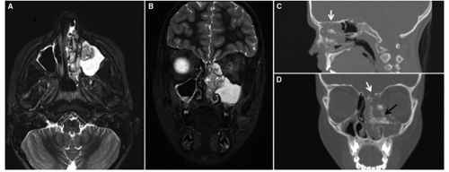
Figure 37.1 Esthesioneuroblastoma in a 9-year-old girl presenting with nasal obstruction and pain. MRI axial (A) and coronal (B) STIR (short tau inversion recovery) images show a mass in the left sinonasal cavity, involving the anterior left ethmoid air cells, the anteromedial aspect of the left maxillary sinus and the left nasal cavity. Sagittal (C) and coronal (D) CT images show thinning of the cribriform plate (white arrow) and the presence of calcifications (black arrow).


CANCER OF THE AERODIGESTIVE TRACT AND RELATED ANATOMICAL SITES


Esthesioneuroblastoma

Esthesioneuroblastoma, also known as olfactory NB (neuroblastoma), represents only 3% to 6% of nasal and sinonasal neoplasms and 0.3% of all upper aerodigestive tract malignancies in adults.5 In children, esthesioneuroblastoma accounts for 25% of cancer of the nasal cavity, second to rhabdomyosarcoma.6 It is considered to be of neural crest origin, and arises from the olfactory neuroepithelium in the superior nasal cavity at the anterior skull base (Fig. 37.1). Esthesioneuroblastoma characteristically occurs after the fifth decade of life, but 7% to 20% of patients present between 10 and 24 years.5,6 These tumors are locally aggressive, but 15% to 25% may present with cervical nodal disease, and 1.5% with distant metastases. The symptoms are related to the local extension of the tumor, initially with unilateral nasal obstruction and hyposmia or anosmia, causing epistaxis and headache as the tumor progresses. More aggressive tumors may extend beyond the nasal and paranasal sinuses and invade the orbits and skull base, cavernous sinuses, and the brain.5,7,8

On pathology, esthesioneuroblastoma presents as a small, round blue cell tumor; cells have indistinct cytoplasmic borders, hyperchromatic nuclei, and infrequent mitoses. Homer-Wright and Flexner-Wintersteiner rosettes are present in 30% to 50% and 5% of cases, respectively.5 The Hyams histopathologic grading system (Table 37.1) is commonly used to characterize the grade of differentiation, which correlates with prognosis.5,7,9 The modified Kadish staging system (Table 37.2), based on anatomic involvement, is traditionally used.5,7


TABLE 37.1 Esthesioneuroblastoma—Hyams Histopathologic Grading System


Grade


Definition


Grade 1


Well differentiated with lobular preservation, prominent fibrillary matrix, no nuclear pleomorphism, Homer-Wright rosettes


Grade 2


Low mitotic index, moderate nuclear polymorphism, fibrillary matrix present, Homer-Wright rosettes


Grade 3


Moderate mitotic index, prominent nuclear polymorphism, low fibrillary matrix, Homer-Wright rosettes, rare necrosis


Grade 4


High mitotic index, anaplasia, marked nuclear pleomorphism, absence of fibrillary matrix and Homer-Wright rosettes, frequent necrosis


Adapted from Bak M, Wein RO. Esthesioneuroblastoma: a contemporary review of diagnosis and management. Hematol Oncol Clin North Am 2012;26:1185-1207.


TABLE 37.2 Esthesioneuroblastoma—Modified Kadish Staging System


Stage


Definition


Stage A


Limited to the nasal cavity


Stage B


Involves the paranasal sinuses


Stage C


Extends outside the sinonasal cavity, including involvement of the base of the skull, orbit, and intracranial cavity


Stage D


Distant metastases at diagnosis


Adapted from Bak M, Wein RO. Esthesioneuroblastoma: a contemporary review of diagnosis and management. Hematol Oncol Clin North Am 2012;26:1185-1207.


Treatment of esthesioneuroblastoma includes surgery and radiation therapy. Surgical resection requires a combined neurosurgical and nasal approach, and radiation therapy is recommended even in the absence of microscopic residual. Survival outcomes range from 60% to greater than 80%. Complete resection, low-grade histology (Hyams grades 1 and 2) and low stage are favorable prognostic factors.5,7,9 While esthesioneuroblastoma is a chemosensitive neoplasm, the use of systemic chemotherapy is traditionally limited to cases with nodal or distant metastases. However, neoadjuvant therapy may facilitate surgery in cases of very advanced disease; in those cases, preoperative radiation therapy may also be indicated.10 The most commonly used regimens are cisplatin-based, although sarcoma therapies such as vincristine, actinomycin D, and cyclophosphamide (VAC) and ifosfamide, vincristine, actinomycin D, and doxorubicin (IVADo) have also been shown to be effective, particularly in children.10 Experience in children is limited; however, available data suggest that while pediatric patients may present with very advanced disease, a multimodal approach with preoperative chemotherapy and radiation therapy results in excellent outcomes.10


Melanotic Neuroectodermal Tumor of Infancy

Melanotic neuroectodermal tumor of infancy is a melanin-containing neoplasm that primarily affects the maxilla during the first year of life.11 More than 80% of the cases are diagnosed in the first 6 months of life.12 This tumor presents as a fast-growing mass with a pseudocapsule due to reactive bone formation. The majority of cases involve the head and neck structures; in more than two-thirds of the cases the tumor arises in the maxilla, followed by the skull and mandible.11,12 Uncommonly reported sites include orbits, meninges, femur, epididymis, ovary, uterus, and mediastinum.13,14 On imaging, these tumors cause compression rather than infiltration of adjacent structures, with local invasion causing tooth displacement, bone destruction, expansion, and remodeling.13 Lesions arising within the skull are seen at the sutures and about half of these occur around the anterior fontanelle. The radiographic appearance is typically that of a well-demarcated radiolucent lesion with bone expansion, entrapping the developing tooth buds.12,13

The pathology of melanotic neuroectodermal tumor of infancy shows a biphasic pattern, with irregular gland-like spaces scattered amid dense fibrous stroma. The gland-like spaces are lined by larger pigmented cells with aggregates of dark, small, round cells resembling neuroblasts. The cells can differentiate into melanoblasts or neuroblasts, and in up to 40% of the cases, the levels of urinary catecholamines may be elevated.11,12

While the presentation is aggressive, with rapid growth and large tumor size, the prognosis is good; patients can be cured with surgery only, and distant metastases have been reported in less than 10% of the cases.12 Surgery is the treatment of choice, and while radical approaches are recommended, it is thought that there is a possible spontaneous involution of residual tumor, as some tumors do not recur despite incomplete resection.15 Most recurrences occur within the first 6 to 18 months, and a repeat surgery with curative intent is recommended. Chemotherapy has also been shown to have a role in cases of recurrent disease and to facilitate surgery; regimens used for the treatment of neuroblastoma or sarcomas have been shown to be effective.16,17


Oropharyngeal Cancer

Oropharyngeal cancer is extremely rare in the pediatric age group; of the 35,000 cases diagnosed annually in the United States, only 0.6% occurred in children younger than 20 years (http://www.seer.cancer.gov/statfacts/html/oralcav.html). The diagnosis of a squamous cell carcinoma in a child should raise the question of an underlying cancer predisposition syndrome. DNA repair defects such as Fanconi anemia,18 dyskeratosis congenita,19 or Bloom syndrome,20 should be considered. Other syndromes with increased incidence of oral cancer include recessive dystrophic epidermolysis bullosa,21 xeroderma pigmentosum,22 and mutations in the connexin 26 gene (Keratitis-ichthyosis-deafness [KID] syndrome).23 Li-Fraumeni syndrome has also been associated with squamous cell carcinomas of the oral cavity.20 Pediatric cancer survivors, particularly those with prior exposure to radiation therapy24 and those with a history of allogeneic hematopoietic stem cell transplant and oral graft-versus-host disease,25 also have an increased incidence of oropharyngeal cancer.

Other risk factors for the development of oral cancer include the use of any tobacco product, heavy alcohol use, certain viral infections such as human papillomavirus (HPV), low consumption of fruits and vegetables, marijuana use, older age, black race, and male sex.26 It has been well documented that the use of smokeless tobacco and cigarette smoking increase the risk of oropharyngeal cancer as well as that of potentially malignant oral lesions such as leukoplakia, erythroplakia, palatal lesion of reverse cigar smoking, oral lichen planus, and submucosal fibrosis.27,28 Of particular concern is the prevalent use of the carcinogenic habit-forming betel nut (areca) and different forms of smokeless tobacco among the South Asian youth.29 Oral HPV infection is strongly associated with the number of recent oral sex partners or open-mouthed kissing partners,30 and oral HPV infection is unequivocally associated
with oropharyngeal cancer regardless of the use of tobacco or alcohol. The incidence of HPV-associated oropharyngeal cancer has significantly increased since 1973, particularly among white males aged 40 to 59 years.31 Approximately 20% to 25% cases of head and neck cancer contain oncogenic HPV, mostly types 16, 31, and 33. In a study of 1,235 children, the prevalence of HPV in the oral cavity was 1.9% and the highest rates were seen in patients younger than 1 year and in those aged between 16 and 20 years.32 HPV-associated oral cancers tend to occur in younger patients of high socioeconomic status, are associated with sexual behavior, more often involve the lingual and palatine tonsils, frequently have poorly differentiated basaloid features, express p16, and have a better prognosis and response to radiotherapy than other head and neck carcinomas.33,34,35 Current preventive strategies that incorporate vaccine programs targeting the adolescent population may prove beneficial in decreasing the incidence of cervical and head and neck HPV-associated cancers.36,37

These tumors are treated using a multidisciplinary approach that often incorporates surgery, radiotherapy, and chemotherapy. For patients with localized disease, an upfront surgical approach with an attempt to achieve negative margins is the standard of care; positive margins require a second surgery or adjuvant radiation. A neck dissection with diagnostic and therapeutic purposes should be considered at the time of local control.38 For patients with advanced disease, neoadjuvant or adjuvant cisplatin-based chemotherapy or chemoradiation is recommended.38


Salivary Gland Tumors

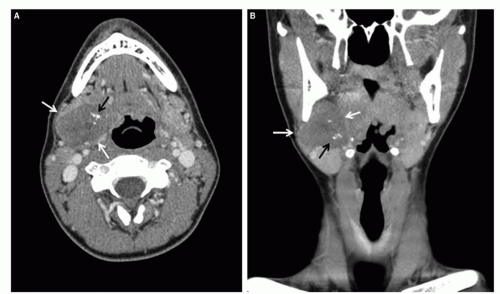
Approximately 5% of salivary gland tumors occur in the pediatric population, primarily in the second decade of life.39 Most of these neoplasms originate in the parotid gland, with 10% to 15% arising from submandibular, sublingual, or minor salivary glands.39,40,41,42,43 Approximately 75% of primary salivary gland tumors are benign, with pleomorphic adenoma predominating (Fig. 37.2).40,41,42 Among the cancerous lesions in children, mucoepidermoid carcinoma accounts for 40% to 50% of all malignant lesions, followed in frequency by acinar cell carcinoma (30% to 35%), adenoid cystic carcinoma (5%), adenocarcinoma (5%), and other histologies.39,44 Compared to adults, children have a higher proportion of mucoepidermoid carcinoma (50% vs. 25%), well or moderately differentiated tumors (88% vs. 49%), and localized disease (76% vs. 50%) as well as a female predominance (60% vs. 40%).39 Survivors of childhood cancer have a 39-fold higher risk than the general population of developing malignant salivary gland tumors, mostly mucoepidermoid carcinomas.45 The risk increases linearly with radiation dose and remains elevated after 20 years.45,46


Figure 37.2 Pleomorphic adenoma of the right parotid gland in a 13-year-old male. Axial (A) and coronal (B) CT images disclose a well-circumscribed right retropharyngeal mass (white arrows) with calcifications (black arrow).

Approximately two-thirds of mucoepidermoid carcinomas in adults have the CRTC1/3-MAML2 fusion, which disrupts the Notch signaling pathway.47 The presence of this fusion is associated with low-grade tumors and in general confers a more favorable prognosis.48,49 However, there are no data on the presence of this characteristic fusion in pediatric mucoepidermoid carcinoma. The AJCC (American Joint Committee on Cancer) system is used to assess the extent of disease.50 Although distant metastases are very rare, nodal disease may occur in up to 50% of patients with high-grade tumors and advanced disease.44

Surgical removal is the treatment of choice when feasible.44,51 For pleomorphic adenomas, a lateral parotidectomy with en-bloc excision of the tumor within the surrounding tissues, preserving facial nerve integrity, is usually indicated; local recurrences in such cases are rare. For malignant tumors, more aggressive surgical approaches are required. Low-grade and early-stage tumors are usually cured with surgery alone. High-grade tumors require a more aggressive surgical approach, including neck dissection, and adjuvant radiation therapy.41,44,51,52 Resection of the facial nerve is controversial and is currently recommended only when there is gross anatomic or histopathologic evidence of neural invasion at the time of the surgery.53 Cisplatin-based chemotherapy is usually reserved for palliation of disease not amenable to either surgery or irradiation or for distant metastatic disease. Overall, the prognosis for children with malignant salivary gland tumors is excellent, with 5-year survival estimates in excess of 90%.39

Sialoblastoma, a very rare congenital salivary gland tumor with primitive embryonic features of salivary gland tissue that most commonly originate in the parotid gland, shows a very aggressive local behavior, although metastases have been reported. Most sialoblastomas can be cured with surgery alone; however, they are chemosensitive, and chemotherapy may facilitate surgery or play a role in cases of recurrent disease.54,55,56


Nasopharyngeal Carcinoma

Nasopharyngeal carcinoma (NPC) is very rare in children; only 1% of all NPC occurs in patients aged less than 19 years, with an incidence in the United States of approximately 1 to 1.5 cases per
million per year.57 Although representing approximately 1% of all pediatric malignancies, it accounts for 35% to 50% of all nasopharyngeal malignancies. NPC originates from the surface epithelium and differs from other head and neck carcinomas by its very distinct histologic, epidemiologic, and biologic characteristics. NPC has an endemic distribution among well-defined ethnic groups, such as inhabitants of some areas of Southeast Asia and in Alaskan Eskimos, where the incidence is 25 to 50 and 15 to 20/100,000 persons per year, respectively.58

Three histologic subtypes of NPC are recognized: Type I, or keratinizing squamous cell carcinoma, which is similar to carcinomas that arise from other sites of head and neck; Type II, or nonkeratinizing squamous cell carcinoma; and Type III, or undifferentiated carcinoma (also called lymphoepithelioma), which is the most common form of the disease. In the western world, NPC occurs sporadically, is usually Type I, and is primarily related to exposure to the classic head-and-neck-cancer risk factors, such as alcohol and tobacco. The endemic form is usually either the Type II or III histologic subtypes. In contrast to the Type I sporadic form, the etiology of the endemic form includes virologic and environmental risk factors, and genetic susceptibility. The Epstein-Barr virus (EBV) plays a key role in the pathogenesis of endemic NPC. Increased levels of IgG and IgA against EBV are frequently observed, particularly in those with undifferentiated NPC.

The anti-EBV serologic profile has been used in the screening, early diagnosis, and monitoring of therapeutic response of NPC in high-incidence areas.59 In addition to serologic evidence, there is also molecular evidence; EBV DNA is present in the NPC tumor cells.59 Furthermore, EBV DNA appears to be clonal, which indicates that it is present within the cell at the time of carcinogenic transformation, thereby suggesting a role for the virus in the transformation event. EBV has also been detected in virtually all nonkeratinizing NPCs, irrespective of geographic origin, and thus appears to be a rate-limiting step in the pathogenesis of these tumors. By contrast, association of squamous-NPC with EBV is much more controversial since EBV DNA is detected in a very small proportion of cases.

In children, NPC presents in a form very reminiscent of the endemic NPC seen in adults, with almost universal Type III histology and EBV positivity. In the United States, studies of pediatric NPC have shown a higher incidence in African Americans and in children of Asian descent.57


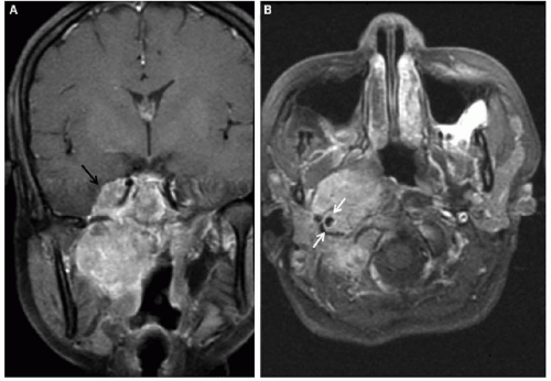
Figure 37.3 NPC in a 16-year-old Asian American female presenting with nasal obstruction, headache, and cervical lymphadenopathy. Coronal (A) and axial (B) T1-weighted images demonstrate a large right nasopharyngeal mass obliterating the right parapharyngeal space. The mass extends posteriorly to the carotid sheath (white arrows), inferiorly to the level of the oropharynx, and superiorly to the cavernous sinus and right middle cranial fossa (black arrow).


Presentation

Childhood NPC usually presents in the second decade of life (median age 13 to 15 years) with a strong male preponderance.57,60,61,62 In the United States, the incidence is higher among African Americans.57 Clinically, NPC has few early signs, such as nose bleeding, congestion, otitis media, or trismus. Local spread to adjacent areas of the oropharynx or invasion of the skull base may occur, the latter resulting in cranial nerve paralysis. Bilateral, bulky cervical lymph node invasion may occur in up to 80% of patients.57,60,61 Compared with adults, children present with more advanced disease, with larger primaries and higher nodal stage.57


Diagnosis and Evaluation

NPC should be always suspected in an adolescent presenting with nasal congestion or epistaxis, headache, and a cervical mass. Nasal endoscopy usually shows a nasopharyngeal mass, and a biopsy of the primary tumor is always recommended; however, a diagnosis can also be made with biopsy of a cervical lymph node in the context of a suggestive nasopharyngeal mass. An MRI (magnetic resonance imaging) of the head and neck, extending to the supraclavicular fossa is recommended, and a CT (computed tomography) may be performed to better define skull base erosion (Fig. 37.3). A CT scan of the chest is also recommended to evaluate for the presence of parenchymal metastases as well as mediastinal nodal disease, which usually develops as extension of the cervical metastases. While the role of PET (positron emission tomography) scan is not clear at the time of diagnosis, it may facilitate the evaluation of response to therapy.63

Undifferentiated NPC is the most frequent type in the pediatric age group, and may mimic a lymphoma. By immunohistochemistry, all types of NPC are immunoreactive with cytokeratin. Additionally, EBER (EBV-encoded RNA) in situ hybridization is positive in all undifferentiated NPCs with 100% of the neoplastic
cells containing the virus. Staging of NPC usually follows the TNM system, which has been shown to be predictive of outcome, and very helpful in defining therapy (Table 37.3).64


TABLE 37.3 AJCC Staging System for Nasopharyngeal Carcinoma


DEFINITIONS


Designation


Definition


T1


Tumor confined to the nasopharynx, or tumor extends to oropharynx and/or nasal cavity without parapharyngeal extension


T2


Tumor with parapharyngeal extension


T3


Tumor invades bony structures of skull base and/or paranasal sinuses


T4


Tumor with intracranial extension and/or involvement of cranial nerves, hypopharynx, orbit, or with extension to the infratemporal fossa/masticator space


N0


No regional lymph node metastasis


N1


Unilateral metastasis in cervical lymph node(s), ≤6 cm in greatest dimension, above the supraclavicular fossa, and/or unilateral or bilateral, retropharyngeal lymph nodes, ≤6 cm in greatest dimension


N2


Bilateral metastasis in cervical lymph node(s), ≤6 cm in greatest dimension, above the supraclavicular fossa


N3


Metastasis in a lymph node(s) >6 cm and/or to supraclavicular fossa


N3a: >6 cm in dimension


N3b: Extension to the supraclavicular fossa


M0


No distant metastasis


M1


Distant metastasis


CLASSIFICATION


Stage


Tumor Spread


Lymph Node Spread


Metastasized


Stage I


T1


N0


M0


Stage IIA


T2a


N0


M0


Stage IIB


T1


T2


T2


N1


N0


N1


M0


M0


M0


Stage III


T1


T2


T3


N2


N2


N0-2


M0


M0


M0


Stage IVA


T4


N0-2


M0


Stage IVB


Any T


N3


M0


Stage IVC


Any T


Any N


M1


Used with permission from American Joint Committee on Cancer. Pharynx. In: Edge SB, Byrd DR, Compton CC, et al, eds. AJCC cancer staging manual. 7th ed. New York, NY: Springer, 2010:41-56.


Nut-Midline Carcinoma

A recently described entity that should be included in the differential diagnosis of nasopharyngeal and mediastinal malignancies in patients of all ages is NUT-midline carcinoma (Fig. 37.4). This is a very rare and aggressive malignancy that is genetically defined (>75% of cases) by rearrangements of the NUT gene on chromosome 15q14. NUT is fused with BRD4 on chromosome 19p13, creating chimeric genes that encode the BRD-NUT fusion proteins. In the remaining cases, NUT is fused to BRD3 on chromosome 9q34 or an unknown partner gene; these tumors are termed NUT-variant.70 The tumors arise in the midline epithelial structures, typically mediastinum and the upper aerodigestive track, and present as aggressive undifferentiated carcinomas, with or without squamous differentiation.71 The outcome is very poor, with average survival less than 1 year. Preliminary data seem to indicate that NUT-variant tumors may have a more protracted course.71

Recommended treatment includes a multimodal approach with systemic chemotherapy, surgery, and radiation therapy. Cisplatin, taxanes, and alkylating agents have been used with some success; however, while early responses are commonly seen, tumors progress early in the course of the disease.71 Preclinical studies have shown that NUT-BRD4 is associated with globally decreased histone acetylation and transcriptional repression that can be restored with histone deacetylase inhibitors. Response to vorinostat has been reported in a case of a child with refractory disease, suggesting a potential role for this class of agents in the treatment of this malignancy.72 BET bromodomain inhibitors represent another promising class of agents currently being investigated for this malignancy. The International NMC (NUT Midline Carcinoma) Registry (http://www.nmcregistry.org), a source of information for physicians and patients, provides diagnostic support and treatment guidelines.


Ameloblastoma and Ameloblastic Carcinoma

Ameloblastoma, which arises from the primitive dental lamina or from odontogenic cysts, is an unusual, locally aggressive odontogenic tumor that accounts for about 10% of all mandibular and maxillary neoplasms.73,74 The disease appears to be more prevalent in African and Asian children with a median age of 14 years at presentation.74 Ameloblastomas can be classified into four groups: (1) solid/multicystic, (2) extraosseous/peripheral, (3) desmoplastic, and (4) unicystic. Multicystic/solid variants have a higher rate of recurrence, whereas unicystic tumors respond favorably to conservative treatment. Peripheral ameloblastomas arise in the gingival area and do not involve bone.73 In African children, there is a predominance of males, the disease often involves the mandible, and there is a higher incidence of the solid/multicystic subtype. In contrast, in children of Western origin, the female sex predominates; the disease is more often unicystic and involves the angle of the mandible.74,75 Clinical symptoms at presentation include facial swelling, the presence of a submucosal
mass, malocclusion, paresthesias, pain, and evidence of unerupted teeth in the affected area.73,75 Analysis of small numbers of tumors suggest that p53, MDM2, and p14 may be responsible for tissue structuring and cytodifferentiation in ameloblastoma.74 In addition, recent reports have documented the existence of a complex interaction that favors tumor formation and is mediated by the secretion of frizzled-related peptide (sFRP2), which impairs bone formation, as well as IL-6 and RANKL, which promote bone resorption.76,77

Solid and multicystic ameloblastomas should be treated by surgical resection that includes a 1-cm margin of normal bone and soft tissue.73,78 Unicystic ameloblastomas can be treated with simple enucleation followed by curettage and physicochemical therapy with either liquid nitrogen or Carnoy’s solution.75,78 Local excision is sufficient for peripheral ameloblastomas. Malignant ameloblastomas have been reported; they often affect the mandible and can metastasize to the lung and lymph nodes.73 One case of metastatic disease has been reported to respond to chemotherapy with carboplatin and paclitaxel.79

Ameloblastic carcinoma is a very rare odontogenic malignancy with features of ameloblastoma and cytologic atypia; the tumor most often affects the mandible and can metastasize to bone, lung, and brain.80


Laryngeal Cancer

Carcinoma of the larynx in children is extremely rare, with fewer than 80 reported cases.81,82 Approximately one-third of children with recurrent respiratory papillomatosis, 50% of whom have received prior radiation therapy, develop laryngeal cancer.81,83,84,85 Recurrent respiratory papillomatosis, which is caused by HPV types 6 and 11, is the most common benign neoplasm of the larynx among children and the second most frequent cause of childhood hoarseness. After changes in voice, stridor is the second most common symptom.86 Infection in children has been associated with vertical transmission during vaginal delivery from an infected mother. The majority of children with the juvenile form of recurrent respiratory papillomatosis have been diagnosed by 5 years of age; younger age at diagnosis is associated with more aggressive disease and the need for more frequent surgical procedures to decrease the airway burden.87 The disease can spread to the tracheobronchial tree in as many as 17% of cases.88 Laser excision is the most commonly used treatment for papillomas. Other therapies have included interferon, acyclovir, retinoic acid, antireflux medications, COX-2 (cyclooxygenase-2) inhibitors, and cidofovir.89 With the implementation of HPV vaccination and the expected decrease of the maternal viral reservoir, the incidence of recurrent respiratory papillomatosis is expected to decrease.87,90

Squamous cell carcinoma of the larynx often presents with dysphagia, dysphonia, stridor, and upper airway obstruction. On laryngeal exam, a polyp or mass may be discovered, making juvenile papillomatosis the presumed diagnosis. The vocal chords are the most common site of involvement in adolescents, followed by supraglottic and subglottic locations.91 The major risk factor for squamous cell carcinoma of the larynx in adolescents is prior radiation for juvenile papillomatosis. Other risk factors include secondhand smoke, asbestos exposure, and a family history of cancer. As in adults, the association with HPV-16 and HPV-18 infection has been reported.91 Guidelines established for the treatment of laryngeal carcinoma in adults should be used for children or adolescents. T1 and T2 tumors (limited to one subsite of supraglottis with normal cord mobility or invasion of mucosa with one adjacent subsite) can usually be controlled with surgery or radiation therapy. Locally advanced disease can be treated with concurrent cisplatin and irradiation with excellent locoregional control and rates of laryngeal preservation.92,93

Rehabilitative efforts should begin with preoperative counseling. An electronic speech device may be used immediately after surgery; approximately 10% of patients develop satisfactory esophageal speech. The electrolarynx transmits sounds from the neck or mouth, with speech from the neck being more easily understood than oral speech. The American Cancer Society offers rehabilitation services for pediatric patients, including information, support, and social outlets, patterned after those used for adults. Long-term attention should be directed toward thyroid size and function for survivors who have received radiation therapy.


CANCER OF THE LUNGS, HEART, AND THYMUS


Thymoma

Thymoma originates within the epithelial cells of the thymus resulting in an anterior mediastinal mass.94,95 It is exceptionally rare in children, with fewer than 50 reported cases96,97,98,99 and accounts for less than 2% of children who presented with mediastinal tumors.99

At the time of initial diagnosis, the tumor is asymptomatic in nearly half of all adult patients and is discovered incidentally during imaging studies of the chest.100 Common presenting symptoms include cough, dyspnea, chest pain, fever, hoarseness, superior vena cava syndrome, dysphagia, and weight loss.97,99 About 40% of adults with thymoma have one or more paraneoplastic disorders101 the most common being myasthenia gravis, which occurs in approximately 30% of patients. This disorder has also been reported in children and is important to recognize it prior to thoracotomy.101 Various other paraneoplastic syndromes have been found to be associated with thymoma. These include pure red cell aplasia, hypogammaglobulinemia, nephrotic syndrome (also in a 16-year-old with thymic carcinoma102), and autoimmune or immune disorders such as scleroderma, dermatomyositis, systemic lupus erythematosus, rheumatoid arthritis, and thyroiditis.

Thymomas are generally located anterior to the great vessels of the mediastinum, which may be displaced posteriorly by the tumor. A prominent organoid histologic pattern has been reported in pediatric thymomas. Although almost all are potentially invasive, metastasis to distant organs or regional lymph nodes is rare. There is no universally accepted staging system for thymic neoplasms although the Masaoka staging system has been widely adopted.103,104 This postsurgical staging system classifies thymomas as either noninvasive (stage I) or invasive (stages II to IV). Five-year survival rates for patients with stage I tumors range from 83% to 100%, whereas only 46% to 70% of patients with stage III or IV tumors survive 5 years.105

Surgery is the preferred modality for both staging and treatment of thymoma.105,106 Radiation therapy is recommended for patients with unresectable or incompletely resected invasive disease.107 Thymomas are generally chemosensitive, with response rates ranging from 30% to 70% and some patients achieving complete responses.106,108 Chemotherapy is generally reserved for patients with advanced-stage disease.107

Because thymomas show high uptake of indium-labeled octreotide, trials using this somatostatin analog have been recently conducted in patients with refractory disease. This combination was evaluated in an Eastern Cooperative Oncology Group Phase II trial in 42 patients; four patients had partial responses to octreotide alone and an additional eight patients responded with the addition of prednisone.109


Lung Cancer

Primary lung cancers are extremely rare in childhood,110,111,112 with only about 50 cases of primary carcinomas reported.113,114 They are most commonly found during adolescence and may be associated with papillomatosis. Patients with recurrent respiratory papillomatosis are at risk of developing lung involvement with lung cancer.115 Most cases are symptomatic, with cough, wheezing,
recurrent pneumonia, or hemoptysis and mediastinal and distant metastases are often present at diagnosis.113,116 Treatment recommendations for children with lung carcinomas should be made in consultation with adult oncologists who are experienced in treating these histologies.
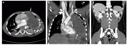


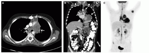
Figure 37.5 DICER1 syndrome. A and B: Six-year-old girl presenting with chest pain and respiratory distress. Family history was significant for thyroid nodules in the father, who underwent thyroidectomy during adolescence. Axial (A) and coronal (B) CT images demonstrate a large left Type III pleuropulmonary blastoma (white arrows). C: Sixteen-year-old female with history of vaginal/cervical rhabdomyosarcoma at 4 years of age. Coronal CT shows bilateral hypoattenuating cystic masses with enhancing septations (black arrows). Partial nephrectomies revealed cystic nephroma. Both patients tested positive for germ-line DICER1 mutations.

Only gold members can continue reading. Log In or Register to continue

Stay updated, free articles. Join our Telegram channel

Aug 25, 2016 | Posted by in ONCOLOGY | Comments Off on Rare Pediatric Tumors

Full access? Get Clinical Tree

Get Clinical Tree app for offline access