Diagnostic Pathology of Pediatric Malignancies


Diagnostic Pathology of Pediatric Malignancies


Timothy J. Triche

John M. Hicks

Poul H. Sorensen


OVERVIEW

Cancer in children and adolescents is distinct from that seen in adults and requires a different approach to diagnosis and treatment. The fact that most pediatric tumors are either of mesenchymal or of neuroectodermal origin, while the majority of adult cancers are of epithelial origin, sets childhood cancer apart.1,2,3,4 In addition, the primary treatment modality in adults is surgery, whereas in children, chemotherapy, in combination with surgery and radiation therapy, constitutes typical front-line therapy. The advent of multimodal therapy protocols brought with them the need to tailor treatment to specific tumor types and subgroups in children. With increasing recognition that even seemingly identical tumors are in fact distinctly different at the genomic level, this trend will only continue. As is occurring in the management of adult cancer, treatment is likely to increasingly focus on “druggable” targets that often occur across disparate tumor types. Receptor kinase inhibitors are popular therapeutics for adult cancer, as many adult tumors display EGFR on their surface and are thus amenable to EGFR inhibitors. In contrast, few childhood cancers display EGFR. Instead, they often display IGFR, rendering them candidates for IGFR inhibitors. Identification of specific gene targets in the treatment of childhood cancer is likely to become a focus of future diagnostics, where simple morphologic categorization will be insufficient to guide therapy with targeted agents like this. The diagnostic workup will likely include methods to document whether a given tumor is a candidate for therapy with a targeted agent. Wholesale categorization by conventional tumor type will likely be insufficient, as only subgroups within such a class will be amenable to such therapy, as illustrated by anaplastic lymphoma kinase (ALK)-amplified, mutated, or fused neuroblastoma, discussed later in this chapter. However, even in such cases, a singular focus on a single target and single agent therapy is unlikely to result in cure; most such patients will ultimately display drug resistance and relapse. The challenge now is to identify alternate signaling pathways in resistant tumors and develop multimodality therapy.5,6 Parallels to the history of the development of chemotherapy itself will not be lost on the readers of this text.7 In this regard, then, childhood cancer is similar to adult cancer: chemotherapy in both cases will increasingly utilize emerging diagnostic methods to identify molecular targets that warrant therapy with a given drug or drugs.

While outcomes have improved dramatically in children over the past several decades, to the current overall nearly 80% survival rate,8,9 there is a substantial toll on affected children, with an eightfold increased risk of serious illness related to treatment, and upward of 40% will experience significant morbidity or mortality due to sequelae of their therapy.10 It is of increasing importance to tailor therapy to meet the minimum needs for successful outcome, when adjusted for known risk factors. This is the reason for the widespread use of risk-adjusted therapy in childhood cancer protocols. Outcome in pediatric cancers is directly linked to treatment protocols developed for a specific cancer. Failure to provide an accurate diagnosis for enrollment on a tumor-specific protocol may result in a less-than-optimal outcome and prognosis. This critical link between a precise diagnosis (including not only diagnosis, but prognostic subgroup, as in leukemia, brain tumors, neuroblastoma, Wilms tumor [WT], and sarcomas) and optimal outcome places considerable demands on the pediatric pathologist to correctly classify tumors using a variety of diagnostic tools. These demands will only increase with the advent of targeted therapeutics that require target identification in the individual patient, as discussed earlier.

Over the past five decades, there has been a concerted and highly organized approach to childhood cancer treatment. The vast majority of children under the age of 16 years in the United States (>90%) are enrolled on NCI-supported Children’s Oncology Group (COG) cooperative group treatment protocols. These protocols have mandated tumor-specific specialized diagnostic methods and biologic marker assessment in conjunction with routine pathologic evaluation. The diagnostic challenge is often greater in childhood cancer, as many pediatric tumors lack morphologic evidence of differentiation that denotes histogenesis, or are of ambiguous lineage. These pediatric neoplasms are often referred to as the “small round cell tumors” (though many are not in fact small or “round,” but all are poorly differentiated or embryonal in character). Many of these tumors may be classified into appropriate diagnostic and treatment categories when a battery of immunohistochemical and molecular diagnostic procedures are applied.11 The recent availability of whole-genome molecular diagnostic techniques has created an unprecedented opportunity to provide important insight into highly specific, objective information on the histogenesis, pathogenesis, and unique genetic defects in a given tumor, and thus the potential use of targeted therapy, or at least less-aggressive conventional therapy with its attendant long-term adverse effects.12,13,14,15 Many of these recently developed molecular diagnostic techniques may not be familiar to all pathologists and oncologists and may be overlooked in the diagnostic workup of pediatric tumors. This chapter seeks to review many of the more useful of these methods with examples of their clinical applications.

With childhood cancer, a well-defined diagnostic workup and standardized diagnostic modalities are required to accurately classify these tumors. Certain tumor types present particular problems in diagnosis (like the “small round cell” or undifferentiated tumors) or in prognostic classification (leukemia, brain tumors, lymphoma, neuroblastoma [NB], rhabdomyosarcoma [RMS]). Certain clinical protocols for nonrhabdomyosarcomatous soft tissue sarcomas provide for risk-adjusted therapy based on identification of prognostic subgroups. The intent of this chapter is to provide useful diagnostic and prognostic guidelines for pediatric tumors. In addition, we discuss new diagnostic methods and technologies that improve diagnostic accuracy and enhance our understanding of certain childhood cancers. These are of particular interest, as they are not based on morphology. Rather, they typically depend on the identification of unique genomic defects that are intrinsic to many of these tumors. The quintessential example is the amplified MYCN gene identified in poor prognosis neuroblastoma over 20 years ago.16 Many more examples will be discussed later in this chapter. Methods, starting with optimal morphologic
diagnosis, the ultimate reference point for all diagnostic procedures, and continuing through immunohistochemistry (IHC), cytogenetics, fluorescence in situ hybridization (FISH), single nucleotide polymorphism (SNP) arrays, polymerase chain reaction (PCR), and whole genome methods, will be discussed in turn. The final portion of the chapter discusses the future implications of the increased availability of novel, often targeted, therapeutics and diagnostic methods on the necessary approach to pediatric cancer diagnosis. It should come as no surprise that diagnostic pathology in the 21st century will bear little resemblance to that in the 20th century.


Childhood Cancer Is Different from Adult Cancer: Age, Ethnicity, and Genetic Factors

Epithelial origin cancers (carcinomas) are unusual in children but common in adults. In contrast, retinoblastoma is associated with young children and unknown in adults.17 It is important to remember that childhood cancer bears little resemblance to adult cancer, as demonstrated by the childhood tumor types illustrated in Figure 8.1. The common carcinomas (breast, lung, colon, prostate) dominate the adult cancer experience but are rarely seen in children. In contrast, mesodermal-derived tumors (leukemia, lymphoma, and sarcoma) and neural tumors (brain and NB) dominate childhood cancer. Pediatric and young adult age groups share certain tumors, such as the Ewing sarcoma family of tumors (ESFTs), osteosarcoma (OS), synovial sarcoma (SS), and RMS.4 These tumors possess the same morphology in both the pediatric and adult populations and are treated with similar protocols.


Age Versus Tumor Type

Many pediatric tumors are associated with specific age ranges (Fig. 8.2). Overall cancer incidence is highest just after birth and declines to its lowest point by 10 years of age. This reflects in utero oncogenesis and growth for many of these blastemal tumors. These neoplasms represent embryonal tumors that resemble primordial cells from specific organ systems, such as RMS (skeletal muscle), WT (developing kidney), and NB (sympathetic peripheral nervous system). After the first decade of life, there is a linear increase in nonembryonal cancers that extends into adulthood. These tumors are distinct from childhood embryonal tumors and common adulthood carcinomas. Although environmental and lifestyle factors play major roles in adult carcinomas, these factors are of no known importance in childhood tumor oncogenesis.


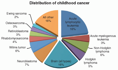
Figure 8.1 Distribution of childhood cancer. The first four slices of this pie chart account for approximately one-third of childhood malignancy and are accounted for by acute lymphocytic leukemia (approximately one-fourth) and acute myelogenous leukemia, non-Hodgkin lymphoma, and Hodgkin disease (the remaining three wedges). The next most common category is brain tumors, which account for approximately 20% of all childhood tumors. This is followed by neuroblastoma and Wilms tumor, which are roughly equal in incidence. Rhabdomyosarcoma, retinoblastoma, osteosarcoma, and Ewing sarcoma account for the remaining four defined wedges of the pie chart. All other tumors account for less than 20% of all childhood tumors in aggregate. Notably absent from the previous is any significant incidence of carcinoma, the most common form of adult cancer.

Specific childhood tumors have distinct age predilections. Figure 8.3 illustrates this connection with peripheral neuroectodermal tumors, such as NB and the Ewing family of tumors (EFTs). Although these tumors affect different age ranges, they may have similar morphologic features that require nonmorphologic diagnostic methods to determine the precise diagnosis (IHC, FISH, and PCR; see later). The patient’s age alone may provide the clue that directs the diagnostic workup and avoids misdiagnosis.

Even within a specific tumor class, age plays an important role. The dominant types of RMS in children are the embryonal and alveolar types. The age at diagnosis between these RMS types is quite different (Fig. 8.4). In this figure, based on data from the Intergroup Rhabdomyosarcoma Study, with both tumor types plotted as a percent of total (y-axis) versus age (x-axis), embryonal rhabdomyosarcoma (ERMS), which is linked to skeletal muscle embryogenesis and development, has a peak incidence shortly after birth. In contrast, alveolar rhabdomyosarcoma (ARMS), which accounts for less than 25% of the total, has a bimodal age distribution quite distinct from ERMS. In older children and adolescents, the alveolar form predominates. This suggests a very different pathogenesis. A solid form of ARMS with the same cytology but lacking an overt alveolar pattern also exists and may be confused with the embryonal type. This has led to a misdiagnosis rate of about 20%.18 Conversely, over 25% of tumors with alveolar morphology lack a characteristic gene translocation and are best considered a form of embryonal RMS.19,20 Without appropriate use of sophisticated diagnostic methods, suboptimal treatment may occur for misclassified types of RMS, as the prognosis for embryonal tumors is much better than that for alveolar type.


Genetic Factors

Genetic factors, such as inherited gene defects, are not uncommon in childhood tumors.21,22,23 The prototypic example is familial versus sporadic retinoblastoma.24 Children with familial tumors possess a constitutional mutation in one RB1 gene allele. With mutation or loss of function of the remaining normal RB1 gene, the affected children develop bilateral and multiple tumors. Sporadic retinoblastoma does not occur bilaterally or as multiple tumors in children with nonfamilial sporadic mutations. Li-Fraumeni syndrome (germline p53 mutation) is another example in which many different tumor types can develop throughout life depending on when the normal p53 allele is lost or mutated.25,26 A similar inherited defect has been reported with ALK in familial neuroblastoma.27 More complex examples such as DICER1 mutations in disparate tumors like pleuropulmonary blastoma and embryonal rhabdomyosarcoma and a host of other tumors have also been reported.28 Perhaps most challenging are syndromes with multiple phenotypes and genotypes, where a single genotype-phenotype as seen in RB1is not observed.21,23 Such syndromes are likely to be more widely recognized in the future, with the advent of whole-genome analysis methods.

Ethnicity is an example of complex cancer susceptibility differences among tumor types. There is no classic Mendelian gene defect, as in retinoblastoma and Li-Fraumeni syndrome, ascribed to ethnicity. Yet to be discovered complex genetic traits are presumed to be responsible. The classic example of the role of ethnicity in tumorigenesis is illustrated with EFTs. This tumor is virtually unknown in individuals of African or (to a lesser extent) Asian descent (Fig. 8.5). In contrast, Ewing sarcoma is more common than osteosarcoma in children and young adults of northern European descent.2,29 Even more surprising, the tumor is most common in Spanish, Israeli (non-Arabic), and Polynesian (Maori, Hawaiian) populations. This has led to comprehensive genomic analysis of susceptibility factors, but a convincing genetic basis for this striking difference remains elusive.30,31 Epidemiologic studies
have documented other less-striking differences in tumor-type incidences among ethnic populations.4,32 Knowledge of a patient’s ethnicity can be a helpful adjunct in diagnosis, particularly with undifferentiated childhood tumors, such as an African American child with a differential diagnosis of NB versus EFT. Such a patient is far more likely to have NB, based on these criteria.


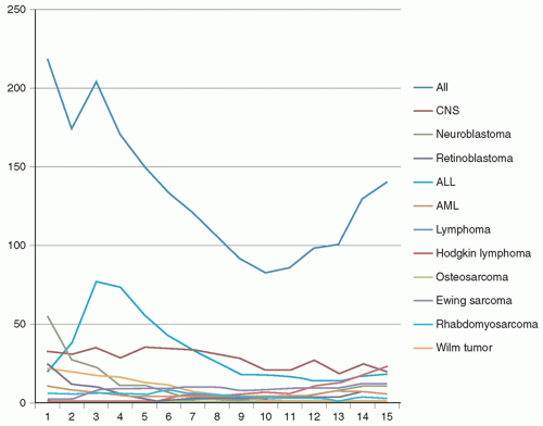
Figure 8.2 Childhood cancer: age incidence. That childhood cancer is largely embryonal in character is immediately evident from this chart of age incidence. The top line, which represents all forms of childhood cancer, shows a nearly linear increase from birth until approximately 10 years of age at which time the slope of the line reverses and begins to climb steadily toward adult-type incidences found later in life, largely due to the appearance of adult-type malignancies (not detailed here). The overall high incidence at birth is largely attributable to neuroblastoma, which generally presents in the first year or two of life. The large peak seen at approximately 3 years of age is due to the peak incidence of the most common form of childhood cancer, acute lymphocytic leukemia. Because of the lesser frequency, all the other tumors seem to be relatively constant in incidence (e.g., brain tumors, the next most common malignancy seen here), but in reality, striking differences in ages are found here as well.


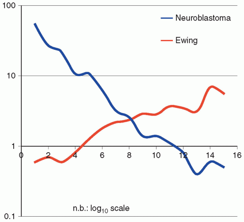
Figure 8.3 Neuroblastoma versus Ewing sarcoma: age-specific incidence. Although the two most common neural tumors in childhood are neuroblastoma and Ewing sarcoma, respectively, they are strikingly different in their incidence and character (age, incidence rate), despite their common phenotype. This is evident from this graph in which neuroblastoma is clearly a largely congenital tumor most common at birth and declining to extreme rarity by the age of 7, versus Ewing sarcoma with a nearly linear increase from birth when it is exceedingly uncommon to the age of 14 when it reaches a maximum in the pediatric age group. From this, one would assume that the genetic origins for the two phenotypically similar tumors are distinctly different. Note that the incidence data are plotted on a log10 scale.


Current Approach to Childhood Tumor Diagnosis

Pathology has been transformed by new diagnostic technology, instruments, and knowledge gained from advances in immunology, molecular biology, genomics, and proteomics during the past few decades. This has changed the methods used to arrive at a diagnosis. No longer is gross examination and light microscopy, in conjunction with clinical information, sufficient to determine a diagnosis that determines therapy, the ultimate purpose of diagnosis in the first place, in many if not most childhood cancers. Almost all childhood tumors require some combination of immunochemical, cytogenetic, FISH, and/or molecular genetic methods to achieve an accurate diagnosis or identification of a prognostic group.11,33,34 These tools have dramatically increased diagnostic precision but also have allowed for improved correlation of diagnosis with clinical behavior, response to therapy, and outcome. Newer genomic methods will further enhance clinically relevant diagnosis and lead to new concepts of tumor classification and targeted “individualized” therapy.35,36,37,38,39,40

Specific tumor types and their optimal diagnostic evaluation are discussed in detail elsewhere. A brief mention of specific examples will suffice to make the point. No child with suspected leukemia, lymphoma, or NB on a COG protocol will fail to have
ancillary diagnostic procedures performed to detect gene translocations, antigen expression, or gene amplification, respectively. The availability of advanced diagnostic procedures common to childhood cancer evaluation is unusual in adult cancer centers, a striking difference often overlooked by adult cancer diagnosticians. This is unfortunate because these advanced diagnostic procedures require special handling (e.g., fresh viable tumor tissue for short-term culture and cytogenetics, fresh frozen tissue for flow cytometry and molecular analysis) that are generally not part of the routine diagnostic workup for adult cancers. In many cases, local institutions lack the facilities to perform these advanced diagnostic studies. In this situation, tissue can still be handled according to prescribed protocols that ensure proper tissue handling and transportation to a central reference laboratory that performs the appropriate studies. The COG, which treats the majority of children with cancer in North America, maintains a central reference facility, the Biopathology Center, in Columbus, Ohio, which receives these specimens from the nearly 250 participating institutions, and offers a number of these advanced diagnostic procedures, as well as central pathology review for many of the treatment protocols. If the primary pathologist is not familiar with the proper protocol or diagnostic procedures required, the opportunity to provide additional diagnostic, treatment, and prognostic information derived from specialized tests may be lost. It is useful to have a well-defined protocol and action plan for childhood tumor diagnosis to avoid loss of critically needed diagnostic information.


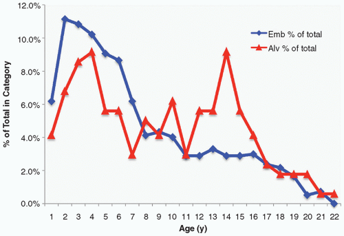
Figure 8.4 Rhabdomyosarcoma: age-specific incidence. Another childhood neoplasm with a striking variation in incidence by age is childhood alveolar rhabdomyosarcoma, which although less common than embryonal rhabdomyosarcoma is nonetheless an important source of fatality in this disease because of its worse prognosis. When plotted as percentage of total cases, alveolar rhabdomyosarcoma shows a bimodal age incidence, the first mode roughly corresponding to the peak age incidence of embryonal rhabdomyosarcoma, and the second mode with no parallel increase in embryonal rhabdomyosarcoma seen at approximately 14 years of age. Studies have suggested that the two modes are in fact genetically distinct as well; the first mode includes mostly PAX7-FOXO1A tumors, while the second is largely PAX3 (see text).


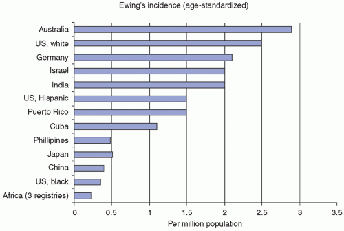
Figure 8.5 Ewing sarcoma family of tumors: ethnicity versus incidence. U.S. SEER data from the National Cancer Institute documents striking differences in the incidence of Ewing sarcoma family of tumors among different ethnic groups. Australia reports the highest incidence on a per-million-population basis, whereas three different African registries report the lowest incidence reported anywhere in the world. U.S. blacks and other Asian populations also have an extremely low incidence of Ewing sarcoma. These observations have strongly suggested a genetic component to Ewing susceptibility, but the genetic predisposing factors have not yet been identified. (Data courtesy of Jonathan Buckley, MD, PhD, Preventive Medicine, Keck School of Medicine, USC.)

With routine handling of childhood tumor tissue as prescribed by the COG (Fig. 8.6), optimal evaluation of childhood tumors will be realized. Many pediatric tumors will not be handled by pediatric pathologists familiar with these protocols. It is important for the clinician to discuss the importance of ancillary tests, the availability of reference laboratories, and tumor protocols with the responsible pathologist prior to tumor biopsy or resection. The diagram (Fig. 8.6) illustrates the single most important principle: Tissue must not be placed into fixative in the operating room! This single oversight is responsible for most lost diagnostic opportunities. This is also the reason why tissue may not be available for many molecular diagnostics and biomedical research. Fixed tissue
has limited utility for many molecular genetic diagnostic procedures and may limit conventional diagnostic techniques, such as immunocytochemistry and PCR. In an attempt to promote nonroutine tissue handling, many childhood cancer protocols specifically request submission of fresh frozen or even viable tumor tissue. Despite these efforts, no protocol ever achieves perfect compliance with these protocol requirements. As a result, many of the necessary diagnostic procedures have been (and continue to be) adapted to formalin-fixed paraffin-embedded (FFPE) tissue. FISH on FFPE tissue for MYCN amplification instead of frozen tissue for DNA extraction and Southern blotting in NB is one example. More recently, so-called whole-genome and next-generation sequencing methods (discussed later in this chapter) have been adapted for use with FFPE tissue.41,42,43,44,45,46,47 The simple fact that the majority of tumor tissue is formalin fixed and embedded in paraffin blocks has forced the development of these methods, to great advantage for pathologists, who otherwise would be precluded from using molecular methods on the majority of cases.


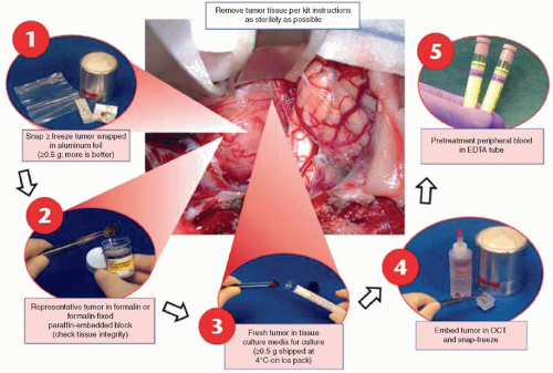
Figure 8.6 Tissue handling diagram. Tissue for optimal pathologic and biologic studies requires specialized handling as summarized in this chart. This diagram, which has been used by participating institutions in the Children’s Oncology Group for over 15 years, has been widely adopted and led to significant improvements in diagnostic accuracy in childhood cancer. Note that fresh tissue, not fixed, is required at the outset, for steps 1, 3, and 4. A pretreatment peripheral blood (for DNA from buffy coat and protein from plasma) is required for step 5, and will allow comparison of tumor versus normal DNA, an increasingly important part of the cancer diagnostic workup (see Molecular Diagnostics section). (Diagram created by Steve Qualman, MD, for Children’s Oncology Group.)

The second stage of tissue handling (Fig. 8.6) is simply precautionary. If tissue is divided into multiple forms for specific use as needed in subsequent diagnostic procedures, nothing is lost. If not, the opportunity to perform diagnostic procedures will be lost. Failure to set aside fresh tissue precludes fluorescence-activated cell sorting (FACS—flow cytometry) analysis, which is necessary for the diagnosis of leukemia and lymphoma. Tissue placed in transport or culture medium for cytogenetics and establishment of tumor cell lines is important. Frozen tissue is perhaps even more important, as all manners of molecular diagnostics (particularly for RNA) are now possible. Small tumor portions in cryopreservative matrix (OCTTM) are invaluable for a “second look” to compare pretreatment versus posttreatment tumor for chemosensitivity and new diagnostic methods, such as gene expression profiling.

If these procedures are followed, all diagnostic modalities available currently and in the foreseeable future may be employed. Because tissue is not always triaged appropriately, diagnostic techniques that provide similar information from fixed and processed tumor tissue have been developed. Cytogenetic analysis for a chromosomal translocation can now be partially substituted by PCR, FISH, and chromogen in situ hybridization (CISH) analyses48 and SNP arrays.49 Gene amplification can be detected by FISH. Even next-generation sequencing (NGS) methods for mutation detection and RNA expression have become available, with certain limitations.41,42,43,44,45,46,47 Antigen retrieval methods allow monoclonal antibody-mediated detection of scant antigens otherwise detectable only by FACS. These alternative methods sometimes contradict the diagnosis determined by conventional methods and may themselves be open to interpretation: What if the PCR is negative? What if the FISH analysis has a high background due to formalin-induced fluorescence that obscures the signal? What if both are performed but the results disagree with one another? What if the immunocytochemical results are equivocal or contradict the molecular genetic data? It is possible to render an appropriate diagnosis in most cases, but it is clear that adherence to a standardized tissue-handling protocol (Fig. 8.6) can help avoid these pitfalls.


The rapid advances in our understanding of the origins and evolution of cancer are largely dependent on the availability of fresh frozen tumor tissue and increasingly with paired normal tissue. The last few years have witnessed remarkable advances in whole-genome DNA and transcriptome sequencing of cancer (discussed in detail later in this chapter), but all such studies benefit from the availability of fresh or frozen tumor tissue. There is an emerging worldwide shortage of suitable tumor specimens to study due to historical methods that require formalin fixation and paraffin embedding. While this remains the foundation of cancer diagnosis, it is no longer sufficient. Clearly, much wider recognition of the need for fresh or frozen tumor tissue will be necessary to meet these emerging needs. At the same time, it is almost certain that there will never be 100% compliance for such requests, which requires the development of molecular testing applicable to FFPE tissue. The general history of molecular biomarkers is to first identify key genomic alterations in optimally handled (fresh or frozen) tissue, then develop testing for these same biomarkers that can circumvent the limitations of FFPE tissue. However, if there is insufficient optimally processed tissue, the reproducibility and utility of such biomarkers will be in doubt. Further, even FFPE tissue must be uniformly processed to obtain consistent results with this universally available diagnostic tissue. Variable time in tissue fixatives, the use of unbuffered fixative, unusual (i.e., heavy metal containing) fixatives, adverse storage conditions, and prolonged exposure to air of unstained sections intended for molecular analysis can each alter the result and introduce uncontrolled variability of the result. This is a problem that has plagued even the rather simple application of prognostic biomarkers for breast cancer.50,51,52 It is even more so a problem for childhood cancer.

A final comment on specimens is appropriate here. While our focus so far has been on primary tumor specimens, in some cases paired with matched normal tissue, there is an emerging need for longitudinal specimens on the same patient. This takes many forms, from re-biopsy of residual or recurrent tumor, to methods that detect circulating tumor cells or cell-free DNA and RNA. One of the challenges pathologists will face in the future is detection of genetic alterations that predict treatment resistance and metastasis. Feasibility of comparing primary tumor with recurrent or persistent circulating tumor cells or even cell-free DNA/RNA is already established at a research level for several cancers.53,54,55,56,57 It s highly likely that this will become a standard methodology for monitoring patient response on clinical trials as well as choice of secondary therapy, as this approach is central to the emerging “precision” or “personalized” medicine paradigm that itself will likely become standard of care in the future.


TABLE 8.1 Top 10 Diagnostic Methods for Tumor Diagnosis


Method


Comment


1.


Light microscopy


Mandatory for all cases


2.


Immunohistochemistry


First-choice ancillary diagnostic; widely used


3.


Molecular genetic: RT-PCR


Most common molecular Dx; now routine in most pediatric hospitals


4.


Molecular genetic: FISH


Rapidly supplanting cytogenetics for many cases with tumors with known genetic abnormalities


5.


Molecular genetic: ISH


Specialized use to date (nontumor; EBV typical)


6.


Special stains


Still needed in limited number of cases; useful, expensive


7.


Electron microscopy


Very limited use to augment light microscopy, but especially useful in undifferentiated pediatric soft tissue tumors (SRCTs)


8.


Cytogenetics


Needed when no suitable FISH probes available or to identify new translocations and karyotypic prognostic factors


9.


Molecular genetic: SKY, CGH


SKY a useful diagnostic; CGH and SNPs for LOH


10.


Molecular genetic: DNA sequencing


Rapidly assuming a critical role in diagnosis and identification of cases amenable to targeted therapy


CGH, comparative genomic hybridization; Dx, diagnosis; FISH, fluorescence in situ hybridization; ISH, in situ hybridization; LOH, loss of heterozygosity; RT-PCR, reverse transcriptase polymerase chain reaction; SKY, spectral karyotyping; SNP, single nucleotide polymorphism.


Interplay of Multiple Diagnostic Techniques in Tumor Diagnosis

The reason for the multiple diagnostic approaches (Fig. 8.6) is that no method alone will suffice for all tumors. Childhood cancer diagnosis is better viewed as a contingency tree. If the initial result suggests a certain diagnosis, then appropriate ancillary diagnostic tests are necessary to confirm the initial diagnostic impression with a high degree of certainty. In most pediatric hospitals, childhood cancers are handled as illustrated in Figure 8.6. After hematoxylin and eosin (H&E) slides prepared from FFPE tissues are examined, the diagnosis is either obvious or requires additional studies. In many cases, tumor protocols mandate special studies, such as MYCN (NMYC) and ploidy studies in NB. Tumor tissue is submitted either at the outset if the diagnosis is equivocal or after the initial studies document an expected or equivocal diagnosis. In the latter instance, failure to follow the tissue-handling guidelines (Fig. 8.6) may seriously compromise diagnosis and oncologic management. If diagnostic uncertainty remains, several methods are available that will lead to a specific diagnosis (Table 8.1). Note that these methods are employed judiciously, such that only the rare diagnostic dilemma will require most or all of these methods. The availability of these methods, especially molecular genetics, has led to accurate diagnoses that are linked to therapeutic protocols.


CHILDHOOD TUMOR DIAGNOSIS


Character of Childhood Cancer

The typical small round cell tumor (SRCT) of childhood either lacks definitive morphologic evidence of lineage or histogenesis or the lineage is ambiguous (spindle cell tumors, undifferentiated tumors). This ambiguity, coupled with the need for definitive diagnosis to establish a treatment regimen, invokes the use of a variety of diagnostic methods. For example, the diagnosis of NB is not sufficient alone. It is necessary to indicate the prognostic subgroup class, as defined by the International Neuroblastoma Study Group (Fig. 8.7).58,59 The copy number status of the MYCN
oncogene, a member of MYC family genes, is tested by FISH and found to be amplified in approximately 20% of NB cases (Fig. 8.8) Amplified MYCN is transcribed and translated, and expressed as an excessive amount of MYCN protein. MYCN protein then forms a heterodimer with MAX (MYC-associated factor X) protein and activates various downstream genes related to cell proliferation, differentiation inhibition, and apoptosis, etc., and results in aggressive clinical behavior.60,61 Recently, MYC (aka C-myc) protein expression has been detected immunohistochemically in some of the undifferentiated NB tumors, especially when the MYCN gene is not amplified. Both MYCN and MYC protein-expressing tumors are often associated with prominent nucleolar formation,62 the putative site of RNA synthesis and accumulation (Fig. 8.9), and patients with those MYC (both MYCN and MYC)
protein-driven NB tumors seem to have a very poor clinical outcome.63 DNA ploidy, 1p LOH, and 11q LOH are also important for diagnosis, treatment, and prognosis.64 SNP arrays have been employed to detect prognostic DNA copy number variation on chromosomes 1, 10, and 17.65,66,67 Each of these factors plays an integral part in determining whether a child with NB will be placed on a high-risk, intermediate-risk, or low-risk protocol. More recently, ALK gene mutations/overexpression, ATRX gene mutation, and LIN28B mutation/aberration have been noted to affect prognosis, independently of other factors.68,69,70 Most recently, a series of reports focusing on NB cells and their microenvironment have been published, which noted that, like other neoplastic diseases, cross-talk between tumor cells and nontumor cells, such as tumor-associated macrophages (TAMs), plays a significant role in promoting metastatic progression of NB tumors.71,72


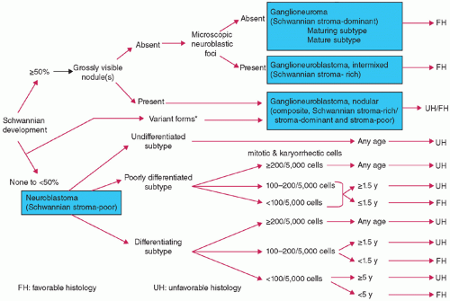
Figure 8.7 International Classification System for Neuroblastoma Prognosis and Diagnosis. Flowchart for prognostic evaluation: There are two groups for the final evaluation of pNTs: they are favorable histology (FH) and unfavorable histology (UH). Prognostic evaluation according to the International Neuroblastoma Pathology Classification is applicable to only those tumors (either primary or metastasis) that are biopsied or surgically excised prior to chemotherapy/irradiation therapy. Those cases with difficulty in making prognostic evaluation can be grouped into “Not Evaluable” category with some note such as “due to a limited amount, limited quality, etc.” Surgical pathology report for the cases whose tumor tissue is obtained after chemotherapy/irradiation therapy should have a clear statement of “Status Posttreatment” in their diagnosis line. (Courtesy: Dr. Hiroyuki Shimada, Children’s Hospital Los Angeles.)


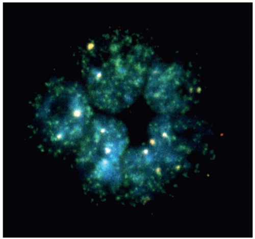
Figure 8.8 MYCN amplification in neuroblastoma detected by fluorescence in situ hybridization. A touch preparation of a neuroblastoma was incubated with a fluorescent probe for MYCN. Here one can readily see hundreds of signals in a tumor rosette, each representing at least one copy of MYCN. This clearly documents MYCN amplification in these tumor cells, denoting a poor prognosis group (MYCN amplified).


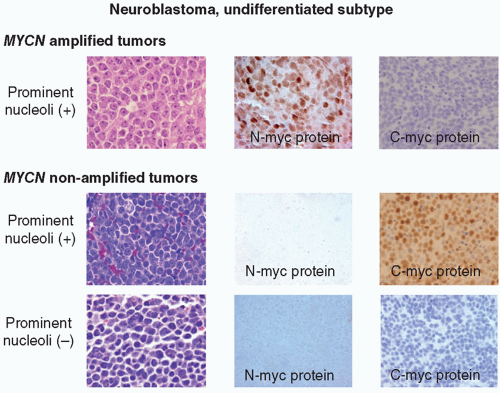
Figure 8.9 Neuroblastoma morphology and MYC protein expression. MYCN-amplified undifferentiated NB tumors often have prominent nucleolar formation and N-myc protein expression. Among MYCN nonamplified undifferentiated NB tumors, those with prominent nucleoli tend to express C-myc protein, and others with typical salt-and-pepper nuclei are negative for both N-myc and C-myc protein expression. (Figure kindly supplied by Dr. Hiroyuki Shimada, op cit.)


TABLE 8.2 Differential Diagnosis of Pediatric Bone and Soft Tissue Solid Tumors


A. Small Round Cell Tumors


  1. Peripheral primitive neuroectodermal tumors (Ewing family tumors)


    • Ewing tumor (classical, atypical, peripheral primitive neuroectodermal tumor)


    • Askin tumor (malignant small cell tumor of the thoracopulmonary region)


    • Malignant ectomesenchymoma


    • Biphenotypic sarcoma


  2. Rhabdomyosarcoma (RMS)


    • Alveolar RMS


    • Embryonal RMS


    • Undifferentiated sarcoma (currently in process of reclassification into nonrhabdomyosarcomatous soft tissue tumors)


  3. Neuroblastoma


  4. Desmoplastic small round cell tumor (DSRCT)


  5. Lymphoma (B-Cell, T-Cell, Null)


    • Anaplastic large cell lymphoma


    • Burkitt’s lymphoma


  6. Clear cell sarcoma of soft parts (malignant melanoma of soft tissues)


  7. Small cell osteosarcoma


  8. Extrarenal monophasic Wilms tumor


  9. Extrarenal rhabdoid tumor


  10. Extraskeletal myxoid chondrosarcoma


B. Spindle Cell Tumors


  1. Congenital infantile fibrosarcoma (CFS)


  2. Adult-type fibrosarcoma (ATFS)


  3. Fibromatosis


    • Infantile fibromatosis (including aggressive fibromatosis)


    • Other forms of fibromatosis


  4. Myofibromatosis (including hemangiopericytoma-like variant)


  5. Synovial sarcoma (SS)


  6. Pleomorphic undifferentiated sarcoma (malignant fibrous histiocytoma-MFH)/primitive sarcoma NOS


  7. Spindle cell rhabdomyosarcoma


  8. Malignant peripheral nerve sheath tumors (including malignant schwannoma, triton tumor)


Small Round Cell Tumors of Childhood

SRCTs (Table 8.2) of childhood refer to the generic “blastic” or “stem cell-like” histopathologic appearance of pediatric tumors that do not declare their histogenesis on routine microscopic examination. The histopathology of the most common SRCTs (EFTs, RMS, NB, lymphoma) is illustrated in Figure 8.10. These tumors are often indistinguishable from each other microscopically. As a
group, they have a primitive or embryonal appearance, often present in misleading clinical locations like bone marrow metastases from an occult primary, and lack specific morphologic features that allow for a precise diagnosis without ancillary methods.73,74


Figure 8.10 Histopathology of typical small round cell tumors. Historically, Ewing sarcoma (A), rhabdomyosarcoma (B), neuroblastoma (C), and lymphoma (D) were difficult to distinguish from one another, particularly when undifferentiated rhabdomyosarcoma or neuroblastoma was encountered, as illustrated here. None of the four tumors show light microscopically discernible evidence of differentiation and therefore histogenesis, which is critical to a diagnosis by light microscopy.

Because these tumors cannot be distinguished readily from one another, other diagnostic methods must be used to establish a reliable diagnosis. SRCTs serve as a model for demonstrating the benefits of a multimodal approach to tumor diagnosis. At a morphologic level, electron microscopy (EM) historically provided evidence of histogenesis among the most common SRCTs (EFTs, NB, lymphoma, RMS) that was not evident by light microscopy. However, ancillary techniques that are not dependent on morphology, most especially IHC, are particularly well suited for providing a precise diagnosis and for prognostic information with these undifferentiated or poorly differentiated tumors, and have correspondingly largely replaced EM. Even more informative methods have emerged in the past few years based on molecular genetic techniques.41,43,44,46,65,66,67,73,75,76 Increasingly, these sophisticated methods offer the potential to confirm the diagnosis while predicting biologic behavior, metastatic potential, tumor recurrence, and sensitivity or resistance to chemotherapy and radiotherapy.77,78,79

A common belief espoused with the advent of “sophisticated” diagnostic methods was that “conventional” diagnostic methods (routine H&E studies) would be eliminated in favor of these methods. That has not proved to be the case. No other method besides routine histopathology returns as much information for so little expenditure of time and money. Further, no “sophisticated” test is reliable if the assay is not performed on representative tumor tissue. That cannot be determined without histopathologic examination of the specimen, a fact that has become all too evident in recent genomic studies of cancer that have documented pervasive failure to account for percent tumor content, or even any tumor at all, in archived specimens that were not quality assured by histopathologic examination.47,80,81 Routine microscopic examination provides information that directs the utilization of appropriate ancillary tests. The discussion that follows is based on the diagnostic workup commencing with routine microscopic evaluation, followed by “more sophisticated” diagnostic methods.


Diagnostic Methodologies

The basic approach to tumor diagnosis employs a variety of diverse methods (Table 8.1).33,82,83 Representative tissue is submitted for formalin fixation, tissue processing, paraffin embedding, and routine H&E staining of the resulting tissue sections. After light microscopic examination of the routinely stained tissue sections,
a differential diagnosis is formulated and a series of special studies are selected to provide a definitive diagnosis. The most commonly used ancillary diagnostic method is immunocytochemistry. Either concurrent with or subsequent to immunocytochemical study, special histochemical stains (periodic acid-Schiff [PAS], reticulin, trichrome), EM, and molecular studies may be initiated. Findings from all of these studies are reviewed and interpreted in concert with the clinical history and diagnostic imaging findings. With the vast majority of SRCTs, this multimodal approach will yield a precise and definitive diagnosis that directs further surgical intervention, oncologic management, follow-up, and prognosis. Using these extensive and coordinated methods, it is rare that diagnoses are modified by additional testing, consultative review by pathologists at other institutions, or by expert pathologist reviewers for COG protocol studies.

Although most tumors require only a few of the procedures listed in Table 8.1, some tumors require all available testing modalities to come to a definitive diagnosis.33,83,84,85 The pathologist plays a critical role in ensuring that adequate tissue is obtained for all diagnostic testing modalities, COG protocols, and institutional research purposes. Appropriate triaging of tumor tissue (Fig. 8.6) by the pathologist is necessary to make certain that representative viable tumor tissue is available for routine light microscopy, immunocytochemistry, EM, cytogenetics, molecular studies, tumor prognostic markers, and COG protocol studies. Fresh tumor tissue must be sent from the operating suite to the anatomic pathology grossing station in a sterile container with saline. The tissue must not be exposed to any fixative. A general schema for handling fresh sterile biopsies or resections of pediatric tumors is to (a) cryopreserve tissue at -70°C with and without cryopreservation matrix for molecular studies; (b) place tumor portions in tissue culture media for cytogenetics, spectral karyotyping (SKY), and comparative genomic hybridization (CGH); (c) perform cytologic imprints (touch preparations) for FISH, CISH, and chimeric translocation detection; (d) fix finely cubed tissue in glutaraldehyde for EM; and (e) submit representative tissue in 10% buffered formalin for routine light microscopic, immunocytochemical, and histochemical analyses. The pathologist is also responsible for determining adequacy of the tumor specimen with respect to amount of tumor tissue submitted, whether viable tumor tissue is submitted, and ensuring that indeed tumorous tissue is submitted and not adjacent reactive tissue. At times, this will require the pathologist to refer to the COG protocols and discuss tissue requirements with the institutional COG clinical research associate and oncologist. For optimal handling of tumor tissue, communication among the surgeon, oncologist, and pathologist is necessary to make certain that tissues are submitted for all required ancillary studies for a particular tumor type. Finally, the pathologist may perform a frozen section or touch preparations on tumorous tissue to determine a preliminary diagnosis for triaging and immediate surgical management purposes. If the tissue procurement scheme (Fig. 8.6) is carried out with pediatric solid tumors, adequate tumor material will be available for diagnosis, oncologic management, prognostic purposes, and oncologic research in the vast majority of cases.


Light Microscopy

FFPE tumor tissue is the most widely studied of all tissues for diagnosis and protocol studies. Attention to optimal fixation, processing, and tissue sectioning are important to allow for optimal light microscopic, immunocytochemical, and histochemical evaluation. This formalin-fixed tissue is also available for DNA extraction to identify tumor-defining translocations by reverse transcriptase polymerase chain reaction (RT-PCR), and tissue can be recovered and subsequently processed for EM if necessary. When the tissue is properly formalin-fixed and processed, light microscopy will provide overwhelming evidence for the precise diagnosis in the majority of cases via immunocytochemical, histochemical, and ultrastructural methods. The need for optimal fixation, processing, and tissue sectioning is illustrated in Figure 8.11. Initial frozen sections of the tumor tissue were of poor quality and could not be interpreted adequately (Fig. 8.11A). With proper fixation, sectioning, and staining of the same tumor tissue block, diagnostic features defining the tumor became readily apparent (Fig. 8.11B).

Optimal fixation is necessary for routine light microscopy, IHC, and DNA extraction for molecular and FISH studies for identifying tumor-defining translocations and prognostic markers (MYCN). Representative portions of the resected tumor (1 to 3 mm in thickness, ˜1.5 cm in maximum width and length) should be submitted for formalin fixation. Tissue portions greater than 3 mm in thickness and greater than 1.5 cm in maximum width and length will not be optimally fixed, resulting in poor tissue preservation with lack of tissue antigenicity and degradation of DNA and RNA. Tissue fixation should be carried out with fresh, commercially available 10% buffered formalin for a minimum of 4 hours and a maximum of 24 hours. Following automated tissue processing to allow for tissue dehydration through graded alcohols and xylene and paraffin permeation, the tissue blocks should be made by embedding the tumor tissue portions in low melting point paraffin, to protect and preserve the antigenicity of the tissue against excessive heat. The production of tissue sections mounted on coated glass slides requires histotechnologists with the ability to cut wrinkle-free 3-micron-thick tissue sections. Placing the tissue sections onto coated glass slides allows for adherence of the tissue sections to the glass infrastructure and allows for immunocytochemistry, in situ hybridization, FISH, in situ PCR, and laser capture microscopic techniques to be performed without loss of tissue sections. The final step in producing high-quality and diagnostically acceptable glass slides of tumor tissue is the staining process. Quality assurance for producing high-quality routine H&E, histochemical, immunocytochemical, and in situ hybridization staining should be an ongoing process in all anatomic pathology laboratories providing diagnostic services with constant comparison with standardized control tissues.


Immunocytochemistry

Remarkable progress has been made in the development of immunocytochemical antibodies, which are capable of defining undifferentiated neoplasms and allowing for definitive diagnosis.33,85,86,87,88,89,90 Rapid development and availability of antibodies of recently discovered antigens identified by genomic and proteomic studies of tumors have become a reality in tumor diagnostics. This has resulted in immunocytochemistry being the most frequently employed diagnostic method for SRCTs (Fig. 8.12A to C).33,86,87,90 In addition, many different antigen-retrieval methods have been identified that allow for unmasking and eliminating formalin cross-linking of tumor antigens in paraffin-embedded, formalin-fixed tissue.33,86,87,90,91 The use of enzymatic digestion (protease, pepsin, trypsin), regulation of pH, antigen-retrieval solutions (citrate buffer, Tris-HCl, EDTA-NaOH), and various methods of heat treatment (microwave, pressure cooker, controlled gentle steaming) have been thoroughly explored and refined to optimize antigen retrieval for numerous antibodies. Technical guidelines for individual antibodies are well delineated by the commercial providers of immunocytochemical products, and many of these companies have developed antigen-retrieval “kits” to produce optimal results with specific antibodies. Antigen retrieval from formalin-fixed tissue has made it possible to achieve immunoreactivity similar to that previously obtained only with immunocytochemistry performed on frozen tissue. This has occurred while reducing background considerably and decreasing false-negative results. With each antibody lot received, it is necessary to determine the concentration of antibody (titration) necessary to provide diagnostic information, while eliminating background and false-positives. Monitoring of accurate immunoreactivity requires that known control tissue be run parallel or on the same glass slides as the patient’s tissue. This technology is readily available and may be performed either manually or with automated instruments in a reproducible manner using FFPE tissues.


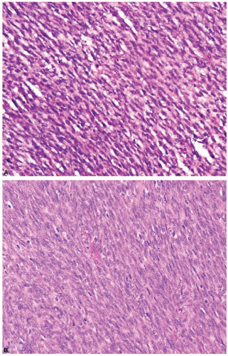
Figure 8.11 Poor (A) and well-done (B) histology of a typical soft tissue tumor. The first panel (A) illustrates the appearance of a soft tissue neoplasm in a 9-month-old infant. The first material is from the frozen section; the second, after formalin fixation and paraffin embedding. The correct interpretation only became apparent with proper fixation and embedding as seen in Panel B, in which the correct identity as congenital fibrosarcoma became evident. Many diagnostic problems in childhood tumor interpretation benefit from optimal quality histology and can be the useful and necessary first step in the choice of other ancillary diagnostics to confirm the impression gleaned from careful scrutiny of a well-prepared hematoxylin and eosin section. (Photomicrographs kindly provided by Dr. Larry Wang, Children’s Hospital, Los Angeles.)

There are numerous antibodies that are available on a commercial and research basis; however, an exhaustive discussion of the specifics of antibodies and immunoreactivity with tumors is beyond the scope of this chapter. Tables 8.3, 8.4, 8.5 provide detailed lists of antibodies that are more commonly employed in pediatric pathology services, along with their common tumor association.18,33,85,87,88,89,90,92,93,94,95,96 The immunoreactivity of an antibody with a specific tumor and the pattern of staining are important (Figs. 8.12A to C). Antigen localization in a tissue depends on several factors, most notably the cellular disposition of the antigen. Three basic patterns are observed: nuclear, cytoplasmic, and cell surface (membranous). Certain tumors may be included or eliminated from consideration based on the pattern of staining. For example, cytoplasmic membrane immunoreactivity with CD99 is characteristic for EFTs and lymphoblastic leukemia/lymphoma (Tables 8.3, 8.4, 8.5). Many different tumors may immunoreact with CD99 in a cytoplasmic or nuclear pattern but not with a cytoplasmic surface membrane pattern. Another example is myogenin immunoreactivity.18,33,85,94,96 This antibody identifies RMS when a nuclear pattern is present; however, diffuse and intense cytoplasmic myogenin staining is found with mast cells. Within a poorly differentiated tumor, mast cells may be frequently present. Cytoplasmic myogenin immunoreactivity of mast cells in such tumors may result in a mistaken diagnosis of RMS. The pathologist has to be aware of and be up to date with the characteristic immunoreactivity and staining patterns of numerous antibodies with a wide variety of tumors to provide accurate diagnoses.

With any tissue to be preserved for possible immunocytochemistry study, attention must be paid to the length of formalin fixation.33,83,85 For tumor antigen integrity, formalin fixation (10% buffered formalin) should be for a minimum of 4 hours and a maximum of 24 hours. Exposure to formalin for a shorter or longer period of time results in significant loss of tumor antigens, making immunocytochemical studies difficult or impossible.

Several antigen-retrieval protocols have been developed that allow for unmasking tumor antigens in FFPE tissue.85,86,90,91,97,98
An antigen-retrieval solution (10 mM citrate buffer at pH 6.0; 100 mM Tris-HCl at pH 8.0; 1 mM EDTA-NaOH at pH 8.0; 5% urea) and heat treatment (microwave, microwave and pressure cooker, autoclave, pressure cooker, water bath, steamer, thermal cycler) allow reversing protein cross-linking by formalin fixation. Optimal antigen-retrieval intensity from FFPE tissue has been achieved with heat treatment at temperatures of 10°C for 20 minutes, 90°C for 30 minutes, 80°C for 50 minutes, and 70°C for 10 hours. After antigen retrieval, enzyme digestion (trypsin, pronase, proteinase K, pepsin) is usually not necessary prior to reaction of the tissue with the antibody but may be needed for certain antibodies. For most antibodies, heat treatment for 20 to 30 minutes at 100°C with either citrate buffer at pH 6.0 or Tris-HCl buffer at pH 7.0 to pH 8.0 are effective for antigen retrieval. The antibody manufacturer provides general guidelines; however, it is necessary to determine the optimal conditions for the antibody in each institution’s immunocytochemistry laboratory. In addition, monitoring for accurate immunoreactivity requires that known control tissue be run parallel, or on the same glass slide as the patient’s tissue, and under the same antigen retrieval conditions. Immunoreactivity may be adversely affected if the tissue sections are prepared sometime in advance of staining. Tissue sections should be prepared and undergo processing for immunostaining without delay to avoid antigen degradation. It is also possible to preserve antigenicity if the tissue sections on glass slides are stored at -70°C until immunocytochemistry is performed.


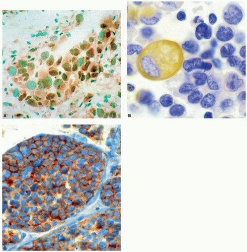
Figure 8.12 Immunocytochemistry by cellular localization. Immunocytochemistry is widely utilized on a daily basis in surgical pathology of adult and childhood tissues. In the case of childhood cancer, many specific classes of antigens are detectable. Illustrated here are three categorical examples: (A) an example of nuclear staining for a transcription factor, MyoD, widely used to detect extremely undifferentiated childhood rhabdomyosarcoma; (B) vimentin, ubiquitous intermediate filament found in all mesenchymal tissues and their tumors counterpart. The staining is localized to the cytoplasm. This antigen is most used to verify antigenic preservation sample and is not specific for any given tumor; (C) surface antigen detection, MIC2 (CD99) in Ewing sarcoma. Here apparent cytoplasmic as well as cell surface localization is detectable. Such antigens routinely show surface staining that is difficult to photograph but easily detected by focusing up and down through the sample under the light microscope. Many of these antigens are also quite labile and not readily detected in routinely processed tissue.

The immunocytochemical results require critical assessment to ensure diagnostic accuracy. These issues have been addressed by many authors in numerous publications, but can be summarized here.18,33,83,85,87,88,89,90,92,93,94,95,96 Possible artifacts include the following:


TABLE 8.3 Immunocytochemical Antibodies of Diagnostic Importance in Childhood Tumors


Antibody


Antigen/CD


Utility


General


Anti-vimentin


Vimentin


Generic anti-intermediate filament; quality control; reactive with most mesenchymal cells


Hematopoietic


Anti-LCA


CD45 (all subunits)


Common leukocyte antigen; no lineage specificity, unlike CD45RA, RB, and RO


Myeloperoxidase


Myeloid cells and tumors


Muramidase


Lysozyme


Myeloid cells and tumors, histiocytic disorders, macrophages, fibrohistiocytic tumors


LeuM1


CD15


T cells, Hodgkin disease


Ki-1


CD30


Hodgkin disease, anaplastic large cell lymphoma


ALK-1


ALK-1


Anaplastic large cell lymphoma, inflammatory myofibroblastic tumor


NPM


P80NPM/ALK


Anaplastic large cell lymphoma


L26


CD20


B cells, rhabdomyoblasts


UCHL


CD45RO


T cells


T3


CD3


T cells; possibly superior to UCHL1


T6


CD1a


Langerhans cell histiocytosis and Langerhans cells


Langerin


CD207


Langerhans cell histiocytosis, Langerhans cells


TdT


Terminal deoxynucleotidyl transferase


Lymphoblastic lymphoma, ALL T cell


EBER-1


Epstein-Barr encoded RNA-1


EBV-associated B-cell lymphoma, Hodgkin lymphoma, lymphoproliferative disease (including posttransplantation), smooth muscle tumors in HIV/AIDS


EBV-LMP-1


EBV-latent membrane protein-1


EBV-associated B-cell lymphoma, Hodgkin lymphoma, lymphoproliferative disease (including posttransplantation), smooth muscle tumors in HIV/AIDS


Fascin


55K2


Dendritic cell sarcoma, dendritic cells


Factor XIIIa


Factor XIIIa


Dendritic cell sarcoma, dendritic cells


Ham 56, KP-1, PGM1, MAC387


CD68


Histiocytic disorders, macrophages, fibrohistiocytic tumors, sinus histiocytosis with massive lymphadenopathy (Rosai-Dorfman disease)


Cathepsin B


Histiocytic disorders, macrophages, fibrohistiocytic tumors


Neural


NSE


Neural antigen, but poorly specific


NB-84


Neuroblastoma


TrkA


Neuroblastoma


PGP 9.5


Neural tumors


TH


Tyrosine hydroxylase


Neuroblastoma


NFTP


Neuronal cells, tumors (e.g., neuroblastoma)


GFAP


Glial cells (gliosis, gliomas)


S-100


S-100A


Nerve sheath tumors, melanoma, neuroblastoma, Langerhans cells


Chromogranin


Neuroblastoma, neural tumors


Synaptophysin


Neural tumors


Calcitonin


Medullary carcinoma of thyroid


P30/32MIC2


CD99, O13, 12E7, HBA71


Ewing sarcoma family of tumors, lymphoblastic lymphoma, lymphocytes, endothelial cells


FLI-1


Ewing sarcoma family of tumors, lymphoblastic lymphoma, lymphocytes


Leu7


CD57


Peripheral nerve sheath tumors, Schwann cells, neuroblastomas


P75NTR


Nerve sheath tumors


Neural cell adhesion molecule


NCAM


Neural tumors, neuroendocrine cells, neuroblastic cells, natural killer T cells, sarcomas


Myogenic


Desmin


Desmin


Myogenic and myofibroblastic tumors


MSA


Muscle-specific actin, HHF5


Myogenic tumors


Myoglobin


Myoglobin


Skeletal muscle tumors


MyoD


MyoD


Skeletal muscle tumors (specific)


Myogenin


Myogenin


Skeletal muscle tumors (specific)


Myf-5


Myf-5


Skeletal muscle tumors (specific)


MRF-4-herculin/myf6


MRF-4-herculin/myf6


Skeletal muscle tumors (specific)


Caldesmon


Myogenic tumors


SMA


Smooth muscle actin


Skeletal muscle, smooth muscle, myofibroblasts, pericytes


Smooth muscle myosin heavy chain


SMMHC


Smooth muscle tumors


Calponin


Smooth muscle tumors


Epithelial


Keratin


Keratins, CAM5.2, AE1/AE3


Carcinoma, synoviosarcoma, germ cell tumors, hepatocellular carcinoma, hepatoblastoma, smooth muscle tumors, epithelioid sarcoma, rhabdoid tumors, atypical teratoid/rhabdoid tumors


EMA


Epithelial membrane antigen


Carcinoma, synoviosarcoma, germ cell tumors, anaplastic large cell lymphoma, rhabdoid tumors, atypical teratoid/rhabdoid tumors


Vascular


Platelet endothelial cell


Adhesion molecule 1


CD31 (PECAM-1)


Vascular tumors, endothelium, epithelioid sarcoma


CD34


Vascular tumors, dermatofibrosarcoma protuberans, epithelioid sarcoma, fibroblastic tumors, gastrointestinal stromal tumors, solitary fibrous tumors, endothelium


Factor VIII


Vascular tumors, endothelium


D240


Lymphatic endothelium, lymphatic tumors and malformations


Ulex


Vascular tumors, endothelium


Type IV collagen


Vascular tumors, vessels, nerve sheath tumors, glomus tumors, endothelium


Laminin


Vascular tumors, nerve sheath tumors, synovial sarcoma, endothelium


Thrombomodulin


Vascular tumors, mesothelial tumors, endothelium


GLUT-1


Congenital infantile hemangiomas


Alpha-1-antitrypsin (chymotrypsin)


Phagocytic cells; histiocytic neoplasms


Alpha-1-fetoprotein


Germ cell tumors, hepatic tumors


Human chorionic gonadotropin


Beta-HCG


Germ cell tumors


PLAP


Placental alkaline phosphatase


Germ cell tumors


Glypican 3


Glypican 3


Germ cell tumors, Hepatoblastoma


HMB45


Melanoma antigen


Melanoma, clear cell sarcoma of soft tissues, angiomyolipoma


MelanA


MART-1


Melanoma, clear cell sarcoma of soft tissues, angiomyolipoma


Tyrosinase


Melanoma, clear cell sarcoma of soft tissues, angiomyolipoma


MTF-1


Micro-ophthalmic transcription factor-1


Melanoma, angiomyolipoma, clear cell sarcoma of soft tissue, melanotic schwannoma


HHV-8


Kaposi sarcoma, B-cell lymphomas (body cavity), Castleman disease (angiofollicular hyperplasia)


c-KIT


CD117


Gastrointestinal stromal tumors, germ cell tumors, mast cells, hematopoietic cells


Osteocalcin


Osteogenic tumors


Osteonectin


Osteogenic tumors


Osteopontin


Osteogenic tumors


WT-1


Wilms tumor 1


Wilms tumor, desmoplastic small round cell tumor, mesothelioma, mesothelial cells


TFE3


Xp11.2 renal tumors, alveolar soft parts sarcoma


INI1/SMARCB1 and SMARCB4


Rhabdoid tumor, epithelioid sarcoma, atypical teratoid/rhabdoid tumor, small cell (undifferentiated) hepatoblastoma, subset of extramesenchymal myxoid chondrosarcoma


TLE1


Synovial sarcoma


Beta-Catenin


Desmoid, hepatoblastoma, myofibroma, solitary fibrous tumor


Proliferation Markers, Tumor Suppressor Genes, and Others


Ki-67


MIB-1


Proliferation marker


Bcl-2


Proliferation marker


Minichromosome maintenance protein-7 (MCM-7)


Proliferation marker


Cyclin-dependent kinases 2, 4, 6


Proliferation marker


BAX


Proliferation marker


TRAIL


Proliferation marker


Survivin


Proliferation marker


P21/waf1


Cell cycle regulation


P16 (INK4a)


Cell cycle regulation


P27 (kip1)


Cell cycle regulation


HMDM2


Cell cycle regulation


Cyclin E


Cell cycle regulation


CD44


Prognostic marker


p53


Tumor suppressor gene


PRb


Tumor suppressor gene


ATM


Tumor suppressor gene


PTEN


Tumor suppressor gene


  • 1. Staining at tissue section edges (trapping) or in crevices, while the tissue away from the periphery and tissue section defects has no significant immunoreactivity.


  • 2. Focal staining due to trapping of reagent in an elevated or depressed region of the tissue. It is possible that the tumor is composed of variable cell types with different immunoreactivity to the antibody, resulting in an island of tumor cells with focal staining; however, the staining pattern should follow the cytoplasmic, nuclear, or membranous pattern characteristic for the particular antibody interaction with a specific tumor type. Aberrant expression of a particular antigen(s) by tumor cells may occur (Table 8.5).


  • 3. Diffusion or “leaking” of antigenic proteins from surrounding normal tissue that has been infiltrated by the SRC T. In particular, false-positive immunoreactivity with myogenic antibodies has been reported when an SRC T has invaded skeletal muscle.


  • 4. Poor techniques, such as incomplete paraffin removal, high-melting-point paraffin, prolonged or excessive heat with antigen retrieval, poorly fixed or necrotic tissue, thick section preparation, endogenous biotin with inadequate peroxidase/biotin blocking, incomplete rinsing of slides, desiccation of tissue sections during processing, inadequate incubation time, chromogen staining too intense, and inappropriate concentration of antibody are all factors that may lead to spurious and misleading immunocytochemical findings and interpretations.

If tumor tissue shows immunoreactivity with an antibody but with an inappropriate staining pattern or evidence of artifacts, the immunocytochemical result is suspect and should be used with caution in formulating a diagnosis. Interpretation of immunohistochemical results depends on the knowledge and expertise of the pathologist.

Immunocytochemical Approach to Small Round Cell Tumors. An immunocytochemical approach to SRCTs, the immunocytochemical profiles of common and relatively infrequent to rare SRCTs, and aberrant immunoreactivity of SRCTs are presented in tabular form (Tables 8.4 and 8.5). On the basis of clinical information, tumor site, cytologic imprints (touch preparations), and frozen section examination, an antibody panel may be ordered immediately following gross examination of the tumor, in order that immunocytochemical studies begin immediately following permanent tissue processing and sectioning. Evaluation of the routinely stained tissue sections the following day may result in additions or deletions to the antibody panel to confirm the diagnostic impression on frozen section evaluation or to eliminate other diagnostic categories from consideration.


TABLE 8.4 Immunocytochemistry Approach to Small Round Cell Tumors of Childhood


Undifferentiated Small Round Cell Tumor Immunocytochemistry Panel


Antibody


Tumor with Immunoreactivity to Antibody


Leukocyte common antigen


Leukemia, lymphoma


NB84


Neuroblastoma


Neuron-specific enolase (NSE)


Neuroblastoma, desmoplastic small round cell tumor


S-100 protein


Neuroblastoma, synovial sarcoma, medulloblastoma (primitive neuroectodermal tumor), peripheral nerve sheath tumor, Ewing sarcoma, liposarcoma, clear cell sarcoma of soft tissues, cutaneous malignant melanoma


Myogenin


Rhabdomyosarcoma


Desmin


Rhabdomyosarcoma, desmoplastic small round cell tumor


Muscle-specific actin


Rhabdomyosarcoma, myofibroma


CD99 (MIC2)


Ewing sarcoma, lymphoma, leukemia


Pancytokeratin


Rhabdoid tumor, synovial sarcoma, germ cell tumors, hepatoblastoma, carcinoma, desmoplastic small round cell tumor


Alpha fetoprotein


Hepatoblastoma, endodermal sinus tumor (yolk sac tumor)


CD1a


Langerhans cell histiocytosis


CD207 (Langerin)


Langerhans cell histiocytosis


CD30 (Ki-1, Ber-H2)


Anaplastic large cell lymphoma


ALK-1


Anaplastic large cell lymphoma, inflammatory myofibroblastic tumor


INI1/SMARCB1 and SMARCA4


Rhabdoid, epithelioid sarcoma, atypical teratoid/rhabdoid tumor, small cell (undifferentiated), hepatoblastoma, subset of extraskeletal myxoid chondrosarcoma


TLE1


Synovial sarcoma


Vimentin


Antigen maintenance determination, rhabdoid tumor, fibrosarcoma, spindle cell tumors, mesenchymal tumors, and absent in neuroblastoma


TFE3


Xp11.2 renal tumors, alveolar soft parts sarcoma


Immunoreactivity of Pediatric Tumors


Neuroblastoma Neuroblastic


Stromal Cell


Cell Surface


NB84


Chromogranin


Protein gene product 9.5


S-100 protein


Leu 7 (CD57)


NSE


Synaptophysin


Microtubule-associated protein (MAP 1/2)


Glial fibrillary protein


TrkA


Dopamine


Peripherin


Vasoactive intestinal protein


Myelin basic protein


NCAM


Neurofilament triple protein


Absence of vimentin


Ganglioside G-D2


Rhabdomyosarcoma


Ewing Sarcoma/Peripheral Primitive Neuroectodermal Tumor


Myogenin


Creatine kinase M subunit


CD99


S-100 protein


Desmin


Titin


NSE


Synaptophysin


Muscle-specific actin


Dystrophin


Beta-2 microglobulin


Neurofilament triple protein


Smooth muscle actin


Calsequestrin


Acetylcholine


Vimentin


Myoglobin


Vimentin


MyoD1


Myf-3, Myf-4


Desmoplastic Small Round Cell Tumor


Rhabdoid Tumor (Renal/Extrarenal)


Synovial Sarcoma


Desmin


Vimentin


EMA


EMA


bcl-2


Cytokeratin


WT-1


Cytokeratin (pancytokeratin)


Cytokeratin


S-100 protein


NSE


Leu-7 (CD57)


Vimentin


Vimentin


TLE1


Epithelial membrane antigen (EMA)


Absence of INI1/SMARCB1 or SMARCA4


Desmoplastic Small Round Cell Tumor


Rhabdoid Tumor (Renal/Extrarenal)


Synovial Sarcoma


Wilms Tumor (Nephroblastoma)


Hepatoblastoma


Tubular Epithelium


Blastema


Stroma


Epithelial


Mesenchymal


Cytokeratin


Vimentin


Vimentin


EMA


Desmin


EMA


W T-1


Desmin


Cytokeratin


Muscle-specific actin


W T-1


S-100 protein


Alpha fetoprotein


S-100 protein


Beta-HCG


CEA


Beta-Catenin


Beta-Catenin


Absence of INI1/SMARC1 or SMARCA4 in Small Cell Type


Glypican 3


Medulloblastoma (Primitive Neuroectodermal Tumor)


Neuron-specific enolase


S-100 protein


Synaptophysin


Neuron-specific


Glial fibrillary acidic protein


Nestin


Tubulin


Enolase


Microtubule-associated protein


Neurofilament triple protein


Chromogranin


Alpha-1-antitrypsin


Leukemia/Lymphoma


Myofibroma/Myofibromatosis


Embryonal Sarcoma of Liver


LCA, CD99 (MIC2)


Smooth muscle actin


Vimentin


Alpha-1-antitrypsin


CD79a, CD 19/20 (B-Cell)


Muscle-specific actin


S-100


Alpha-1-chymotrypsin


CD3/CD4/CD8/CD45RO (T cell)


Collagen, Beta-Catenin


Vimentin


ALK-NPM, CD30, epithelial membrane antigen (anaplastic large cell lymphoma)


Germ Cell Tumors


Endodermal Sinus Tumor


Embryonal Carcinoma


Germinoma


Choriocarcinoma


Alpha fetoprotein


Placental alkaline phosphatase


Placental alkaline phosphatase


Beta-human chorionic gonadotropin


Cytokeratin


Cytokeratin


Placental alkaline phosphatase


Cytokeratin


Cytokeratin


Intratubular Germ Cell Neoplasia


PLAP, p53


Pleuropulmonary Blastoma


Mesenchymal Chondrosarcoma/Chondrosarcoma


Vimentin


S-100 protein


Desmin


Vimentin


Muscle-specific actin, DICER1


CD99


Langerhans Cell Histiocytosis


Small Cell Osteosarcoma


Malignant Peripheral Nerve Sheath Tumor


CD1a


Vimentin


Leu 7 (CD57)


CD207 (Langerin)


pRb


Glial fibrillary protein


S-100 protein


Collagen type IV


Factor XIIIa


S-100 protein


CD68


Myxoid Liposarcoma/Myxoid Lipoblastoma


Dysplastic Nevus/Cutaneous Melanoma/Clear Cell Sarcoma of Soft Tissues


S-100 protein


S-100 protein


HMB-45


Melan-A


Tyrosinase


MTF-1


Proliferation Markers


Mib-1 (Ki-67)


p15, p16


Cyclin-dependent kinases


PCNA


Cyclins (A, B, D1, D2)


Bromodeoxyuridine


p53


Bcl-2


WAF/p21/Cip1


pRb


BAX


Caspases


p105


Immunocytochemistry plays an important role in allowing the pathologist to place a relatively undifferentiated SRCT into a diagnostic category (Tables 8.3, 8.4, 8.5).18,33,82,83,85,88,89,90,92,93,94,95,96,99,100,101,102,103,104,105,106,107,108,109,110 When dealing with pediatric tumors, it becomes apparent that there is considerable cross-reactivity among various neoplasms and antibodies (Tables 8.3, 8.4, 8.5). For example, CD99 (MIC2) has been touted as the “Ewing sarcoma marker”; however, it is quite obvious that this antibody is not specific to EFTs alone (Tables 8.4 and 8.5). In fact, several SRCTs of childhood immunoreact with CD99, but Ewing sarcoma has a characteristic cytoplasmic membrane-staining pattern, whereas other SRCTs more typically have a diffuse cytoplasmic staining pattern. This emphasizes that basing a diagnosis solely on immunocytochemistry, without consideration of clinical information, and histopathologic and ultrastructural features is fraught with problems.

The initial antibody panel (Table 8.3) for an undifferentiated SRCT would include markers to evaluate myogenic, neural, lymphoid, hematopoietic, germ cell, neural crest, and mesenchymal origins. Pediatric tumors with epithelial differentiation are less likely and usually are of a lesser diagnostic challenge. The typical initial panel may include myogenin and desmin (RMS), NB84 (NB), leukocyte common antigen (leukemia, lymphoma), CD99 (ES), vimentin (rhabdoid tumor, antigen preservation confirmation), and alpha-fetoprotein (germ cell tumors, hepatoblastoma). Expansion of the antibody panel (Tables 8.3, 8.4, 8.5) may be necessary when the immunoreactivity is limited for these markers, or aberrant immunoreactivity (Table 8.5) is present. This panel is for undifferentiated SRCTs and those that display features of a specific tumor type require only a limited panel of antibodies to confirm the suspected diagnosis.18,33,82,83,85,88,89,90,92,93,94,95,96

The most common SRCTs include NB, EFTs, RMS, and non-Hodgkin lymphoma (NHL)/lymphoid leukemia (Table 8.4).73,74,85,89,93,96,106,108 Although most of these tumors present with a certain degree of differentiation and provide diagnostic evidence for their classification on routine histopathologic examination, several tumors will be undifferentiated or poorly differentiated or possess features that overlap with other SRCT categories. With these tumor types, immunocytochemistry is particularly useful in arriving at an accurate diagnosis. NB expresses several antigens that typically immunoreact with several monoclonal or polyclonal antibodies (Table 8.4).73,74,85,89,93,96,106,108 In particular, NB84, neuron-specific enolase (NSE), PGP9.5 and chromogranin are identified to a high level and support the diagnosis of NB. With differentiation, intermediate filaments associated with neural development become expressed (neurofilament triple proteins, microtubule associated protein, myelin basic protein). The pathologist should be aware that megakaryocytic leukemia immunoreacts with NB84. NB84-positive megakaryocytic leukemia, particularly involving the liver, may be mistaken for NB, especially in children thought to have stage IV and stage IV-S NB. Megakaryocytic leukemia may be separated by immunocytochemical study from NB (platelet glycophorin A, CD61, CD43). EFTs demonstrates a range of neural differentiation (Table 8.4). Classic Ewing sarcoma lacks neural differentiation and usually expresses only CD99 and vimentin. Atypical Ewing sarcoma undergoes initial neural differentiation and immunoreacts with CD99 and usually one to two neural markers (NSE, chromogranin, synaptophysin, S-100 protein). Peripheral primitive neuroectodermal tumor (PPNET) possesses pseudorosettes, reacts with CD99, and expresses several neural proteins. As noted previously, lymphoblastic leukemia and lymphoma express membranous CD99 staining identical to that for ESFTs. With leukemic infiltration of soft tissues or extranodal lymphoma, a diagnosis of Ewing sarcoma may be made incorrectly. Myogenic differentiation is the hallmark of RMS (Tables 8.3, 8.4, 8.5). Many different muscle precursor antibodies are available and may be needed for diagnosis because of the variable degree of differentiation of myoproteins in this tumor (Tables 8.3 and 8.4). Myogenin, polyclonal desmin, myoD1, and muscle-specific actin are expressed in more than 90% of RMSs, whereas myoglobin is expressed in about three-fourths of these tumors. It is interesting to note that high levels of myogenin are expressed preferentially in the alveolar subtype when compared with ERMS. This may be particularly useful in discriminating between a solid alveolar pattern and an embryonal pattern. Less than 10% of RMSs immunoreact with smooth muscle actin. In a subtype of RMS named undifferentiated sarcoma, vimentin may be the only tumor marker identified. RMS may be particularly troublesome, because CD99 (Ewing sarcoma marker), CD19 (B-cell lymphocyte), CD20 (B-cell lymphocyte), and NSE (neural marker) may also be expressed. This may lead to erroneous diagnoses of B-cell leukemia, B-cell lymphoma, Ewing sarcoma, and NB in some cases of RMS that do not express the expected myogenic markers. Flow cytometry of an undifferentiated or poorly differentiated RMS may lead to the diagnosis of B-cell leukemia or lymphoma due to CD19 and CD20 cell surface antigen detection. NHLs and lymphoid leukemias immunoreact with leukocyte common antigen (CD45) and either a B-cell marker (CD19/CD20) or a T-cell (CD3/CD4/CD8/CD45RO) marker. Typically, these lymphoid neoplasms are not a particular diagnostic problem. However, anaplastic large cell lymphoma (ALCL) may resemble a solid tumor and not immunoreact with any lymphoid antibodies but display CD30, cytokeratin, or epithelial membrane antigen, either alone or in combination. More recently, an antibody to the protein product (ALK-1, ALK-NPM) of the characteristic translocation in ALCL (ALK-NPM) has been cloned and is commercially available to assist with the diagnosis of ALCL.

Less common SRCTs (Table 8.4) include desmoplastic small round cell tumor (DSRCT), small cell (undifferentiated) osteosarcoma, small cell hepatoblastoma, blastemal predominant WT, malignant peripheral nerve sheath tumor (MPNST), SS, and rhabdoid tumor.73,74,85,89,93,96,106,108 Although these tend to be infrequent to rare SRCTs, these neoplasms may be confused with the more common SRCTs, such as NB, Ewing sarcoma, RMS, lymphoma, and leukemia. DSRCT characteristically is polyphenotypic, and
this feature is determined by immunocytochemical and ultrastructural investigations (Tables 8.4 and 8.5). Typically, this tumor expresses desmin in a “dot-like,” “globoid” or Golgi-like cytoplasmic pattern, NSE, and cytokeratin. Several other neural, myogenic, and epithelial antigens may also be expressed. Of particular interest is the immunoreactivity with tumor suppressor antibody WT-1. The Ewing sarcoma marker, CD99, may also be expressed in a limited number of cases. In the absence of desmin and cytokeratin staining, the misdiagnosis of extraosseous Ewing sarcoma could be rendered. Typically, small cell (undifferentiated) osteosarcoma (Tables 8.4 and 8.5) reacts with only two readily available antibodies, vimentin and retinoblastoma protein, and it may react with
p53. Aberrant immunoreaction with smooth muscle actin, Leu7 (CD57), and S-100 protein may confuse this neoplasm with other poorly differentiated peripheral nerve sheath tumors or mesenchymal sarcomas. The small cell variant of hepatoblastoma (Tables 8.4 and 8.5) may be confused with metastatic NB or primary hepatic NB in stage IV-S disease. This tumor has several overlapping features with NB in that both may immunoreact with NSE and chromogranin. In contrast, small cell hepatoblastoma should express cytokeratin and epithelial membrane antigen, and may also immunoreact with alpha-fetoprotein, beta-human chorionic gonadotropin (HCG), carcinoembryonic antigen (CEA), and alpha-1-antitrypsin. Interestingly, small cell hepatoblastoma has been shown to lack nuclear INI1/SMACRB1 expression, similar to rhabdoid tumor.111 Blastemal predominant WT (Tables 8.4 and 8.5) may be somewhat difficult to diagnose if the presence of a kidney tumor is not known, and metastatic disease at another site, such as the lung, is considered to be a primary lesion by the clinician. This tumor may have a very undifferentiated appearance and may mimic any of the SRCTs morphologically. Immunocytochemical studies of blastemal WT should demonstrate pancytokeratin and vimentin expression. In addition, more than 40% of WTs will immunoreact with WT-1 antibodies. MPNST (Tables 8.4 and 8.5) may also resemble SRCTs; however, it will typically express markers associated with peripheral nerve derivation, such as S-100 protein and Leu7. It may also immunoreact with HMB-45 and epithelial antibodies. Aberrant immunoreactivity to desmin, muscle-specific actin, and CD68 may confuse this tumor with other SRCTs. It is also well known that rhabdomyoblastic cells may be seen in MPNSTs (triton tumors), and this tumor could be confused with a poorly differentiated spindle cell RMS or malignant ectomesenchymoma. Synovial sarcoma (Tables 8.4 and 8.5) tends to express the epithelial markers, cytokeratin and epithelial membrane antigen, and the intermediate filament, vimentin. In addition, bcl2 and CD99 may also be identified. Confusion with peripheral nerve sheath tumors may occur when SS immunoreacts with S-100 protein. An aberrant expression of CD99 may be seen in this tumor. In poorly differentiated SS, there may be unexpected expression of Leu7 (CD57), nerve growth factor receptor, CD56, type IV collagen, and neurofilament triple protein. There may be a gray zone between poorly differentiated SS and MPNST that can only be resolved definitively by molecular studies for the translocation characteristic for SS [t(X:18), SYT/SSX]. Immunocytocytochemical expression of TLE1 in SS has been proposed as a unique marker of SS, but this has been recently questioned.112 Extrarenal rhabdoid tumor (Tables 8.4 and 8.5) may mimic RMS; however, this tumor typically expresses cytokeratin and vimentin in a particular pattern, and lacks nuclear expression of IN1/SMARCB1.113 The cytoplasm is engorged with intermediate filaments that displace the nucleus toward the periphery. Rhabdoid tumors may express desmin and muscle-specific actin, whereas RMS may aberrantly immunoreact with pancytokeratin and epithelial membrane antigen. The characteristic intermediate filament pattern by immunocytochemistry and EM is useful in differentiating these tumors from one another.


TABLE 8.5 Immunocytochemical Workup for Undifferentiated Childhood Tumor


Antibody Panel


Tumor Type


Myogenin


Rhabdomyosarcoma, DSRCT


Desmin


Rhabdomyosarcoma, DSRCT


NB84


Neuroblastoma


Chromogranin


Neuroblastoma, DSRCT


Leukocyte common antigen


Lymphoma, leukemic infiltrate


CD99 (MIC2)


Ewing sarcoma family of tumors


Alpha fetoprotein


Hepatoblastoma, germ cell tumors


Pancytokeratin


Rhabdoid, DSRCT


INI1/SMARCB1 and SMARCA4


Rhabdoid Tumor, epithelioid sarcoma, atypical teratoid/rhabdoid tumor, small cell (undifferentiated) hepatoblastoma, subset of extraskeletal myxoid chondrosarcoma


TLE1


Synovial sarcoma


Vimentin


Antigen preservation, rhabdoid tumor, fibrosarcoma, myofibroma, spindle cell tumors, mesenchymal tumors, but absent in neuroblastoma


TFE3


Xp11.2 renal tumors, alveolar soft parts sarcoma


Antibodies for Defining Cell of Origin


Myogenic


Desmin, myogenin, MyoD1, muscle-specific actin


Neural


NB84, NSE, S-100 protein


Hematopoietic/lymphoid


LCA, myeloperoxidase


Germ cell


α-fetoprotein, PLAP, β-HCG, keratin


Neural crest


S-100 protein, HMB-45, CD99, NCAM


Mesenchymal


Vimentin, smooth muscle actin


Aberrant Immunoreactivity


Vimentin


Neuroblastoma (absence of vimentin)


Cytokeratin


Ewing sarcoma family of tumors, rhabdomyosarcoma, lymphoma, lymphoid leukemia


CA-125


Desmoplastic small round cell tumor


Desmin


Ewing sarcoma family of tumors, malignant peripheral nerve sheath tumor, rhabdoid tumor


Muscle-specific actin


Ewing sarcoma family of tumors, malignant peripheral nerve sheath tumor, rhabdoid tumor


Smooth muscle actin


Small cell osteosarcoma


Epithelial membrane antigen


Ewing sarcoma family of tumors, rhabdomyosarcoma, lymphoma, lymphoid leukemia


NB84


Ewing sarcoma family of tumors, desmoplastic small round cell tumor, megakaryocytic leukemia


S-100 protein


Rhabdomyosarcoma, small cell osteosarcoma


Neuron-specific enolase


Rhabdomyosarcoma, rhabdoid tumor


TrkA


Ewing sarcoma family of tumors, rhabdomyosarcoma


CD99


Rhabdomyosarcoma, desmoplastic small round cell tumor, synovial sarcoma


CD19/20


Rhabdomyosarcoma


Leu7 (CD57)


Rhabdomyosarcoma, small cell osteosarcoma


CD68


Rhabdomyosarcoma, malignant peripheral nerve sheath tumor


LeuM1 (CD15)


Desmoplastic small round cell tumor


CD 34


Malignant peripheral nerve sheath tumor


Certain SRCTs express antigens based on their degree of differentiation.73,74,85,89,93,96,106,108 For example with RMS, many different muscle precursor antibodies are available and may be needed for diagnosis because of the variable degree of myogenic differentiation in these tumors (Tables 8.3, 8.4, 8.5). Myogenic regulator gene proteins (myogenin, myoD1, myf-3, and myf-4) will be expressed as nuclear antigens at an earlier phase of muscle protein differentiation than those associated with later cytoplasmic myogenic maturation (myoglobin). As noted previously, myogenin, myoD1, polyclonal desmin, and muscle-specific actin are expressed in more than 90% of RMSs, whereas myoglobin is expressed variably (29% to 78%). In contrast, less than 10% of RMSs immunoreact with smooth muscle actin.

Cytogenetic, Tumor Suppressor Proteins, and Proliferation Markers. Several antibodies capable of indirectly detecting cytogenetic translocations and tumor suppressor proteins have become available for FFPE tumor tissues (Tables 8.3 and 8.4). Recently, antibodies (Alk-1, p80) to the chimeric protein produced by the translocation [t(2;5), ALK-NPM] associated with ALCL may provide a means for expedited diagnosis via immunocytochemistry.108,114 The mutated tumor suppressor, WT-1, has been identified in 40% of WTs and in a large proportion of DSRCTs. TP53 protein overexpression in several SRCTs, including RMS and WT, has been associated with unfavorable histology, recurrences, metastatic disease, and decreased survival.95 Overexpression of the retinoblastoma gene protein (pRb) may be seen in SRCTs and may have diagnostic value in certain tumors, such as small cell (undifferentiated) osteosarcoma. FLI-1 and EWS antibodies are also available for immunocytochemical, FISH, and CISH studies and may be helpful in the diagnosis of ESFTs.107

Many proliferation markers associated with the cell cycle have unfavorable prognostic significance.33,85,115,116,117,118 The overexpression of MIB1 (Ki-67), PCNA, bcl-2, p15, p16, and cyclin-dependent kinases are associated with higher grades and stages of tumors, as well as unfavorable outcomes. In the future, semiquantitative and more rigorous quantitative analysis of tumor suppressor gene products and cell cycle proliferation markers may become a standard of care. Prognosis in spindle cell sarcomas is also linked to the expression levels of the cyclin-dependent kinase inhibitor p27 (Kip1) and cyclin E. Decreased metastasis-free survival (odds ratio 21.3) has been determined when there is low expression of p27 and high expression of cyclin E. Survivin, an apoptosis inhibitor, may be helpful in predicting outcome. With nonmalignant tumors, survivin is not expressed. With malignant tumors, detection of survivin and increased expression levels are associated with a more aggressive clinical course and are independent negative predictors of survival in patients with soft tissue sarcomas.

Expression of certain markers may provide a means to predict favorable outcome.33,82,83,85,93,115,116,117,118 For example, CD44-positive tumors are associated with improved survival in soft tissue sarcomas (odds ratio of 3.1). This cell surface marker has been shown to be an independent predictor of survival. Also, TrkA overexpression with NBs is known to be associated with improved survival, in contrast to decreased survival noted with MYCN amplification in neuroblastic tumors.

Tissue Microarrays for Immunocytochemical Analysis. During the past decade, technology has been developed that allows for the creation of tissue microarray (TMA) blocks containing several hundred tumor samples and controls in a single paraffin block.84,119,120,121,122,123,124 Using a punch biopsy method, tumor tissue can be removed precisely from a “donor” paraffin tissue block and placed in a “recipient” paraffin tissue block. This allows for rapid analysis of many different tumor types with tissue cores of representative tissues from donor blocks, arrayed into a recipient block. The tissue cores are mapped for specific tumor type identification and data acquisition purposes. Such TMA blocks allow for parallel testing such as (a) routine, immunocytochemical, and in situ hybridization staining; (b) DNA and RNA detection for genetic profiling; (c) FISH for genetic markers; and (d) in situ PCR for specific genes. Up to 200 markers per block may be analyzed because each block will provide up to 200 consecutive 3-mm tissue sections. An example of a TMA provided by the pediatric Cooperative Human Tissue Network (CHTN) consisting of more than 100 cores of childhood RMS of various types that have been reacted with the alveolar type-specific antibody AP2b is shown in Figure 8.13. Both the low-magnification picture of the glass slide and the individual cores, labeled by type, confirm the type-specificity of the antibody. With one slide and one incubation with antibody, it is possible to validate a candidate antibody for potential diagnostic use. The pathologist plays a pivotal role in the development of these TMAs. It is necessary to select representative tumor tissue from donor tissue blocks. Because of heterogeneity within the tumor tissue block, the pathologist may select several different areas of the tumor for analysis.


Figure 8.13 Tissue microarray (TMA). This TMA with more than 100 cores of childhood rhabdomyosarcoma (RMS) and related control tissues has been reacted with an antibody (TFAP2β) that reacts only with the fusion-positive (e.g., PAX-FOXO1A, PAX 3 or 7) alveolar rhabdomyosarcoma (ARMS). The ARMS fusion-positive cores are highly reactive; embryonal RMS and fusion-negative ARMS are not, as is also true of other tumors on the array and normal control tissues such as skeletal muscle. Thus, with one slide, an antibody can be evaluated for its specificity and sensitivity, as shown here. (Courtesy of Mike Anderson, Children’s Hospital, Los Angeles, Keck School of Medicine, USC.)

Of interest is the fact that TMAs provide valid and reliable information regarding the lesional tissue as a whole.84,119,120,121,122,123,124 It has been shown that 92% of known gene amplifications for a specific tumor type are found when at least 25 cases per tumor type are utilized. In addition, only 4% of tissue cores are lost during the sectioning process. Because of this loss, it is recommended that either three cores or two cores from each sample be included in a TMA when using 0.6- or 1.5-mm cores, respectively. It is possible to perform an incredible amount of research in a very short time period. A single marker has been performed on 532 kidney tumor samples over a 3-day period. In a single study, a total of 2,317 tumor specimen cores placed into five recipient TMAs were evaluated by immunocytochemical means for numerous antibodies within a 4-hour time period, and FISH was completed for several tumor markers within a 6-day period. This illustrates well the utility of this technique in defining tumors and obtaining data in a rapid manner.

It is quite obvious that the FFPE tissue blocks from tumors provide a wealth of information that may be “mined” in a relatively short time frame when TMAs are created. The development of tumor-specific TMAs may allow for rapid comparison of the expression of many different antibodies that may be potentially useful in making a diagnosis; directing oncologic management; and predicting metastatic potential, recurrence, and long-term survival. In addition, with TMAs containing a variety of tumors, it is possible to test new antibodies to determine which tumor types react with the antibodies and determine if these antibodies will be helpful in diagnosis and directing clinical management and predicting prognosis. The added benefit with these TMAs is that FISH, CISH, in situ PCR, and molecular studies may also be performed. This opens a large arena for expanding knowledge regarding rare tumors and more conventional tumors, when frozen tissues are not available for in-depth study.


Special Stains

The success of immunocytochemistry and EM has largely replaced the use of special stains in diagnostic surgical pathology. A few remain useful and warrant at least a brief comment here. The basic special stains in common use, especially the connective tissue stains (trichrome, pentachrome, reticulin), are employed most commonly to detect fibrous supporting stroma. This can be useful in determining whether a given tumor is a stroma-producing tumor (sarcoma). It can also be useful in distinguishing certain types of sarcoma. At one extreme, hemangiopericytoma (HPC) generally produces an obvious stroma when stained with a silver reticulin stain, whereas the EFTs do not. This provides a simple method of distinguishing between these two tumors, because occasionally they resemble one another to a striking degree. These simple special stains may direct the ancillary molecular pathology studies. It will become apparent in the subsequent molecular diagnostic section that this new technology has become the gold standard for diagnosis in certain tumor types. However, when suitable tissue for molecular studies is not available, a connective tissue stain can be quite helpful in supporting the diagnostic impression of a stroma-poor sarcoma, such as EFT as opposed to HPC, or even a small cell osteosarcoma.

The “older” diagnostic literature refers to the value of PAS in the diagnosis of childhood tumors.125 It has been shown that some tumors that are supposed to accumulate PAS-positive glycogen do not have detectable PAS-positive glycogen, whereas other tumors that should not contain glycogen have identifiable glycogen on PAS staining. This illustrates that special stains may also give false-negative or false-positive results. It should be noted that formalin fixation and processing extract more than 70% of the glycogen within a cell. If preservation of cytoplasmic glycogen is a goal, it is necessary to use alcohol tissue fixation and tissue processing that contains no formalin. With the current practice of pathology, separate alcohol fixation and dedicated automatic tissue processors
for maintaining cytoplasmic glycogen are not currently a standard of practice. The utility of the PAS stain in tumor diagnosis has lost much of its sensitivity and specificity in the face of far more tumor-specific diagnostic methods like IHC and molecular genetic methods. Still, a strongly positive PAS stain in a suspected Ewing tumor versus lymphoma versus neuroblastoma is still strong evidence for a diagnosis of Ewing tumor. When EM is not available, CD99 results are equivocal, molecular methods yield conflicting or no result, and histopathology is equivocal, a strong positive PAS stain can lend credence to a diagnosis of Ewing sarcoma over the alternatives.

Finally, myeloperoxidase activity demonstrated on tissue sections (von Leder stain) can be an invaluable diagnostic adjunct in the rare case of suspected granulocytic sarcoma (chloroma). Often, these tumors present in unusual clinical settings, such as periorbital masses, preceding overt peripheral blood abnormalities suggestive of myeloid leukemia.126 The distinction from more conventional solid tumors of soft tissue like Langerhans cell histiocytosis, lymphoma, RMS, and NB (especially periorbital) is generally easily made, because the Leder stain is highly specific for myeloperoxidase activity. Although antibodies against myeloperoxidase have been developed, the Leder stain remains the procedure of choice.


MOLECULAR GENETIC EVALUATION OF CHILDHOOD CANCER


Molecular Genetic Diagnostic Techniques

Molecular genetic diagnostic techniques refer to several distinct methods currently used to examine the genomic status of cells. These include (a) conventional cytogenetics, (b) FISH, (c) SKY, (d) array CGH, or, more commonly now, SNP microarray, also known as CMA, or chromosomal microarray, and the closely related MIP (molecular inversion probe) assay,127 (e) PCR (conventional, quantitative, array-based, and digital methods76,128,129), and, most recently, a host of DNA or cDNA sequencing methods covering selected genes for mutational hot spots (so-called gene panels by NGS), the coding genome, or exome, termed WES, and the corresponding mRNA transcriptome, or polyA RNA seq (so called because messenger RNA is always polyadenylated and thus easily captured for sequencing from a complex mixture of ribosomal, mitochondrial, transfer, and other noncoding RNAs that do not originate in the exome). Virtually all these methods are now applicable to fresh or FFPE tissue or cells, but the results are usually demonstrably better with fresh tissue, as discussed earlier, due to fixation-induced DNA and RNA fragmentation that reduces long strands either from megabases or kilobase size to 100 to 200 base fragments, thus limiting the target range possible by these methods. PCR specifically must use primers that encompass less than 150 bases typically or the reaction is likely to fail on FFPE-derived DNA or RNA. In other cases, fixation can be an advantage, as with FISH, which requires DNA or RNA immobilization for a successful result. A significant problem in this case is that many routinely processed tissues are actually poorly fixed, resulting in loss of target DNA or RNA during subsequent hybridization and wash steps. Thus, it is important to control the quality of fixation if tissue is fixed, and it is always desirable to have fresh or frozen tissue, such as from the OCT block that remains after a frozen section evaluation.

Only gold members can continue reading. Log In or Register to continue

Stay updated, free articles. Join our Telegram channel

Aug 25, 2016 | Posted by in ONCOLOGY | Comments Off on Diagnostic Pathology of Pediatric Malignancies

Full access? Get Clinical Tree

Get Clinical Tree app for offline access