The Nonrhabdomyosarcoma Soft Tissue Sarcomas


The Nonrhabdomyosarcoma Soft Tissue Sarcomas


Sheri L. Spunt

Lynn Million

Cheryl Coffin


INTRODUCTION

Although soft tissue sarcomas (STSs) account for fewer than 1% of all cancers in the general population, they are considerably more common in children, representing approximately 7% of cancers in patients younger than 20 years of age.1 Rhabdomyosarcoma (RMS) is by far the most common pediatric STS, comprising nearly half of these tumors in childhood, and is discussed in Chapter 31. The remaining diseases, collectively known as the nonrhabdomyosarcoma soft tissue sarcomas (NRSTSs), are a heterogeneous group of neoplasms of presumed mesenchymal origin. More than 30 distinct NRSTSs have been described, some of which are more common in infants and children than in adults. These tumors demonstrate a wide range of biologic behavior, making accurate diagnosis critical to treatment selection. In the first section of this chapter, we provide an overview of NRSTS, including epidemiologic and clinicopathologic features as well as general treatment considerations. In the second section, we discuss the more common NRSTS subtypes seen in infants and children, focusing primarily on their distinguishing features. We recommend that the reader with an interest in a particular histologic subtype first read the general section and then the specific subsection of interest.

The information presented in this chapter is drawn from both the adult and pediatric published data. Because NRSTSs occur more frequently in adults, there is more information available about their natural history and treatment in adult populations. Caution is needed when extrapolating findings in adults to pediatric populations, however. The distribution of histologic subtypes differs in adults and children2 (Table 32.1); thus, analyses of prognostic factors and treatment outcomes must be interpreted within the context of these observations. In certain NRSTS subtypes, infants and young children are known to have biologically less aggressive disease.3 Treatment considerations also differ in children because certain therapeutic interventions may affect normal growth and development or may lead to greater late effects when administered in childhood.


TABLE 32.1 Distribution of Histologic Subtypes of Soft Tissue Sarcoma Derived from the Surveillance, Epidemiology, and End Results Database in Children and Adults in the United States, 1993 to 2002


Rank


Subtype


%


Subtype


%


Pediatric (age < 20 y)


Adult (age < 20 y)


1


RMS


41.3


Kaposi sarcoma


27.0


2


DFSP


8.4


Leiomyosarcoma


13.7


3


Synovial sarcoma


7.7


Malignant fibrous histiocytoma


10.1


4


Sarcoma, NOS


5.4


Liposarcoma


8.0


5


Malignant fibrous histiocytoma


4.9


DFSP


6.5


6


Fibrosarcoma


4.5


Sarcoma, NOS


5.5


7


MPNST


3.4


Carcinosarcoma


4.9


8


Liposarcoma


2.8


GIST


3.7


9


Epithelioid sarcoma


2.0


Hemangiosarcoma


2.5


10


Leiomyosarcoma


1.8


Spindle cell sarcoma


2.3


NOS, not otherwise specified. Reprinted with permission. © 2006 American Society of Clinical Oncology. All rights reserved. Spunt SL, Pappo AS. Childhood nonrhabdomyosarcoma soft tissue sarcomas are not adult-type tumors. J Clin Oncol 2006;24:1958-1959.


EPIDEMIOLOGY

In 2014, approximately 12,000 new cases of STS will be diagnosed in the United States, and about 700 of these cases will be in patients younger than 20 years (Fig. 32.1).4 The incidence of specific STS histologies varies by age (Table 32.1). In infants, RMS, infantile fibrosarcoma, and malignant rhabdoid tumor are the most common entities. Adolescents more frequently have synovial sarcoma and malignant peripheral nerve sheath tumor (MPNST). There is a slight male predominance of STSs in both adults and children.1 Black children may have a slightly higher incidence rate than white children (rate ratio, 1.17:1), with the largest difference observed in 15- to 19-year-olds (rate ratio, 1.33:1).1 This is in distinction to Ewing sarcoma, which is particularly uncommon in black children.

Few patients with NRSTS are found to have an underlying genetic predisposition. Patients with Li-Fraumeni syndrome, a rare autosomal-dominant cancer predisposition syndrome characterized by germline mutations of the p53 gene, are at increased risk for the development of STSs.5 Germline mutation of the RB gene, seen in children with hereditary retinoblastoma, is a risk factor for the development of STSs, especially leiomyosarcoma.6 An excess of various STSs has also been observed in patients with
Werner syndrome. Patients with neurofibromatosis type 1 (NF-1) have a significantly increased risk of developing MPNST, with a lifetime risk estimated to be in the 6% to 13% range.7 Leiomyosarcoma occurs at greater than background levels in children with acquired immunodeficiency syndrome (AIDS); Epstein-Barr viral infection is thought to play a causal role in the development of this cancer. Desmoid fibromatosis has been reported to develop in about 10% of patients with familial adenomatous polyposis (FAP); the risk is greater for those with APC gene mutations that occur 3′ of codon 1444 and for those who undergo abdominal surgery.8


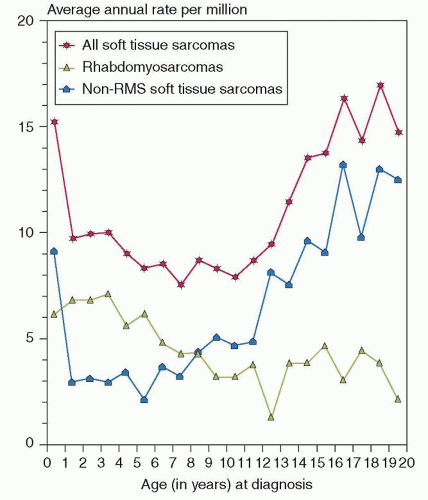
Figure 32.1 Graph shows the annual incidence of STSs by year of age using data from 1976 to 1994. RMS is more common from 1 to 8 years of age, whereas NRSTS is more common in infants younger than 1 year and in children older than 8 years. (From Gurney JG, Young JL, Roffers SD, et al. Soft tissue sarcomas. In: Ries LAG, Smith MA, Gurney JG, et al., eds. Cancer incidence and survival among children and adolescents: United States SEER Program 1975-1995. National Cancer Institute, SEER Program. NIH Pub. No. 99-4649. Bethesda, MD, 1999.)

Secondary sarcomas of both soft tissue and bone have been well documented following ionizing radiation from therapeutic radiation therapy and atomic bomb exposure.9 The risk of developing a secondary sarcoma (including soft tissue and bone sarcomas) among 14,374 participants in the Childhood Cancer Survivor Study was more than ninefold higher than in the general population.10 Multivariate analysis indicated that a primary diagnosis of sarcoma, a history of other secondary neoplasms, and treatment with radiotherapy (RT) or higher doses of anthracyclines or alkylating agents were associated with an increased risk of developing secondary sarcoma. Occupational exposure to vinyl chloride is causally related to angiosarcoma of the liver, and exposure to chlorophenols and dioxin is associated with a greater risk of STSs generally. Chronic lymphedema has been identified as a risk factor for lymphangiosarcoma.


PATHOLOGY

The classification of soft tissue tumors was revised in the fourth edition of the World Health Organization Classification of Tumors of Soft Tissue and Bone, published in 2013.11 This provides current information about the pathology and genetics of NRSTSs, grouped according to their resemblance to mature non-neoplastic human tissues. More specific details of the pathology of soft tissue tumors in children and adolescents are covered in recent reviews.12,13 Although grading according to either the Pediatric Oncology Group (POG) system or the French Federation of Cancer Centers (FNCLCC) system is applied to sarcomas, nonmalignant soft tissue neoplasms are also separated into several groups: benign, locally aggressive, and rarely metastasizing.11,12,14,15 Several locally aggressive and rarely metastasizing tumors that do not meet criteria for true sarcomas were included in the original POG grading system, and grading criteria for NRSTSs vary to some extent between the POG and FNCLCC grading systems. Nonetheless, retrospective data indicate that the two systems correlate fairly well with outcome,16 and a prospective analysis is ongoing from Children’s Oncology Group (COG) study ARST0332, which evaluated outcomes in patients under 30 years of age treated according to a risk-based strategy for NRSTS.

The optimal time for a comprehensive pathologic, cytogenetic, and molecular analysis of a soft tissue tumor is when the specimen is fresh. This allows sampling for histology, immunohistochemistry, electron microscopy, cytogenetics, molecular diagnostics, flow cytometry, and other procedures as indicated. It is important to assess both microscopic and macroscopic margins of excised tumors. Specific guidelines for pathologic evaluation and reports are available for sarcomas and are widely used in both synoptic template and text formats.17 Fresh, frozen, or formalin-fixed and paraffin-embedded tissue can be used for genetic tests including fluorescence in situ hybridization and reverse transcriptase-polymerase chain reaction (RT-PCR), and Table 32.2 summarizes genetic abnormalities useful as diagnostic adjuncts for NRSTSs.11,12,17 Current evidence indicates that grading is more reliable when performed on incisional biopsies than needle biopsies.18

Resection specimens for treated sarcomas are evaluated pathologically to determine whether residual neoplasm is present. The utility of grading histologic treatment effects using a method similar to that for treated osteosarcoma is as yet unproven for the NRSTSs. More data on this will be forthcoming from COG ARST 0332.


CLINICAL PRESENTATION

Because of their rarity, NRSTSs may not be suspected by the treating clinician. In a retrospective study of children with cancer, those with STSs experienced a median lag time of 9.5 weeks between symptom onset and establishment of the diagnosis.19 A painless mass is the most common clinical presentation, although impingement on normal structures may produce pain or other symptoms. NRSTS may arise in any part of the body, but the extremities and trunk are the most common sites. Systemic symptoms such as fever, night sweats, and weight loss are rare, although occasionally they are seen in patients with widely metastatic disease. Non-islet cell tumor-induced hypoglycemia, a rare paraneoplastic phenomenon, has been reported in a variety of NRSTSs, including solitary fibrous tumor, leiomyosarcoma/gastrointestinal stromal tumor (GIST), and fibrosarcoma. Elevated levels of pro-insulin growth factor II have been implicated in the pathogenesis of this complication.

Only a small proportion of patients with NRSTS have metastatic disease at the time of diagnosis, and the lung is most commonly involved.20 Regional lymph node involvement is unusual except in certain histologic subtypes such as epithelioid sarcoma and clear cell sarcoma.21 Bone, liver, subcutaneous, and brain metastases have been reported in a small proportion of children with metastatic NRSTS; bone marrow involvement is exceedingly rare.20


TABLE 32.2 Genetic Aberrations in Childhood Nonrhabdomyosarcoma Soft Tissue Sarcomas


Tumor


Cytogenetics


Genes Involved


ASPS


t(X;17)(p11.2;q25)


ASPSCR1-TFE3


Angiomatoid fibrous histiocytoma


t(2;22)(q33;q12)


EWSR1-CREB1


t(12;22)(q13;q22)


EWSR1-ATF1


t(12;16)(q13;p11)


FUS-ATF1


Clear cell sarcoma


t(12;22)(q13;q12)


ATF1-EWSR1


t(2;22)(q32.3;q12)


CREB1-EWSR1


DFSP


t(17;22)(q22;q13)


COL1A1-PDGFB


Desmoid-type fibromatosis


5q21 loss; trisomies 8,20


APC; CTNNB1


DSRCT


t(11;22)(p13;q12)


EWSR1-WT1


Epithelioid sarcoma


22q11-12 alterations


SMARCB1


Extraskeletal mesenchymal chondrosarcoma


t(11;22)(q24;q12)


Unknown


Extraskeletal myxoid chondrosarcoma


t(9;22)(q22-23;q11-12)


EWSR1-NR4A3


t(9;17)(q22;q11.2)


NR4A3-RBP56


t(9;15)(q22;q21)


NR4A3-TCF12


Giant cell fibroblastoma


t(17;22)(q22;q13)


COL1A1-PDGFB


Infantile fibrosarcoma


t(12;15)(p13;q25) Trisomies 8, 11, 17, 20


ETV6-NTRK3


IMT


Translocations involving 2p23 and other chromosomes


ALK, various fusion partners


Leiomyosarcoma


Deletion of 1p


Low-grade fibromyxoid sarcoma


t(7;16)(q33; p11)


FUS-CREB3L2


t(11;16)(q13; p11)


FUS-CREB3L1


MPNST


Complex


Myoepithelioma


t(6;22)(p21;q12)


EWSR1-POU51


t(1;22)(q23;q12)


EWSR1-PBX-1


Myxoid liposarcoma


t(12;16)(q13;p11)


FUS-DDIT3


t(12;22)(q13;q12)


DDIT3-EWSR1


t(12;22;20)(q13;q12;q11)


DDIT3-EWSR1


Myxoinflammatory fibroblastic sarcoma


t(1;10)(p22-31; q24-25)


FGF8 and TGFBR3 deregulation


Synovial sarcoma


t(X;18)(p11.23;q11)


SS18-SSX1, SS18-SSX4


t(X;18)(p11.21;q11)


SS18-SSX2


t(X;20)(p11.2;q13.3)


SS18L-SSX1


t(5;18)(q11;q11)


SS18-Unknown


Undifferentiated embryonal sarcoma of liver


t(11;19)(q13;q13.4)


MALAT1-MHLB1


Undifferentiated round cell sarcoma


t(4;19)(q35;q13)


CIC-DUX4


t(10;19)(q26;q13)


CIC-DUX4


X chromosome inversion


BCOR-CCNB3


PROGNOSTIC FACTORS

Because few prospective studies have been conducted in children with NRSTS,22,23,24 most information about predictors of outcome in childhood NRSTS is derived from retrospective, single-institution analyses.20,25,26 The factors that most clearly influence survival in pediatric NRSTS are:


  • Extent of disease (nonmetastatic vs. metastatic)


  • Histologic grade (low vs. high)


  • Size of the primary tumor (≤5 cm vs. >5 cm)


  • Extent of surgical resection (resected vs. unresected).

These factors are also key predictors of survival in adults with STSs.15 On the basis of these four factors, patients may be grouped into high-, intermediate-, and low-risk groups (Fig. 32.2). Those in the high-risk category have metastatic disease. These patients have a dismal survival rate of 15% at best, and most die of progressive metastatic disease. Those in the intermediate-risk category include patients with nonmetastatic but unresectable tumors and those with nonmetastatic tumors that are both high grade and more than 5 cm in maximal diameter. Survival in this patient cohort is approximately 50%. Patients with unresectable tumors, regardless of histologic grade, usually die of local tumor progression, whereas
those with large, high-grade tumors tend to develop fatal distant disease dissemination. Patients in the low-risk category include those with nonmetastatic, resectable tumors that are either high grade and less than 5 cm in maximal diameter or low grade (any size). These patients have a long-term survival estimate of approximately 90%.


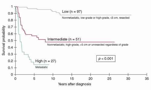
Figure 32.2 Kaplan-Meier survival distributions of patients treated at St. Jude Children’s Research Hospital according to risk group. (Figure constructed using data from Spunt SL, Poquette CA, Hurt YS, et al. Prognostic factors for children and adolescents with surgically resected nonrhabdomyosarcoma soft tissue sarcoma: an analysis of 121 patients treated at St. Jude Children’s Research Hospital. J Clin Oncol 1999;17:3697-3705; Spunt SL, Hill DA, Motosue AM, et al. Clinical features and outcome of children with unresected non-rhabdomyosarcoma soft tissue sarcoma [NRSTS]. J Clin Oncol 2002;20:3225; and Pappo AS, Rao BN, Jenkins JJ, et al. Metastatic nonrhabdomyosarcomatous soft-tissue sarcomas in children and adolescents: the St. Jude Children’s Research Hospital experience. Med Pediatr Oncol 1999;33:76.)

Recently, a relationship between body size and tumor size has been documented in pediatric patients, arguing in favor of considering the relative tumor size as it relates to body size when analyzing prognostic factors in children with NRSTS.27 Other factors that may contribute to survival outcomes but that are not known to be independent of the other prognostic features include microscopic surgical margin (in resected tumors), primary site (visceral sites seem to have a worse outcome), and older age (age ≥10 years has been reported to be an adverse prognostic factor in unresected tumors). Other predictors of survival in adults with STS including anatomic location (upper extremity vs. lower extremity), tumor depth (superficial vs. deep), and histologic subtype have not been shown to be prognostically important in pediatric NRSTS.

A nomogram that estimates the likelihood of 12-year sarcoma-specific death has been developed by Memorial Sloan-Kettering Cancer Center based on an analysis of 2,136 adult patients prospectively followed at that institution.28 The nomogram utilizes histologic grade, tumor size, depth, site, histology, and age to predict outcome. An effort to validate this nomogram in pediatric patients has been undertaken, but this effort was limited by the small number of patients available for study.29

Prognostic factors for local tumor control in pediatric NRSTS are not well delineated. However, the extent of surgical resection plays a key role. Patients who undergo a gross total tumor resection have a cumulative incidence of local recurrence in the 15% range, whereas those who present with unresectable tumors experience local tumor recurrence or progression in about 45% of cases.20,25,26 The data on other predictors of local tumor control are contradictory, most likely due to inadequate numbers of patients available for analysis. However, in patients with resected NRSTS, large tumor size, invasiveness, intra-abdominal primary site, the presence of microscopic residual tumor, and avoidance of RT have been associated with poorer local control in univariate analyses.20,26 In patients with initially unresected tumors, univariate analyses have identified female sex, age more than 10 years, a diagnosis of MPNST, and lower doses of RT to be correlated with the risk of local recurrence.20,25


STAGING

Although there is a standardized staging system for pediatric RMS, no staging system has been prospectively validated for pediatric patients with NRSTS. In adults, the most commonly employed system is the one developed by the American Joint Committee on Cancer. This system is used for all STSs, with the exception of GIST, dermatofibrosarcoma protuberans (DFSP), Kaposi sarcoma, and infantile fibrosarcoma. This system recognizes three distinct histologic grades, which in combination with size, depth, and extent of nodal and distant metastatic disease are the primary determinants of clinical stage.


GENERAL TREATMENT CONSIDERATIONS

The goals of therapy are to achieve long-term disease control with minimum morbidity in both the short and long term. Decision making about the treatment approach is often complex and requires balancing the risks of disease recurrence against the risks of serious complications that may occur years after treatment finishes. Late effects are of particular concern in young children, whose potential survival after successful therapy is long and who often experience the most substantial long-term toxicities. In adults, treatment at a high-volume center is an independent predictor of improved survival.30 Children with NRSTS should be treated at a center of excellence by a multidisciplinary team of experts that can accurately weigh the benefits and burdens of therapy before making recommendations about treatment. Members of the treatment team should have sarcoma expertise and should include a pediatric surgeon and surgical subspecialists, a radiation oncologist, and a pediatric oncologist. Key support services include pathology, diagnostic imaging, rehabilitation (particularly physical and occupational therapy), nutrition, and psychosocial services (social work, child life, psychology/psychiatry, chaplain). Since the treatment approach depends on the diagnosis, review of the pathology by an expert pediatric STS pathologist should be strongly considered whenever the diagnosis is uncertain. Because so little is known about ideal therapy for pediatric NRSTS, patients should be entered on prospective clinical trials whenever possible.

Although the overall approach to treatment of children with NRSTS is similar to that for adults, important differences exist. First, the distribution of histologic subtypes of STS differs in adults and children (Table 32.1),2 so caution must be exercised when extrapolating treatment recommendations from adult to children. The most common STSs in childhood differ from those in adulthood, so responses to therapy may in some cases differ. Although some histologic subtypes appear to have a similar behavior in children and adults, certain tumors behave differently. The best example of this phenomenon is fibrosarcoma, which is considerably less aggressive in young children.3 Tumor biology may differ in
children and adults, in some cases leading to differences in treatment recommendations. For example, fewer than 15% of pediatric GISTs harbor KIT or PDGFA gene mutations, whereas these mutations occur in about 90% of adult GIST. Based on these findings, upfront treatment for most pediatric patients is surgery alone, whereas most adults receive tyrosine kinase inhibitor therapy.31

Local control measures such as surgery and RT pose unique challenges in children because of their incomplete growth and development. Tumor resection may result in greater deformity or disability due to the lesser amount of surrounding normal tissue in young children. Limb-sparing procedures in younger children are more complex due to the small and variable size of the patients. Young children are generally more physically active than adults, so activity-related complications after limb-sparing surgery are more common. Due to their incomplete linear growth, most children require interventions to increase the size of the prosthesis. The use of expandable modular prostheses and noninvasive expandable prostheses is a promising technique, although data on the long-term structural integrity of these prostheses are limited.

The long-term side effects of RT in children are often considerably greater than in adults because the doses required to treat NRSTS impair normal tissue growth, leading to problems such as limb length discrepancy, joint dysfunction, and cosmetic deformity. Chemotherapy carries risks as well, depending on the agents used and their cumulative doses. The combination of ifosfamide and doxorubicin, the most common regimen utilized in pediatric NRSTS, may produce infertility, cardiomyopathy, renal dysfunction, and secondary neoplasia. Some of these complications, such as cardiomyopathy, are more severe in young children, in whom growth and development are incomplete. Infertility, which may be of little concern to an older adult, may have a significant impact on long-term psychosexual functioning and lifestyle. Further, the long potential lifespan of pediatric patients may increase the likelihood that a given patient will develop a long-term complication. The risk of second malignant neoplasms, in particular, increases with time since exposure. Thus, the chances of experiencing a second cancer after a given treatment are considerably higher for children than for older adults.


Surgery

Surgery is a critical component of effective treatment for pediatric NRSTS. In retrospective case series from two large pediatric centers, patients whose tumors were grossly excised had an estimated 5-year survival exceeding 85%, whereas patients whose tumors were unresectable had an estimated 5-year survival in the 55% range.20,25,26 The effect of surgical resection is similar in adults.32

When feasible, a complete surgical resection should be performed with the goal of excising the primary tumor and all metastases with margins sufficient to prevent local recurrence. In the event that microscopic disease remains after surgery, adjuvant RT produces adequate local tumor control. However, primary re-excision should be considered whenever feasible to achieve widely negative margins.33 If the tumor cannot be widely resected with acceptable morbidity at the time of initial diagnosis, neoadjuvant chemotherapy and/or RT should be considered.34,35


Definitive Surgery

The goal of the definitive operation is to excise the tumor en bloc without violating the pseudocapsule and achieving an adequate margin. Removal of the entire muscle compartment is almost never required. Amputation may be considered for extremity tumors that cannot be grossly excised otherwise and for very young children in whom RT may produce long-term complications more severe than amputation. Examples of the appropriate use of amputation include ray resection for epithelioid sarcoma of the finger or toe and above-knee amputation for an infant with a large tumor encasing the neurovascular bundle around the knee.

What constitutes an adequate margin for local tumor control without RT remains controversial. Although it may be feasible to remove a large cuff of normal tissue around the tumor in an adult, such a wide margin may be impossible to achieve in a small child. Evidence in adults treated without adjuvant RT suggests that although a margin of 1 cm results in 100% local tumor control, those whose closest margins were less than 1 cm still had an 87% 10-year local control rate.36


Primary Re-excision

It is not infrequent for a patient with NRSTS to be referred to the treatment center after an excisional biopsy elsewhere. The initial surgical procedure may not have been performed anticipating a malignancy, and frequently the tumor has been “shelled out” through the pseudocapsule, leaving microscopic disease behind. In some cases, the status of the surgical margins is unclear due to uncertainty about the surgeon’s findings or suboptimal handling of the surgical specimen. In a pediatric study looking at unplanned excisions of NRSTS, residual tumor was found in nearly half of re-excision procedures performed after the initial surgeon reported that clear margins had been obtained.37 Given the high percentage of patients with residual tumor after an unplanned excision, pretreatment re-excision should be performed whenever it is uncertain whether the tumor was adequately excised.


Lymph Node Sampling and Dissection

The overall incidence of regional lymph node metastasis for most NRSTS is low, so lymph node sampling should be considered only in certain histologic subtypes. In analyses of adults with STSs, clear cell sarcoma and epithelioid sarcoma were the only entities with an incidence of regional lymph node metastases consistently exceeding 15%.21 Because treatment differs if nodal involvement is present, sentinel lymph node biopsy or lymph node sampling should be considered for patients with these tumors. Lymph node sampling may also be considered for patients with vascular sarcomas (angiosarcoma, lymphangiosarcoma) since some studies have documented high rates of nodal involvement. Although a relatively high frequency of lymph node metastases was reported in synovial sarcoma in early published series, more recent studies have documented a rate of only 1.4% to 3.6%.21

Sentinel lymph node biopsy may be performed at the time of upfront biopsy or definitive surgery, and is performed by injecting the tumor bed with a combination of technetium-labeled sulfur microcolloid and isosulfan blue dye (Lymphazurin). After injection, a radioisotope detector is used to localize the sentinel node, and an incision is made over the localized area to identify the blue node(s) with a high radioisotope count that represent the nodes
draining the tumor. These lymph nodes are excised and then thinly sectioned for review by the pathologist.

The optimal management of pediatric patients with involved lymph nodes is uncertain due to the small number of reported cases. Both formal lymph node dissection and adjuvant RT may be considered.


Resection of Pulmonary Metastatic Disease

The lung is the most common site of distant metastatic disease in NRSTS. Although fewer than 20% of patients with pulmonary metastases become long-term survivors, pulmonary metastasectomy can lead to long-term survival. Factors that have been associated with a greater likelihood of long-term survival in multivariate analyses in adults include unilateral involvement, late onset of pulmonary metastases (>1 year from initial diagnosis), complete resection of pulmonary metastases, repeated resections of pulmonary metastases, and greater than 2-year interval between extrapulmonary recurrences.38,39


RADIATION THERAPY

RT is an adjuvant to surgery in many cases of pediatric and adolescent NRSTS with the goal of improving local control while preserving limb or organ function. Preoperative RT is considered for unresectable sarcomas to facilitate surgical resection, whereas postoperative RT is selectively administered to prevent local recurrence for high-grade, large (>5 cm) tumors. Careful consideration should be given to the radiation dose and treatment volume to minimize latent normal tissue injury including the development of radiation-induced malignancies. RT should be delivered at an institution with experience in radiation effects on normal tissues, including growth and fertility. The recently completed COG NRSTS trial that evaluated a risk-based treatment approach in patients under 30 years of age (ARST 0332) forms the basis for contemporary guidelines for those who benefit from neoadjuvant and adjuvant RT. Prognostic factors including tumor size and histologic grade, presence of metastases, and, most importantly, whether the tumor is resectable are critical to make informed decisions on who benefits from RT.20,25,26,40,41,42,43


Postoperative RT

Retrospective analyses of prognostic factors in pediatric NRSTS have shown that grade is one of the most important factors predicting local recurrence after surgery and likelihood of death. Patients with low-grade NRSTS rarely develop distant metastases. Therefore, local recurrence of a low-grade tumor after surgery is usually not life threatening. A management strategy of careful observation despite close or positive margins is often adopted in an attempt to avoid or delay the administration of RT. Preservation of limb/organ function and excellent overall survival is possible with re-excision and consideration of radiation at the time of recurrence. An exception to this strategy might be if the location of the primary tumor would require an amputation or other mutilating procedure if the tumor does recur locally. In distinction, high-grade sarcomas carry an increased risk for both local recurrence and distant metastases. Local recurrences are difficult to control as these sarcomas are typically infiltrative with rapid growth patterns. Retreatment is often associated with significant morbidity (amputation or loss of organ function), and local recurrence may place the patient at a greater risk for distant recurrence. The benefits of optimizing local control in this setting outweigh the risks associated with RT.

The only United States multicenter trial for resectable pediatric NRSTS was conducted through the POG from 1986 to 1993. Postoperative RT was recommended only for patients with microscopic residual disease. The analysis suggested that among 80 patients, 8 patients had a local recurrence (12.5%) with at least 5 occurring in patients with negative margins who did not receive RT; the majority of these were high-grade tumors.24

St. Jude Children’s Research Hospital reviewed 88 children with resectable NRSTS for risk of local recurrence in relationship to tumor grade and width of the surgical margin. Of 14 patients with a <1 cm surgical margin and high-grade tumors, local recurrences were noted only in patients who did not receive postoperative RT. Of 20 patients with >1 cm negative surgical margin and a high-grade tumor, none received postoperative RT and 4 recurred. This report confirmed the importance of grade, and also established that the surgical margin width, even if negative, should be assessed since it may influence the risk of local recurrence and therefore the approach to local treatment.41

Spunt et al. extended the St. Jude Children’s Research Hospital experience assessing prognostic factors for pediatric patients presenting with resectable NRSTS. Of 81 patients who underwent complete resection, only 10 received adjuvant RT with a median dose of 54.9 Gy (range 26.5 to 60.4 Gy). The 5-year estimated cumulative incidence of local failure was 12.8%. Factors associated with local recurrence were microscopic residual disease, large tumor size, and intra-abdominal disease, whereas improved local control was associated with the use of RT.

University of Florida reported the long-term outcome of 95 pediatric and young adults with resectable NRSTS treated over a 34-year period with surgery and adjuvant RT. The overall local failure rate was 12% at 5 years. Patients with a negative margin had a 6% local recurrence rate at 5 years compared with 27% when the margin was close or positive. Of note, the authors reported that all patients who developed a local failure ultimately died of disease.43

Tumor size and invasiveness may influence whether there is a benefit to adjuvant RT. A multivariate analysis of synovial sarcoma in childhood and adolescence compiled from four large research organizations showed that there was not a statistically significant local recurrence-free survival benefit whether or not patients received RT. The authors hypothesize that the small number of local recurrences or possible mis-staging could contribute to the noted lack of benefit from RT.42

A treatment strategy designed to avoid RT in young patients with localized completely resected synovial sarcoma was conducted from 1984 to 2003 through the International Society of Pediatric Oncology Malignant Mesenchymal Tumor Study. There were three local recurrences (15%), all occurring in patients who did not receive postoperative RT. All were alive without disease at the study report, leading the authors to conclude that RT could be omitted for completely resected synovial sarcomas as retreatment for local failure may be successful.44

Another common NRSTS in the pediatric and young adolescent age group is MPNST. Italian and German studies between 1975 and 1998 showed a lower local recurrence rate after complete resection with adjuvant RT (17%) as compared to omission of RT (36%). For microscopic margins, the local recurrence rate with and without RT was 45% and 68%, respectively. The radiation dose in this study population was 45 Gy using a hyperfractionated treatment with 1.6 Gy given twice daily. This series suggests that local control is a critical problem, even for completely resected disease, and suggests an inherent biologic resistance to current therapy.45

Age may influence the decision to recommend adjuvant RT, particularly in very young children where the latent long-term risks associated with the addition of RT may outweigh the benefit of such therapy.42 Other considerations for postoperative RT include resection with microscopic or gross residual disease, unplanned resection where re-excision to confirm the margin status is not feasible, and uncertainty about the surgical margins.


Preoperative RT

Preoperative RT is indicated for initially unresectable NRSTS. Although anatomical location is an important factor in determining
resectability, most of these tumors will be of high grade, large, and invasive that would likely require RT based on pretreatment evaluation. The intent of preoperative RT is to shrink tumors and facilitate limb- or organ-sparing resection. The advantage of preoperative RT over postoperative RT is that the total dose delivered may be lower by 10% to 15% and the volume irradiated smaller, potentially resulting in fewer long-term effects on normal tissues. Another theoretical benefit to preoperative RT is that much of the irradiated tissue is resected, therefore potentially further reducing late effects of ionizing irradiation, particularly second cancers. Local treatment failure is a major challenge for patients with initially unresectable as compared with resected tumors.25 Spunt et al. showed a 44% cumulative incidence of local failure in 27 patients treated before 1987 where lower radiation doses (39.6 Gy) were thought to be a contributing factor; higher radiation doses (59.4 Gy) resulted in lower local recurrence rates (20%). Ferrari et al. reported the pooled results of over 300 patients with unresected pediatric NRSTS from United States and Europe research groups, and concluded that local progression or recurrence was the major cause of treatment failure, with RT correlating with a better outcome.46


Radiation Dose and Volumes

The radiation doses used in the recent COG NRSTS trial (ARST0332) were lower than those used for adults as were the margins encompassing the tumor volume and were chosen based on concerns regarding side effects of RT, the potential synergy of combined modality therapy including neoadjuvant chemotherapy, and the broad range of ages of patients who might be enrolled on the study.

A prospective single-institution study tested whether a smaller margin for expansion on the gross tumor volume (GTV) as compared to what is recommended for adults was feasible without compromising local control. Of 32 pediatric and young adults with a high-grade NRSTS, 27 received adjuvant RT. Using a 2-cm expansion around the GTV and delivering a median cumulative postoperative radiation dose of 60 Gy (range 41.4 to 70.4 Gy) or a preoperative dose of 45 Gy (range 45 to 50.4Gy), the 3-year cumulative local recurrence rate for patients who underwent a marginal or complete resection was <4%. There were no failures in the patients with clear surgical margins. Those who failed locally did so within the high-dose radiation volume, suggesting that limited-margin RT is an effective strategy to employ in pediatric patients with NRSTS. This strategy has been employed by the COG, and is expected to confer less normal tissue injury including a possible reduction in secondary cancer induction.47

For resectable tumors, the postoperative initial radiation field (PTV1) defined by the tumor volume on preoperative MRI or computerized tomography (CT) imaging, the tumor bed defined by operative report, surgical clips, and any potential occult tumor spread encompassed by at least a 2-cm margin is treated to 45 Gy. A field reduction (PTV2) is contoured to encompass the highest-risk region of close/negative surgical margins, and is treated to 55.8 Gy total dose. For patients with microscopically positive margins or gross residual disease, a higher radiation dose is recommended. In the case of very young children, lower doses may be considered. However, there is no evidence that NRSTS are more sensitive to RT in younger children as compared with adolescents or adults.

For unresected tumors, preoperative radiation (45 to 50 Gy) is delivered to the initial target volume (PTV1) defined by preoperative imaging and should include potential occult tumor spread and biopsy site encompassed by at least a 2-cm margin. If chemotherapy is given concurrently with preoperative RT, the lower dose of 45 Gy is recommended. A boost to the site of close/positive margins should be considered in intraoperative or postoperative settings.


RT for Treatment of Metastases

The role of radiation therapy for STS metastases must be individualized. In general, RT is reserved for unresectable metastases. However, the number of sites and location of metastases will determine whether palliative RT might be of benefit. For example, patients with three or fewer sites might benefit from conformal RT if the anticipated toxicity is deemed acceptable. Whole-lung or -liver radiation is not recommended for diffuse metastases. Sophisticated conformal techniques such as stereotactic radiosurgery are an excellent way to limit normal tissue toxicity.


RT Technical Factors

External-beam radiation therapy (EBRT) is the most common type of RT used to treat pediatric sarcomas. Photons are a form of ionizing irradiation delivered by a linear accelerator, which can target tumors below the skin surface. There are many contemporary methods to deliver photon irradiation with intensity-modulated radiation therapy (IMRT) being the most conformal and allows sculpting of the radiation dose around target volumes while maximally protecting normal tissues/organs. Refinements in IMRT delivery such as sophisticated arc-based methods include helical tomotherapy and volumetric modulated arc therapy (VMAT). VMAT confers highly conformal dose delivery to target volumes, rapid delivery of radiation, and less radiation exposure to normal tissues. Proton therapy is also a type of EBRT that requires a specialized particle accelerator to generate a proton beam. The advantages of protons compared with photons are less scatter radiation dose in the normal tissues and less radiation dose beyond the desired depth of penetration. The primary goal of either type of EBRT is to deliver the most precise coverage to the tumor volume while protecting the adjacent tissues and organs leading to fewer normal tissue side effects including secondary cancers.

Effective immobilization techniques provide the ability to deliver extremely conformal treatment, and include the use of anesthesia in very young children. Conformal radiation therapy relies on acquiring three-dimensional (3D) images, such as with CT, while the patient is in the treatment position. Additionally, sarcomas located in the chest or abdomen may shift with diaphragmatic motion during radiation therapy. Accounting for respiratory movement with 4D imaging allows for further refinements in the precise delivery of radiation to the target volume. Sophisticated computer planning systems are used to contour tumor volumes and critical organs at risk. Diagnostic scans, including MRI or PET CT, can be fused and digitally formatted to align with the treatment planning scan for more precise contouring of the target volumes. To ensure that the treatment plan is accurately reproduced, image-guided radiation therapy (IGRT) is necessary for IMRT plans. IGRT is a process of frequent 2D and 3D imaging, during a course of radiation treatment, to direct radiation therapy utilizing the imaging coordinates of the actual radiation treatment plan, enabling physicians to make precise adjustments in the radiation field in relation to the patient position. These technologic advances work in concert to achieve improved tumor localization and meet the critical objective of protecting normal tissues from unnecessary irradiation.

Brachytherapy can be used as a boost after EBRT in an effort to deliver high doses of radiation to a focal area within the operative bed when clear surgical margins are difficult to achieve. At the time of surgery, removable catheters are placed in the operative bed and 5 to 6 days after surgery loaded with high-dose rate or low-dose rate radioactive sources. Wound complications are reported to be higher in the implanted site using brachytherapy.48 Intraoperative radiation therapy is also a technique to boost margins that in the surgeons estimate will likely be positive. EBRT is delivered while the operative bed is exposed. The major advantage of this technique is that critical structures can be moved out of the
radiation beam to allow direct high-dose, single-fraction RT to be delivered to the tumor bed with minimal toxicity.49 All of these contemporary technologies contribute to keeping the radiation therapy dose as low as possible, hence minimizing normal tissue exposure.


CHEMOTHERAPY

The role of systemic therapy in pediatric NRSTS has not been well studied. Since 1986, only three prospective pediatric clinical trials that accrued a total of 182 patients have been completed and published in North America.22,23,24 European STS clinical trials routinely included certain subtypes of NRSTS, but these trials were designed for the treatment of RMS and outcomes for NRSTS were not commonly reported.40,50

In 2007, the Children’s Oncology Group opened ARST0332, a clinical trial for patients under 30 years of age with NRSTS that was designed to assess a risk-based treatment strategy. The trial closed in 2012 after enrolling about 550 eligible patients. During a similar timeframe, the European Soft Tissue Sarcoma Group (EpSSG) launched its own NRSTS study with separate guidelines for synovial sarcoma and “adult type” STSs. Both the COG and EpSSG trials utilized an ifosfamide and doxorubicin systemic chemotherapy regimen for intermediate- and high-risk patients. The results of both of these studies, which should help clarify the role of cytotoxic chemotherapy in pediatric NRSTS, will be available in the next few years.

The clearest indication for chemotherapy in pediatric NRSTS is unresectable tumor, since few patients are cured in the setting of gross residual disease.32 Chemotherapy may also have a role in patients with resected tumors who are at high risk for metastatic tumor recurrence, such as those with large, high-grade tumors. The most active cytotoxic chemotherapy agents overall are doxorubicin and ifosfamide. In pediatric patients, overall response rates are in the 40% range.22 Unfortunately, these drugs have considerable short- and long-term toxicities, further complicating the decision about when chemotherapy is warranted. Certain histologic subtypes (for example, desmoid fibromatosis) respond to other drug combinations. Histology-specific regimens are discussed further in the sections focused on specific histologic subtypes.


Adjuvant Therapy—Adult Trials

The role of adjuvant chemotherapy for adults with resected STS is controversial because chemotherapy has limited efficacy and considerable toxicity. The Sarcoma Meta-Analysis Collaboration (SMAC) conducted a meta-analysis of outcome for more than 1,500 adults included in 14 randomized controlled clinical trials conducted in the 1970s and 1980s comparing local therapy alone to local therapy plus chemotherapy.51 This analysis showed that although chemotherapy significantly lengthened the local, distant, and overall recurrence-free interval (p = 0.016, p = 0.0003, p = 0.0001, respectively), the hazard ratio (HR) for overall survival was not significantly affected (HR, 0.89; 95% confidence interval [CI], 0.76 to 1.03; p = 0.12). Various disease features, including primary site, histology, grade, and size, did not appear to influence the likelihood of survival benefit from chemotherapy. There was also no evidence that the addition of other agents to doxorubicin produced a different outcome than doxorubicin alone. This meta-analysis has been criticized for including tumors at all anatomic locations and of all histologic grades, which may have diluted the observed benefits of chemotherapy. Furthermore, only one of the trial chemotherapy regimens included ifosfamide, which is widely acknowledged to be among the most active agents in adult STSs. A later update included the 14 trials in the SMAC analysis as well as 4 additional studies that incorporated ifosfamide.52 Included were six studies that used single-agent doxorubicin, five studies that used ifosfamide and an anthracycline (with or without dacarbazine), and seven that used doxorubicin in combination with agents other than ifosfamide. Patients receiving adjuvant chemotherapy had a small but statistically significant reduction in the risk of local recurrence, distant recurrence, and overall recurrence, and a slight but statistically significant improvement in survival. The absolute risk reduction was 4% for local recurrence, 9% for distant recurrence, and 10% for recurrence overall. Adjuvant chemotherapy significantly reduced the risk of death, with an HR of 0.77 (95% CI, 0.64 to 0.93; p = 0.01). This translated to an absolute risk reduction of 6%, or a 40% versus 46% risk of death. There was some evidence that ifosfamide-containing regimens were superior to those without ifosfamide. The absolute reduction in risk of death was 11% for ifosfamide-containing regimens (p = 0.01) but only 5% for doxorubicin alone (p = 0.07).

Interpreting the findings of adjuvant chemotherapy studies in adult STSs for a pediatric patient population is difficult. The distribution of histologic subtypes of STSs differs in children,2 so outcomes after chemotherapy in children may not reflect the adult experience. Indeed, some data suggest that synovial sarcoma, one of the most common pediatric NRSTS, may respond better to ifosfamide than other types of STS more common in adults.53 Children have a potentially longer life span than adults, so minor improvements in overall survival in a pediatric patient population may lead to substantially more years of life saved. However, children may also be more likely to experience long-term complications from chemotherapy exposure than adults because their growth and development are incomplete and because they have a longer life span. Whenever possible, adjuvant chemotherapy for pediatric NRSTS should be administered in the context of a clinical trial so that further data can be collected about its risks and benefits in these young patients.


Adjuvant Therapy—Pediatric Trials

In children, there has only been one prospective randomized trial of adjuvant chemotherapy for NRSTS. From June 1986 through May 1991, the POG evaluated the benefit of adjuvant chemotherapy (vincristine, doxorubicin, cyclophosphamide, and dactinomycin) compared with observation in surgically resected pediatric NRSTS.24 Of the 81 eligible patients, only 30 accepted randomization, and no evidence of improved outcome was observed in the subgroup that received adjuvant chemotherapy. Five-year survival and event-free survival estimates were significantly worse for patients who received adjuvant chemotherapy; however, this is likely the result of an excess of patients with high-grade lesions who received adjuvant chemotherapy.

More recently, COG study ARST0332 treated patients with all stages of NRSTS who were under 30 years of age with a risk-based treatment approach. The risk stratification approach and treatment plan are illustrated in Figure 32.3. In this study, adjuvant chemotherapy was administered only to patients with high-grade tumors larger than 5 cm in maximal diameter who had undergone an upfront gross tumor resection. Other patients at high risk for distant disease dissemination (i.e., those with unresected, high-grade tumors >5 cm in maximal diameter and those with metastases at the time of diagnosis) received neoadjuvant combined chemotherapy and RT. The chemotherapy regimen that was tested in this clinical trial was doxorubicin (75 mg/m2/cycle) and ifosfamide (9 g/m2/cycle). A similar clinical trial in Europe, sponsored by the European Pediatric Soft Tissue Sarcoma Study Group, utilizes a virtually identical ifosfamide/doxorubicin regimen but slightly different criteria for patient selection. The findings of these two clinical trials, which should be reported in the next few years, will provide considerable data about the value of adjuvant chemotherapy in patients at high risk for metastatic tumor recurrence.


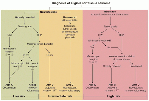
Figure 32.3 Risk group and treatment assignment algorithm for Children’s Oncology Group protocol ARST 0332, “Risk-Based Treatment for Non-Rhabdomyosarcoma Soft Tissue Sarcomas (NRSTS) in Patients Under 30 Years of Age.” (Used with permission, James Anderson, Children’s Oncology Group.)


Therapy for Advanced Disease—Adult Trials

Because chemotherapy is relatively ineffective for STS, it is often difficult to judge which patients with advanced disease will benefit from systemic therapy. The clearest indication for chemotherapy in advanced disease is the presence of an unresectable tumor that may become resectable with chemotherapy-induced shrinkage. A retrospective analysis of 488 adults with unresectable or metastatic STS showed that 45% derived clinical benefit (defined as an objective response or ≥ 6 months of stable disease) from chemotherapy.54 The median time to progression of the entire cohort was 3 months. However, the median duration of response in those who responded to chemotherapy was 9 months, and the median duration of stable disease among those for whom this was the best response was 6 months. Combination chemotherapy produced a better response rate than single-agent chemotherapy (47% vs. 25%, p < 0.001). On multivariate analysis, favorable prognostic factors for survival included age less than 40 years, synovial sarcoma or liposarcoma histology, the absence of bone metastases, and the use of combination chemotherapy. This analysis suggests that chemotherapy may provide significant patient benefit even if therapy does not produce a cure.

There are ample data on the activity of single agents for adult STS. Doxorubicin has been a mainstay of STS chemotherapy since the 1970s. More recent studies have confirmed the modest activity of doxorubicin as a single agent, with response rates in the 25% range. Dose intensification of doxorubicin does not seem to improve the rate of response or the likelihood of long-term survival significantly.55 A variety of alkylating agents have been used for adult STS. Dacarbazine has been used since the 1970s when a single-agent response rate of 17% was seen in adult STS patients. The rate of response to single-agent cyclophosphamide approaches 10%. Temozolomide, an oral prodrug of the active metabolite of dacarbazine, has demonstrated response rates below 15%. Ifosfamide has produced responses in approximately 20% to 30% of newly diagnosed adults with STS. There is some evidence of an ifosfamide dose-response relationship, although ifosfamide dose escalation in combination with doxorubicin did not improve the rate of response or disease-free survival.56 A randomized comparison of single-agent doxorubicin to two different schedules of single-agent ifosfamide showed no difference in progression-free survival, but toxicity was greater on the ifosfamide arms.57 Gemcitabine has limited efficacy overall in STS in adults, but appears to be relatively more effective in leiomyosarcoma.58

Combination chemotherapy for STS in adults dates back to the 1970s. In general, combination chemotherapy regimens have demonstrated higher response rates, although these have been accompanied by greater toxicity and, in many cases, no clear survival advantage. The earliest combination, doxorubicin and dacarbazine, demonstrated response rates that exceeded those observed with single-agent doxorubicin therapy. Cyclophosphamide and vincristine were added to doxorubicin and dacarbazine in CYVADIC. However, this regimen failed to produce a higher response rate, a longer duration of response, or a longer median survival than single-agent doxorubicin. MAID (mesna, doxorubicin, ifosfamide, and dacarbazine) chemotherapy proved superior to doxorubicin/dacarbazine in terms of response rate and time to progression. Dacarbazine was omitted from MAID to permit dose intensification of ifosfamide and doxorubicin. This two-drug combination produced responses in about two-thirds of patients. However, a randomized study of doxorubicin in combination with 6 or 12 g/m2/cycle of ifosfamide showed no benefit of the higher
dose of ifosfamide in terms of disease-free or overall survival.56 Finally, the combination of gemcitabine and docetaxel has produced modest rates of response.59


Therapy for Advanced Disease—Pediatric Trials

Few studies have been conducted on pediatric patients with advanced NRSTS. In the 1980s, the POG evaluated the role of dacarbazine in the context of multiagent chemotherapy with vincristine, doxorubicin, cyclophosphamide, and dactinomycin for patients with unresected or metastatic NRSTS.23 The addition of dacarbazine did not improve the response rate (44% with vs. 56% without, p = 0.4), or 4-year event-free survival (26.4% with vs. 36% without). A subsequent POG phase II study of vincristine, ifosfamide (9 g/m2/cycle), and doxorubicin (60 mg/m2/cycle) for unresectable or metastatic NRSTS demonstrated similar findings.22 Forty-one percent of patients experienced a complete or partial response, and the 3-year progression-free survival was 60% ± 10% for unresectable disease and 14.3% ± 9% for metastatic disease.

The recent COG study (ARST0332) for patients younger than 30 years with newly diagnosed NRSTS utilized a combined chemotherapy and RT approach for patients with intermediate- and high-risk disease (defined as a high-grade tumor >5 cm, unresectable disease, or metastatic disease [low-grade metastatic tumors were excluded]). Those with tumors excised at study entry received dose-intensive chemotherapy (ifosfamide 9 g/m2/cycle and doxorubicin 75 mg/m2/cycle) and 55.8 Gy of adjuvant RT starting at week 4. Those whose primary tumor has not been resected received 4 cycles of the same chemotherapy regimen along with 45 Gy of neoadjuvant RT starting with cycle 2. Doxorubicin was omitted in cycles 3 and 4 during RT. After a delayed surgical resection, an RT boost was given on the basis of the margin status (10.8 Gy for microscopic residual disease and 19.8 Gy for gross residual disease). All intermediate- and high-risk patients received a cumulative dose of 375 mg/m2 of doxorubicin and 54 g/m2 of ifosfamide. Results of this study, which closed in 2012, are expected soon. A similar study conducted by the EpSSG will provide additional information.


Toward Targeted Therapies in NRSTS

The discovery of specific genetic changes and unique pathways involved in the pathogenesis of various STSs is gradually changing the therapeutic approach to these diseases. Several tyrosine kinase inhibitors have demonstrated activity in STSs. The PALETTE study documented that pazopanib produced a lengthening of progression-free survival in adults with metastatic nonadipocytic STS after failure of standard chemotherapy (from 1.6 to 4.6 months), leading to FDA approval for treatment of NRSTS in adults after failure of prior chemotherapy.60 Sunitinib and cediranib have shown promising activity in alveolar soft part sarcoma (ASPS),61,62 and sorafenib has demonstrated activity in angiosarcoma.63 Imatinib mesylate has been successfully used to treat metastatic DFSP due to its effect on PDGFRB signaling.64

Other newer agents include trabectedin, bevacizumab, IGF-1R antibodies, mammalian target of rapamycin (mTOR) inhibitors, and tubulin inhibitors. Data on these agents in pediatric patients are very limited, although several have completed phase I and II testing. Trabectedin is a novel marine compound that binds the DNA minor groove and blocks cell cycle progression and transcription of inducible genes. It has been shown to be active in various STSs, particularly liposarcoma and leiomyosarcoma.65 In myxoid liposarcoma, evidence suggests that this agent causes adipocyte differentiation by targeting the FUS/CHOP-mediated transcriptional block. Bevacizumab, a recombinant humanized antibody against vascular endothelial growth factor, has shown promising activity in vascular sarcomas such as angiosarcoma and epithelioid hemangioendothelioma.66 The combination of cixutumumab (an IGF-1R antibody) and temsirolimus (an mTOR inhibitor) produced a 31% rate of freedom from progression at 12 weeks in adults with chemotherapy-refractory STSs.67 A phase II study of eribulin mesylate, a non-taxane inhibitor of microtubule function, had documented activity in adipocytic sarcoma and leiomyosarcoma.68


SELECTED SPECIFIC DISEASES (LISTED BY BIOLOGIC POTENTIAL AND IN ALPHABETICAL ORDER)


Benign Soft Tissue Tumors

Most soft tissue masses in children are benign and encompass a range of developmental, malformative, reactive, and neoplastic proliferations, with vascular lesions among the most frequent of the mesenchymal neoplasms.12 The current World Health Organization Classification of soft tissue tumors includes morphologic categories based on resemblance to mature non-neoplastic tissue types, to the extent possible, and tumors coded as benign in behavior account for a large proportion of the entities.11 Among the benign soft tissue tumors of childhood, various types of hemangiomas and vascular malformations, lymphatic malformations, lipoblastoma, neurofibroma, the fibromatoses, and nodular fasciitis are potential clinical or pathologic mimics of more aggressive neoplasms. Although a detailed discussion of these benign neoplasms is beyond the scope of this chapter, it is important to be aware of their capacity for rapid growth, large size, and alarming clinical appearance in some situations, to avoid diagnostic and therapeutic pitfalls.


INTERMEDIATE, LOCALLY AGGRESSIVE SOFT TISSUE TUMORS

The World Health Organization defines intermediate, locally aggressive soft tissue tumors as neoplasms with the capacity for recurrence and/or aggressive growth confined to the original anatomic site, but without a capacity for metastasis. Those that are likely to be encountered in children and adolescents are summarized in this section and in Table 32.3.


Desmoid-Type Fibromatosis


Epidemiology

A population-based study in Finland estimated the incidence of desmoid tumors to be 2.4 to 4.3 cases per million annually. The median age at presentation is in the fourth decade and females are affected about twice as often as males.69 In pediatric patients, desmoid tumors have been described in infancy but adolescents account for the majority of cases.70,71 Pediatric desmoid fibromatosis does not seem to have a gender predominance. Desmoid tumors usually occur sporadically, although they are seen in about 10% of patients with FAP.8 In patients with FAP, risk factors for development of a desmoid tumor include surgery, an APC gene mutation 3′ of codon 1444, and a positive family history.


Biology and Pathology

Desmoid-type fibromatosis in Gardner-type FAP have inactivating mutations of the APC gene of chromosome 5q21, and sporadic desmoids may harbor mutations of APC or the gene encoding beta-catenin, CTNNB1.11 The presence of 45 F CTNNB1 in sporadic desmoid-type fibromatosis has been associated with an inferior recurrence-free survival. The tumor is composed of fascicles of elongated fibroblastic cells embedded in a usually abundant collagen matrix, which may have areas with broad keloidal collagen


bands.11 Delicate regularly distributed vessels with perivascular edema are distinctive. The tumor cells express vimentin and variable muscle-specific and smooth muscle actin, and up to 75% display nuclear beta-catenin reactivity.


TABLE 32.3 Clinical Features, Treatment Approach, and Outcome of Intermediate and Malignant Childhood Soft Tissue Sarcomas


Diagnosis


Key Clinical Features


Treatment


Outcome


Intermediate, Locally Aggressive


Desmoid fibromatosis


Most common in adolescents


Usually arises in extremities/body wall


May be multifocal


Develops in 10% of patients with FAP


Observation, since some spontaneously resolve


Surgical resection


Systemic chemotherapy when symptomatic and resection not feasible


Generally favorable although recurrent head/neck and visceral tumors may have a more aggressive clinic course


Giant cell fibroblastoma


Predilection for males


Dermal or subcutaneous location typical


Surgical resection


Good, although may recur locally


Kaposiform hemangioendothelioma


Most common in infancy


Usually arises in head/neck and extremities


Large and deep tumors may be associated with Kasabach-Merritt syndrome


Surgical resection when feasible


Steroids for enlarging, unresectable tumors


Steroids plus vincristine for unresectable tumors associated with Kasabach-Merritt syndrome


Generally favorable


Lipofibromatosis


Most common in infants and young children


Male predominance


Usually arises in extremity or body wall


15% of cases are congenital


Surgical resection


Generally favorable


Myofibroma


Most occur in infancy


May be congenital


Rare familial cases reported


Solitary form: male predominance, head/neck or trunk location


Multifocal form: female predominance, visceral involvement in 25%


Observation, since some spontaneously resolve


Surgical resection


Systemic therapy for those with extensive visceral involvement


Generally favorable, although deaths reported in infants with extensive visceral involvement


Intermediate, Rarely Metastasizing


Angiomatoid fibrous histiocytoma


Slight female predominance


Usually arises in subcutaneous tissues of extremities/trunk, often near lymph nodes


Systemic symptoms (fever, anemia, malaise) have been reported


Surgical resection


Generally favorable


DFSP


Significantly more common in blacks


Usually arises in subcutaneous tissues


Most common in extremities and body wall


Surgical resection (consider Mohs surgery)


Imatinib mesylate for unresectable or metastatic disease; consider for microscopic residual disease after maximal surgery


RT effective for microscopic disease after maximal surgery, but may be more associated with more side effects than imatinib mesylate therapy


Generally favorable


GIST


Strong female predominance


Associated with Carney triad, Carney-Stratakis dyad, NF-1


Some cases are familial


Usually arises in stomach or small intestine and may be multifocal


Metastasizes to liver and peritoneal surfaces


Surgical resection with lymph node sampling if feasible


Observation for unresectable/metastatic tumors if minimally symptomatic; surgical debulking or tyrosine kinase inhibitor therapy if treatment required


Depends on extent of disease and resectability


Infantile fibrosarcoma


Nearly always presents before 2 years of age


May be congenital


Usually arises in deep tissues of the extremities or trunk


Often grows very rapidly


Surgical resection if feasible


Vincristine/actinomycin D chemotherapy and consider delayed surgical resection for unresectable or metastatic tumors


Generally favorable, although rapid tumor growth may produce morbidity and mortality


IMT


Occurs throughout childhood


Usually arises in soft tissues of body cavities and viscera


20% have associate syndrome of fever, weight loss, malaise, growth failure, anemia, thrombocytosis, elevated erythrocyte sedimentation rate, and polyclonal hypergammaglobulinemia


Surgical resection when feasible


Consider ALK inhibitors for ALK-rearranged tumors


Anecdotal responses to nonsteroidal anti-inflammatory agents and steroids


Limited data on outcome


Low-grade myofibroblastic sarcoma


Most commonly arises in the head and neck region


Surgical resection


Generally favorable, although may recur locally


Myoepithelioma


Most commonly arises in the extremities and limb girdles


Surgical resection


Cytotoxic chemotherapy for unresectable or metastatic tumors


Limited data on outcome


Myxoinflammatory fibroblastic sarcoma


Predilection for the distal extremities


Surgical resection


Limited data on outcome


Plexiform fibrohistiocytic tumor


Occurs throughout childhood, including in infancy


Usually involves superficial tissues


Most commonly arises in the upper extremity or shoulder


Surgical resection


Generally favorable, although may recur locally


Solitary fibrous tumor


Most commonly arises in the head and neck in childhood


Surgical resection


Limited data on outcome


Malignant


ASPS


Female predominance


No cases reported in children under 2 years of age


Most commonly arises in the extremities


Metastasizes to the lung most commonly


Clinical course often indolent; even those with extensive lung metastases may survive for decades


Surgical resection


RT for microscopic residual disease


Resistant to cytotoxic chemotherapy; agents targeting angiogenic pathways may be useful


Good for those with nonmetastatic disease. Outcome poor for metastatic disease, although clinical course is often indolent


Angiosarcoma


Typically presents as rapidly enlarging mass, often with overlying ulceration


May arise in soft tissues or viscera; can be multifocal


Surgical resection, consider lymph node sampling


If lymph nodes involved, lymph node dissection


RT for microscopic or gross residual disease after maximal surgery


Chemotherapy for unresectable or metastatic disease


Generally poor, except for nonmetastatic resectable tumors


Clear cell sarcoma of soft tissue


Male predilection in childhood


Most commonly arises in extremities


May be painful


Propensity for regional nodal spread


Distant metastases usually involve the lung


Surgical resection, including lymph node sampling


If lymph nodes involved, lymph node dissection


RT for residual disease after maximal surgery


Chemotherapy only for unresectable tumor to facilitate surgery


Generally poor, except for nonmetastatic, resectable tumors


DSRCT


Most common in adolescents


Strong male predominance


Commonly arises in the abdomen or pelvis, with invasion into solid organs and serosal dissemination


Metastasizes to regional lymph nodes, liver, kidney, lung, bone, and bone marrow


Surgical resection, either at initial diagnosis or after induction chemotherapy


Chemotherapy


RT


Autologous stem cell transplantation may lengthen survival


Consider hyperthermic peritoneal perfusion with cisplatin chemotherapy for peritoneal metastases


Generally poor, except for nonmetastatic, resectable tumors


Embryonal sarcoma of the liver


Usually presents with abdominal pain and distension; obstructive jaundice is unusual


May be multifocal


Serum alpha-fetoprotein usually normal or only slightly elevated


Surgical resection, including liver transplantation when necessary


Chemotherapy


Generally favorable for nonmetastatic, resectable tumors treated with chemotherapy. Poor for those with metastatic or unresectable disease


Epithelioid hemangioendothelioma


Most common in older children and adolescents


Most commonly arises in the deep soft tissues of the extremity and liver


Most hepatic tumors are multifocal


Metastasizes to regional lymph nodes, lung, liver, and bone; metastases may develop many years after the diagnosis


Surgical resection, including liver transplantation when necessary


Transarterial chemoembolization may control disease while awaiting liver transplant


Rare responses to systemic therapy (doxorubicin, 5-fluorouracil, interferon α2b, bevacizumab, sorafenib)


Generally favorable for those with resectable disease


Epithelioid sarcoma


Strong male predominance


Occurs from infancy through adolescence


Predilection for the distal extremities


Metastasizes to regional lymph nodes and lung


Surgical resection and lymph node sampling


If lymph nodes involved, lymph node dissection RT for microscopic residual disease after surgery


Chemotherapy for unresectable or metastatic disease


Generally favorable for small tumors that are grossly excised. Poor outcome for those with large tumors and those with unresectable or metastatic disease


Fibrosarcoma (adult-type)


Low-grade fibromyxoid sarcoma is the most likely subtype to be seen in children


Arises most commonly in extremities, body wall, and head/neck region


The lung is the most frequent site of metastasis


Surgical resection


RT for microscopic residual disease after maximal surgery, although RT may be avoided in low-grade tumors


Chemotherapy, with or without RT, for unresectable or metastatic disease


Outcome depends on tumor grade, size, presence or absence of metastases, and extent of surgery


Leiomyosarcoma


May occur in immunosuppressed patients (AIDS, solid organ or bone marrow transplant), in whom it may be multifocal


May arise in virtually any anatomic location


Most common site of metastasis is the lung


Surgical resection


RT for microscopic residual disease after maximal surgery


Consider chemotherapy and RT for unresectable or metastatic disease


Outcome depends on tumor grade, size, presence or absence of metastases, and extent of surgery


Liposarcoma


More common in females and adolescents


Most commonly arises in the deep soft tissues of the extremities


Lymph node and distant metastases are uncommon at initial diagnosis


Surgical resection


RT for microscopic residual disease after maximal surgery


Consider chemotherapy and RT for unresectable or metastatic disease


Outcome depends on tumor grade, size, presence or absence of metastases, and extent of surgery


MPNST


25% of cases associated with NF-1


Often arises in association with major nerve trunks, so may be painful


Most commonly arises in the trunk or extremities


The lung is the most common site of metastasis


Surgical resection


RT for microscopic residual disease after maximal surgery


Consider ifosfamide-containing chemotherapy and RT for unresectable or metastatic disease


Outcome depends on tumor grade, size, presence or absence of metastases, and extent of surgery


Malignant rhabdoid tumor


Most common in infancy and early childhood


Male predominance


Associated with germline SMARCB1 mutation in about one-third of cases


Arises most commonly in the kidney; visceral and head/neck sites predominate


May be multifocal


Most frequent sites of metastasis are lymph nodes and lung


Surgical resection


RT


Chemotherapy


Generally poor, especially in fetal and neonatal cases


Synovial sarcoma


Occurs from infancy through adulthood


Arises most frequently in extremities (lower more often than upper)


Lymph node metastases are uncommon


The lung is the most common site of metastasis


Surgical resection


RT for microscopic residual disease after maximal surgery and for unresectable disease


Chemotherapy for large, high-grade tumors and for those with unresectable or metastatic disease


Outcome depends on tumor grade, size, presence or absence of metastases, and extent of surgery


Undifferentiated sarcoma


Occurs throughout childhood


Arises most commonly in the soft tissues of the body wall


Surgical resection


RT for microscopic residual disease after maximal surgery and for unresectable disease


Chemotherapy for large, high-grade tumors and for those with unresectable or metastatic disease


Outcome depends on tumor grade, size, presence or absence of metastases, and extent of surgery

Only gold members can continue reading. Log In or Register to continue

Stay updated, free articles. Join our Telegram channel

Aug 25, 2016 | Posted by in ONCOLOGY | Comments Off on The Nonrhabdomyosarcoma Soft Tissue Sarcomas

Full access? Get Clinical Tree

Get Clinical Tree app for offline access