Purine Antimetabolites


Purine Antimetabolites


Kenneth R. Hande


Guanine Analogs

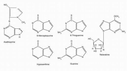
Three guanine analogs, azathioprine, 6-mercaptopurine (6-MP), and 6-thioguanine (6-TG), continue to be used in treatment of a variety of clinical conditions nearly 60 years after their initial development. 6-MP is employed as primary therapy for adults and children with acute lymphoblastic leukemia (ALL), childhood acute myeloid leukemia, and childhood lymphoma.1 6-TG is given for remission induction and maintenance therapy of acute myelogenous leukemia. Azathioprine, a prodrug of 6-MP, is widely used as an immunosuppressant for inflammatory bowel diseases, rheumatologic illnesses, and organ transplantation. These three drugs are closely related in structure (Fig. 11-1), metabolism, mechanism of action, and toxicity. Because of their similarities, they will be discussed together in this section. The key pharmacologic features of these drugs are summarized in Tables 11-1, 11-2 and 11-3.


Mechanism of Action

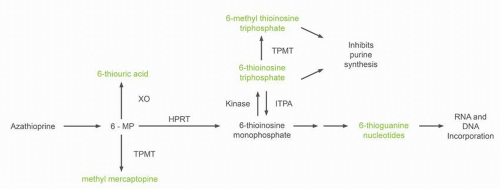
6-MP is a structural analog of hypoxanthine with a substitution of a thiol for the naturally occurring 6-hydroxyl group (Fig. 11-1). 6-MP undergoes extensive hepatic and cellular metabolism after dosing.2 Several competing routes for drug metabolism are present, some leading to inactive metabolites and others leading to active metabolites. 6-MP is activated intracellularly by the enzyme hypoxanthine-guanine phosphoribosyl transferase (HGPRT) to form 6-thioinosine monophosphate (TIMP). Sequential metabolism of TIMP to thioguanine nucleotides, such as 6-deoxy-thioguanosine triphosphate (6-TGTP), then occurs. 6-TG nucleotides are incorporated into DNA and RNA (Fig. 11-2). The quantity of 6-MP metabolite present in DNA correlates with cytotoxicity.3 Incorporation of 6-TGTP into DNA triggers programmed cell death by a process involving the mismatch repair pathway.4,5 Cytotoxicity depends on (a) incorporation of 6-TG into DNA, (b) miscoding during DNA replication, and (c) recognition of the abnormal base pairs by proteins of the postreplicative mismatch repair system. Methylation of 6-MP via conversion of TIMP to methyl thioinosine triphosphate may also contribute to the antiproliferative properties of the thioguanines through inhibition of de novo purine synthesis by methylmercaptopurine nucleotides.6

6-TG is converted to 6-thioguanylic acid (TGMP) by HGPRT.7 Conversion of 6-TG to TGMP requires two fewer enzymatic conversions than is necessary for the activation of 6-MP. Since methyl thiol inosine monophosphate is not formed following 6-TG administration, the effect of 6-TG on inhibition of purine metabolism is less than that of 6-MP. As a deoxynucleoside triphosphate, 6-TG acts as a substitute for DNA polymerase, and, as TGMP, it is subsequently incorporated into RNA and DNA. Incorporation of fraudulent nucleotides into DNA is believed to be the primary mechanism of 6-TG cytotoxicity,8 triggering apoptosis by a process involving the mismatch repair pathway, similar to 6-MP.5 Significantly higher cellular concentrations of thioguanine nucleotides are seen after 6-TG administration than with 6-MP.9

Azathioprine (Fig. 11-1) is rapidly cleaved in plasma by nonenzymatic mechanisms to 6-MP and methyl-4-nitro-5-imidazole derivatives (Fig. 11-2). Although incorporation of false nucleotides into DNA and inhibition of purine synthesis by 6-MP ribonucleotides are the probable mechanisms for cytotoxicity, the mechanism by which azathioprine and mercaptopurine modify immune response is likely different. Azathioprine inhibits T-lymphocyte activity to a greater extent than B-lymphocytes. 6-Thioguanine triphosphate (6-TGTP) binds to and inhibits Rac1, a small GTP-binding protein. Rac proteins play a major role in T cell development, differentiation, and proliferation. The activation of Rac1 targeted genes, such as mitogen-activated protein kinase (MEK), NFk BI, and bcl-XL, is suppressed by azathioprine leading to the mitochondrial pathway of apoptosis.10


Clinical Pharmacology


6-Mercaptopurine

6-MP is commercially available in 50-mg tablets, which also contain the inactive ingredients corn and potato starch, lactose, magnesium stearate, and stearic acid. An intravenous preparation of 6-MP has been formulated for research purposes. 6-MP is relatively insoluble and unstable in alkaline solutions.

Plasma 6-MP, 6-TG, and metabolite concentrations as low as 0.1 μM can be measured using high-performance liquid chromatography techniques.11 Accurate kinetics of oral and intravenous preparations have been determined. Following oral administration of commonly used 6-MP doses (75 mg/m2), peak plasma concentrations of 0.3 to 1.8 μM are seen within a mean of 2.2 hours.12 The volume of distribution exceeds that of total body water (0.9 L/kg). There is little penetration into the cerebrospinal fluid (CSF). With high-dose oral 6-MP (500 mg/m2), plasma 6-MP concentrations of 5 to 12 μM are achieved.13 In human leukemic cell culture models, concentrations of 1 to 10 μM are cytotoxic. Following intravenous dosing, the half-life of 6-MP is 50 to 100 minutes and plasma concentrations of 6-MP reach 25 μM and CSF concentrations 3.8 μM.14 Only weak protein binding is noted with 6-MP (20% bound).

Oral absorption of 6-MP is incomplete and highly variable.15 At a dose of 75 mg/m2, 6-MP, mean 6-MP bioavailability is 16% (range, 5%
to 37%).16 Clearance occurs primarily through two routes of metabolism. 6-MP is oxidized to the inactive metabolite, 6-thiouric acid, by xanthine oxidase (Fig. 11-2). 6-MP also undergoes S-methylation by the enzyme, thiopurine methyltransferase (TPMT), to yield 6-methyl mercaptopurine. The intestinal mucosa and liver contain high concentrations of the enzyme xanthine oxidase. The low bioavailability of 6-MP is the result of a large first-pass effect as drug is absorbed through the intestinal wall into the portal circulation and metabolized by xanthine oxidase. The use of concomitant allopurinol (an inhibitor of xanthine oxidase) increases 6-MP bioavailability fivefold.17 Allopurinol does not alter the plasma kinetics of intravenously administered 6-MP, although more 6-MP and
less thiouric acid are excreted in the urine following allopurinol therapy.18 Methotrexate, often used with 6-MP in maintenance treatment of ALL, is a weak inhibitor of xanthine oxidase. Concomitant use of methotrexate results in a small increase in the bioavailability of 6-MP. However, the modest increase in bioavailability is thought not to be clinically significant.15 The plasma concentration versus time profile of 6-MP differs in the same patient when studied on repeated occasions.19 High-dose, oral 6-MP (500 mg/m2) has been used in an attempt to saturate the first-pass metabolism of 6-MP, thereby increasing bioavailability.13 Even at a dose of 500 mg/m2 6-MP, xanthine oxidase is not saturated and no improvement in bioavailability is seen. Food intake and oral antibiotics reduce the oral absorption of 6-MP.20,21 The drug should be taken 1 to 2 hours after eating.


FIGURE 11-1 Structures of the naturally occurring purines guanine and hypoxanthine and related antineoplastic agents 6-mercaptopurine, 6-thioguanine, azathioprine, and nalarabine.


TABLE 11.1 Key features of mercaptopurine


Factor


Results


Mechanism of action


  1. Primary: incorporation of metabolites into DNA causes miscoding during DNA replication. Correlates with cytotoxicity


  2. Secondary: inhibits de novo purine synthesis; incorporated into RNA


Metabolism


Activation: conversion to thiopurine nucleotides


Catabolism: to 6-thiouric acid by xanthine oxidase


Catabolism: to 6-methylthiopurine by TPMT


Pharmacokinetics


t1/2: 50 min


Poor (<25%) and variable oral bioavailability


Elimination


Metabolism, by xanthine oxidase and TPMT


Drug interactions


Allopurinol decreases mercaptopurine elimination. Concomitant use of these two drugs requires dose reduction (75%).


Mesalamine, sulphasalazine, and olsalazine inhibit TPMT increasing thiopurine toxicity.


Toxicity


  1. Myelosuppression


  2. Mild gastrointestinal (nausea, vomiting)


  3. Hepatotoxicity including veno-occlusive disease


  4. 4. Immunosuppression


Precautions


  1. Dose reductions with allopurinol


  2. Persons with genetic deficiency of TPMT will have significantly increased toxicity (genetic screening available to test for TPMT deficiency).


TABLE 11.2 Key features of 6-TG


Factor


Results


Mechanism of action


Incorporation of fraudulent nucleotides into DNA


Metabolism


Activation: conversion to thiopurine nucleotides


Catabolism: to 6-thioxanthine by guanase


Catabolism: to 2-amino-6-methyl thiopurine by TPMT


Pharmacokinetics


t1/2: 90 min


Poor (15%-40%) and variable bioavailability


Elimination


Hepatic metabolism


Drug interactions


None well defined


Toxicity


  1. Myelosuppression


  2. Mild gastrointestinal (nausea, vomiting)


  3. Rare hepatotoxicity


Precautions


Increased toxicity in individuals with genetic deficiency of TPMT


TABLE 11.3 Key features of azathioprine


Factor


Result


Mechanism of action


Similar to mercaptopurine


Metabolism


Rapidly converted to 6-MP by nonenzymatic mechanisms


Pharmacokinetics


See mercaptopurine


Elimination


Rapid metabolism to 6-MP with subsequent elimination similar to 6-MP


Drug interactions


Allopurinol decreases elimination. Concomitant use with allopurinol requires azathioprine dose reductions (≥75%).


Toxicity


  1. Myelosuppression


  2. Gastrointestinal (nausea, vomiting)


  3. Rare hepatotoxicity


Precautions


  1. Dose reduction with allopurinol required


  2. Increased toxicity in individuals with genetic deficiency of thiopurine methyltransferase


As previously mentioned, two catabolic pathways for 6-MP metabolism exist that significantly affect drug activity: one via xanthine oxidase (just discussed) and a second via TPMT. Patient-to-patient variation in TPMT activity results in significant changes in 6-MP metabolism and drug toxicity. As seen in Figure 11-2, TPMT catalyzes the S-methylation of 6-MP to a relatively inactive metabolite, 6-methyl mercaptopurine (6CH3MP). TMPT also metabolizes thioinosine monophosphate into methyl thioinosine monophosphate, a molecule that can inhibit de novo purine biosynthesis. The frequency distribution of TPMT activity in large population studies is trimodal.22 One in two hundred to three hundred subjects has absent enzyme activity; 10% of the population has intermediate activity, and the rest have high enzyme activity. A reciprocal relationship between TPMT activity and the formation of 6-thiopurine nucleotides has been demonstrated. Over 20 nonsynonymous variations in the TMPT gene have been identified, 17 of which have reduced TMPT activity.23 Among these genetic variations, three (TPMT*2, TPMT*3A, and TMPT*3C) account for 90% of patients with low or intermediate TMPT activity. The proteins formed by TPMT*2 and TPMT*3A have degradation half-lives of 15 minutes compared to a half-life of 18 hours for wild type TPMT.24 Lymphoblasts from individuals homozygous or heterozygous for a variant TMTP gene have lower TMPT activity than lymphoblasts homozygous for the normal gene.25 Patients with low TPMT activity are susceptible to 6-MP and 6-TG-induced myelosuppression. Genetic testing using PCR-based methods can now identify TPMT-deficient and heterozygous patients.26 This test, as opposed to direct measurement of TPMT activity in red blood cells, is not affected by prior blood transfusions to the patient. In 2004, the FDA suggested TMPT testing for patients developing myelosuppression with standard dose 6-MP with subsequent dosage modifications for TMPTdeficient patients.27 Patients with absence of TMPT activity should have the dose of 6-MP reduced, but not the dose of other chemotherapeutic agents. It has been suggested that children with ALL should receive only 5% to 10% of a standard mercaptopurine dose
if they are homozygous for a variant TMPT gene and 65% if they are heterozygous.28


FIGURE 11-2 Mechanism of activation and catabolism of azathioprine and 6-MP. Active metabolites are noted in red. Inactive metabolites are indicated in blue. 6-MP, 6-mercaptopurine; HGPRT, hypoxanthine-guanine phosphoribosyl transferase; ITPA, inosine triphosphate pyrophosphatase; XO, xanthine oxidase; TPMT, thiopurine methyltransferase.

Several studies,29,30 but not all,16 have suggested that children with high TPMT activity are at greater risk of disease relapse as a result of decreased drug activation. In a large Scandinavian study in children with ALL, the risk of relapse following therapy was 18% for patients with wild type TPMT versus 6% for patients heterozygous or deficient in TMPT activity (P = 0.03).31 Despite a lower probability of relapse, patients with low TPMT activity did not have superior survival (P = 0.08), perhaps due to an increased rate of drug toxicity including the development of secondary malignancies.

While screening for TMPT variants with decreased enzymatic activity identifies individuals at risk for mercaptopurine toxicity, the association is not perfect. Toxicity still occurs in individuals with wild-type TMPT. Individuals heterozygous for TPMT*2, *3A and *3C can have a wide range of TPMT activity. Mercaptopurine metabolism is not dependent on a single gene (Fig. 11-2). Inosine triphosphate pyrophosphatase (ITPA) catalyzes the hydrolysis of inosine triphosphate (ITP) to inosine monophosphate which protects cells from the accumulation of potentially harmful nucleotides. A polymorphism in the ITPA gene occurring in roughly 10% of individuals results in a 25% decrease in enzyme activity in heterozygous individuals.32 ITPA inactivates the potentially toxic thioinosine triphosphate metabolite from patients receiving mercaptopurine or azathioprine (Fig. 11-2). In children with ALL who have had their mercaptopurine dose adjusted based on TMPT phenotype, the ITPA mutant allele has been associated with an increase incidence of febrile neutropenia33 and myelosuppression.34 However, an association of ITPA polymorphism with drug toxicity has not been seen in all studies.32


6-Thioguanine

6-TG is available as 40-mg tablets for oral use. An intravenous preparation is investigational. As with 6-MP, the absorption of 6-TG in humans is variable and incomplete (mean bioavailability is 30%; range: 14% to 46%).35 Peak plasma levels of 0.03 to 5 μM occur 2 to 4 hours after ingestion; the median drug half-life is 90 minutes but with wide variability reported.36 Intravenously administered 6-TG has been evaluated. Clearance of drug (600 to 1,000 mL/min/m2) appears to be dose dependent, suggesting saturation of clearance at doses over 10 mg/m2/h.37 Plasma concentrations of 4 to 10 μM can be achieved.

The catabolism of 6-TG differs from that of 6-MP. Thioguanine is not a substrate for xanthine oxidase. Thioguanine is converted to 6-thioinosine (an inactive metabolite) by the action of the enzyme, guanase. Because thioguanine inactivation does not depend on the action of xanthine oxidase, allopurinol will not block the detoxification of thioguanine. In humans, methylation of thioguanine, via TPMT, is more extensive than methylation of 6-MP. The product of methylation, 2-amino-6-methylthiopurine, is substantially less active and less toxic than thioguanine.

Azathioprine

Azathioprine is a prodrug of mercaptopurine. About 90% of an administered dose of azathioprine is non-enzymatically converted to 6-MP by sulfhydryl-containing compounds such as glutathione or cysteine. The metabolic pathways following conversion to mercaptopurine are identical to those just described for 6-MP.38 In transplant patients taking 2 mg/kg/d azathioprine, peak 6-MP plasma concentrations (Tmax < 2 hours) are low (75 ng/mL) and plasma drug half-life is short (1.9 hours).39 Plasma 6-MP concentrations exceed those of azathioprine within an hour of drug administration. Loss of renal function does not alter the plasma kinetics of either azathioprine or 6-MP. 6-MP was not detected in significant quantities in breast milk of women taking azathioprine and plasma concentrations of thiopurine metabolites were not found in infants breast-fed by women taking azathioprine.40


Toxicity


6-Mercaptopurine

The dose-limiting toxicity of 6-MP is myelosuppression, occurring 1 to 4 weeks following the onset of therapy and reversible when the drug is discontinued. Platelets, granulocytes, and erythrocytes are all affected. Weekly monitoring of blood counts during the first 2 months of therapy is recommended. Myelosuppression following 6-MP therapy is related to TPMT phenotype. Most patients (65%) with excessive toxicity following 6-MP or azathioprine administration have TMPT deficiency or heterozygosity.41

6-MP is an immunosuppressant. Immunity to infectious agents or vaccines is subnormal in patients receiving 6-MP. Gastrointestinal mucositis and stomatitis are modest. Approximately one quarter of treated patients experienced nausea, vomiting, and anorexia. Gastrointestinal side effects appear to be more common in adults than in children. Pancreatitis is seen in 3% of patients with long-term therapy, with rash, fever, or joints pains seen in 2%.41 Three types of hepatotoxicity can bee seen with thiopurine therapy.42 A small percentage of patients have transient, asymptomatic elevation in transaminases, which return to normal with follow-up and do not require dose alterations. Thiopurines may induce a several cholestatic jaundice that may not regress. Thiopurine therapy should be discontinued in this situation. Thiopurines may cause endothelial cell injury with raised portal pressures (VOD or veno-occlusive disease). The development of hepatotoxicity, in contrast to myelosuppression, is not associated with TPMT polymorphisms43 but is correlated with the dose of 6-MP given and with the formation of methylated metabolites of 6-MP (but not with 6-TG nucleotide formation).44

At very high doses (>1,000 mg/m2), the limited solubility of 6-MP can cause precipitation of drug in the renal tubules with hematuria and crystalluria.45 Thioguanine therapy does not appear to markedly increase a patient’s rates of developing a second malignancy.46 However, an increased risk of myelodysplastic syndrome or AML was found in children treated with mercaptopurine for ALL who had reduced TMPT activity.47


Thioguanine

As with 6-MP, the primary toxicity of 6-TG is myelosuppression.48 Blood counts should be frequently monitored because there may be a delayed effect during oral drug administration. Higher doses result in mucositis. Thioguanine produces gastrointestinal toxicities similar to 6-MP but less frequently. Jaundice and hepatic VOD have been reported more frequently (11% incidence) with 6-TG than with mercaptopurine or azathioprine.49,50 At present, 6-TG should be used only as second-line thiopurine therapy. There is suggestive
data that VOD may be caused by the formation of methylmercaptopurine nucletides.51


Azathioprine

Adverse effects from azathioprine are similar to those seen with 6-MP. These effects include leukopenia, diarrhea, nausea, abnormal liver function tests, and skin rashes. Frequent monitoring of the complete blood count is warranted throughout therapy (weekly during the first 8 weeks of therapy). Molecular testing for TMPT may be a cost-effective way of identifying the 10% of the population at high risk for toxicity.32

A hypersensitivity reaction, generally characterized by fever, chills, severe nausea, diarrhea, hypertension, and hepatic dysfunction, has been reported.52 The mechanism for the hypersensitivity reaction is unclear. Chronic azathioprine therapy results in a 3% per patient-year incidence of myelotoxicity (1% severe). The cumulative risk of infectious complications among azathioprine myelosuppressed patients is 6.5%.53 Patients homozygous and heterozygous for mutant TPMT are at high risk for toxicity and dose modification.54


Use and Drug Interactions

6-MP is a standard component of maintenance therapy for ALL. It has little role in therapy of solid tumors or remission induction in myeloid leukemias. 6-MP is also used to treat inflammatory bowel disease. 6-TG should be used only rarely as second- or third-line therapy for leukemias and lymphomas given its greater incidence of hepatic toxicity. Azathioprine is used as an immunosuppressant in preventing rejection of organ transplants and in the therapy of illnesses believed autoimmune in character (such as lupus, rheumatoid arthritis, and ulcerative colitis). Thiopurines are considered the standard of care for the treatment of Crohn’s disease.

As previously mentioned, allopurinol inhibits the catabolism of 6-MP and increases its bioavailability.17 Oral doses of 6-MP and azathioprine should be reduced by at least 75% in patients also receiving allopurinol. Combined use of standard dose azathioprine (or 6-MP) with allopurinol will result in life-threatening toxicity.55 Methotrexate causes a modest increase in 6-MP bioavailability but not to an extent significant enough to warrant dosage reduction.15 Methotrexate increases 6-MP plasma concentrations slightly but antagonizes thiopurine metabolite disposition in leukemia blasts resulting in lower thioguanine nucleotide incorporation.56 Olsalazine, mesalazine, and sulfasalazine are inhibitors of TPMT and can increase the toxicity of mercaptopurine or azathioprine.57 Other agents including furosemide and various nonsteroidal anti-inflammatory agents can inhibit TPMT in vitro but the clinical importance of this finding remains unclear.


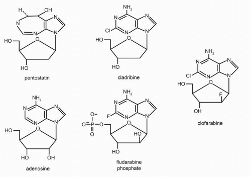
FIGURE 11-3 Chemical structure of adenosine analogs.


Adenosine Analogs

Adenosine analogs with documented clinical utility are fludarabine, pentostatin, cladribine (2′-chlorodeoxyadenosine), and clofarabine (Fig. 11-3). Key pharmacologic features of the adenosine analogs are listed in Tables 11-4, 11-5, 11-6 and 11-7. Adenosine arabinoside is cytotoxic in vitro but in man is quickly inactivated by the enzyme adenosine deaminase (ADA). Substitution of a halogen (fluorine in fludarabine or chlorine in cladribine) at the two position of deoxyadenosine produces molecules which are resistant to the action of ADA.


Fludarabine (Fludara)

Fludarabine (or 9-β-D-arabinofuranosyl-2-fluoroadenine monophosphate) is a monophosphate analog of adenosine arabinoside (Fig. 11-3). The monophosphate moiety results in aqueous solubility allowing intravenous administration.58 Key features of fludarabine are summarized in Table 11-4.


TABLE 11.4 Key features of fludarabine


Factor


Result


Mechanism of action


  1. Incorporation into DNA as a false nucleotide


  2. Inhibition of DNA polymerase, DNA primase, and DNA ligase


  3. DNA chain termination


Metabolism


  1. Rapid dephosphorylation in plasma to 2-fluoro-ara (F-are-A)


  2. Activation of F-ara-A to F-ara-ATP (the active metabolite) within cells


Pharmacokinetics


  1. Rapid dephosphorylation to 2-F-ara-A


  2. t1/2 2-F-ara-A = 6-30 h in plasma; intracellular t1/2 of F-ara-ATP = 15 h


Elimination


Primarily renal excretion of 2-F-ara-A


Drug interactions


Increases cytotoxicity of cytarabine and cisplatin


Toxicity


  1. Myelosuppression


  2. Immunosuppression with resulting infections


  3. Neurotoxicity at high doses


  4. Rare: interstitial pneumonitis and hemolytic anemia


Precautions


Dose reduction needed for patients with renal failure


Mechanism of Action

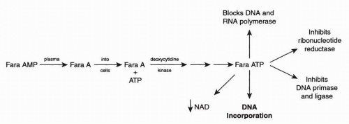
After intravenous administration, fludarabine is rapidly and completely dephosphorylated in plasma to the nucleoside, 9-β-D-arabinofuranosyl-2-fluoroadenine (F-ara-A) (Fig. 11-4).59 F-ara-A enters cells via carrier-mediated transport60 and is phosphorylated to its active form, F-ara-ATP. All cytotoxic mechanisms of action of fludarabine require the presence of fludarabine triphosphate (F-ara-ATP).61 F-ara-ATP inhibits several intracellular enzymes important in DNA replication including DNA polymerase, ribonucleotide reductase, DNA primase, and DNA ligase I. In addition, fludarabine is incorporated into DNA. Once incorporated, F-ara-AMP is an effective DNA chain terminator,62 primarily at the 3′ end of DNA. The amount of fludarabine incorporated into DNA is linearly correlated with loss of clonagenicity. Excision of the 3′-terminal F-ara-AMP does not easily occur, and the presence of this false nucleotide leads to apoptosis.


FIGURE 11-4 Activation of fludarabine.

Although the effects of fludarabine on DNA synthesis account for its activity in dividing cells, fludarabine is also cytotoxicity in diseases with very low growth fractions such as chronic lymphocytic leukemia (CLL) or indolent lymphomas. This raises the question as to how an “S-phase” agent is active in nondividing cells.63 The specific mechanism(s) by which fludarabine induces cell death among quiescent cells remains under investigation, but several proposed mechanisms of action include fludarabine’s ability to inhibit RNA polymerases by incorporation into RNA, depletion of nicotinamide adenine dinucleotide (NAD) with resultant decrease in cellular energy stores, and interference with normal DNA repair processes.64 The most compelling evidence suggests fludarabine, after incorporation into DNA triggers apoptosis during the DNA repair process.65


Clinical Pharmacology

Following intravenous administration, parent drug (2-F-ara-AMP) undergoes rapid (2 to 4 minutes) and complete conversion to F-ara-A through the action of 5′ nucleotidase present in erythrocytes and endothelial cells. With commonly used doses of 25 to 30 mg/m2 fludarabine, peak plasma F-ara-A concentrations of 1 to 5 μmol/L are achieved within 5 minutes.66 Wide variations in terminal drug half-life (7 to 33 hours) and area under the curve (AUC) are found. Drug clearance is linear, with no change with repeated doses. Peak F-ara-ATP concentrations in circulating leukemic cells are achieved 4 hours after intravenous fludarabine administration. F-ara-ATP has a relatively long intracellular half-life (15 hours), which may account for the efficacy of a daily administration schedule.61 A linear relationship exists between plasma F-ara-A concentrations and intracellular F-ara-ATP in leukemic cells.67

F-ara-A is excreted primarily in the urine (50% to 60%) with no metabolites detected.61 Patients with renal impairment, compared to patients with normal kidney function, have a significant decrease in clearance of 2-F-ara-A (ClT = 51.82 ± 6.70 versus 73.53 ± 3.79 mL/min/m2).66 Lichtman et al.68 have proposed that patients with a
creatinine clearance (Clcr) of greater than 70 mL/min per 1.73 m2 should receive 25 mg/m2/d for 5 days of fludarabine, and patients with Clcr of 30 to 70 mL/min per 1.73 m2 should receive 20 mg/m2/d for 5 days, and those with Clcr of less than 30 mL/min per 1.73 m2 should receive 15 mg/m2/d for 5 days fludarabine. Aronoff et al.69 suggests a 25% dose reduction for a Clr of 10 to 50 mL/min and a 50% dose reduction for a Clcr less than 10 mL/min. The FDA package insert suggests a 20% dose reduction for Clcr of 30 to 70 mL/min and avoiding use in patients with a Clr less than 30.

Oral administration of fludarabine has been evaluated, and an oral preparation is available.70 The AUC of F-ara-A increases linearly with increasing oral dose. Absorption is not affected by meals.71 An oral dose of 40 mg/m2/d should provide similar systemic drug exposure to an intravenous dose of 25 mg/m2/d. Mean bioavailability averages 50% to 55% with large interpatient (30% to 80%), but minimal intraindividual variability.


Toxicity

The primary dose-limiting toxicities of fludarabine are myelosuppression and infectious complications resulting from immunosuppression.57 Toxicity is similar with oral and intravenous preparations.70 Reversible leukopenia and thrombocytopenia have been reported following fludarabine administration with a median time to nadir of 13 days (range, 3 to 25 days) and 16 days (range, 2 to 32 days), respectively. Grade 3 neutropenia (nadir <1,000/mm3 at standard doses of 25 mg/m2/d for 5 days) is seen in 25% to 30% of patients with Grade 3 thrombocytopenia (platelet nadirs <50,000/mm3) in 5% to 10%.72 Myelosuppression is more common when fludarabine is combined with other chemotherapeutic drugs, including rituximab.73 Up to 25% of patients treated with fludarabine will have a febrile episode. Many will be fevers of unknown origin but one third will have a serious documented infection.

Fludarabine is immunosuppressive, inhibiting signal transduction important in lymphocyte activation.74 Therapy is associated with an increased risk of opportunistic infections. CD4 and CD8 T-lymphocytic subpopulations decrease to levels of 150 to 200/mm3 after three courses of therapy.75 Lymphopenia may persist for over 1 year. The most frequent infectious complications are respiratory. Infections with Cryptococcus, Listeria monocytogenes, Pneumocystis carinii, cytomegalovirus, herpes simplex virus, Varicella zoster, and mycobacterium, organisms associated with T-cell dysfunction, are seen.76

Only gold members can continue reading. Log In or Register to continue

Stay updated, free articles. Join our Telegram channel

May 27, 2016 | Posted by in ONCOLOGY | Comments Off on Purine Antimetabolites

Full access? Get Clinical Tree

Get Clinical Tree app for offline access