Bleomycin and Other Antitumor Antibiotics


Bleomycin and Other Antitumor Antibiotics


Bruce A. Chabner


In a search for new antimicrobial and antineoplastic agents, Umezawa et al.1 isolated a number of small glycopeptides from culture broths of the fungus Streptomyces verticillus. The most active antitumor agent found was, in fact, a mixture of peptides now known as bleomycin, a drug with important activity against Hodgkin’s disease, testicular cancer, malignant pleural effusions, and cancers of the cervix and penis. Bleomycin combined with vinblastine, etoposide, and cis-diamminedichloroplatinum cures most patients with germinal neoplasms of the testis.2 The drug has attracted great interest because of its unique biochemical action, its virtual lack of toxicity for normal hematopoietic tissue, and its unfortunate ability to cause pulmonary fibrosis. Its primary pharmacologic and pharmacokinetic features are shown in Table 16-1.


Structure and Mechanism of Action

The bleomycins are a family of peptides with a molecular weight of approximately 1,500 (Fig. 16-1). All contain a unique structural component, bleomycinic acid, and differ only in their terminal alkylamine group. Because of their unusual structure, catalytic properties, and important antitumor activity, the bleomycin antibiotics have been the subject of intensive basic and clinical investigation. Bleomycin A2, the predominant peptide, has been prepared by total chemical synthesis, as has a series of analogs.3 More than 100 additional bleomycin-like antitumor antibiotics have been isolated or synthesized, but none has yet emerged as a superior drug.

The clinical mixture of bleomycin peptides is formulated as a sulfate salt, and its potency is measured in units (U) of antimicrobial activity. Each unit contains between 1.2 and 1.7 mg of polypeptide protein. The powdered clinical mixture is stable for at least 1 year at room temperature and for 4 weeks after reconstitution in aqueous solution at 4°C.

The multiple glycopeptides found in the clinical preparation of bleomycin have been separated and purified by high-performance liquid chromatography (HPLC).4 The predominant active component, comprising approximately 70% of the commercial preparation, is the A2 peptide. The other bleomycins differ only in their terminal amine. The native compound isolated from S. verticillus is a blue-colored Cu(II) coordinated complex, although the peptide will complex in vitro with other metals including iron, cobalt, zinc, and manganese, in their various valence states. The Co(III) complexes are essentially inert with respect to biologic activity and the exchangeability of their bound metal and thus have been candidates for tumor localization, especially with cobalt 57 (57Co). Unfortunately, the half-life of 57Co is 270 days rather than the desired several hours or days common for clinically useful diagnostic agents. Among endogenous metals, bleomycin has the highest affinity for Cu(II); bleomycin has a fourfold greater affinity for reduced Cu(I) than for Fe(II).5 In initial clinical trials with Cu(II)·bleomycin, patients experienced profound phlebitis, and the white apobleomycin was soon adopted for clinical use. Nevertheless, after systemic administration, bleomycin appears to speciate rapidly with Cu(II) removed from plasma proteins.6 The Cu(II)·bleomycin complex is internalized
through a poorly described endocytotic system that may include discrete plasma membrane proteins of 250 kD.7,8 Most investigators believe that the Cu-bleomycin is a prodrug and that cleavagecompetent bleomycin is Fe(II) speciated. Considerable chemical evidence regarding DNA damage produced by Fe(II)·bleomycin exists to support this hypothesis.5 The primary model was first outlined by Umezawa’s group6 and is schematically represented in Figure 16-2. Cu(II) associated with intracellular Cu(II)·bleomycin is reduced, possibly by intracellular cysteine-rich proteins, to Cu(I), which is released, and the apobleomycin quickly complexes with the more abundant Fe(II).9 Nuclear translocation of the Fe(II)·bleomycin complex proceeds with subsequent chromatin damage. The metal coordination chemistry of bleomycin has been the subject of considerable attention, and primarily on the basis of studies using electron paramagnetic resonance (EPR),10 crystallography,11 and Fe(II) surrogate metals such as Cu(II), a square-pyramidal complex, as indicated in Figure 16-1, is most favored.5 Six distinct moieties are required for this metal coordination complex, and the N-1 of the pyrimidine, the N of the imidazole, and the secondary amine are undisputed participants.5 Debate still exists about the arrangement of the remaining ligands, and this presumably will be resolved with further structural analyses. Although many aspects of the cellular scheme found in Figure 16-2 are biologically and chemically appealing, several questions remain unanswered, including the identity of the sulfhydryl-rich reductant for Cu(II), the recipient and fate of the Cu(I), the intracellular source of Fe(II), and the mode by which the metal-bound bleomycin translocates the plasma membrane and the nucleus. Therefore, others12,13 have developed a rival hypothesis to explain the antitumor activity and fate of bleomycin. Even though Cu(II)·bleomycin is not highly cytotoxic,14 persuasive arguments for a potential functional role of Cu(I)·bleomycin in the biologic actions of this antineoplastic agent have been presented.5,13 Thus the widely embraced concept that Fe(II)-complexed bleomycin is the only biologically relevant species may require revision.


TABLE 16.1 Key features of bleomycin pharmacology


Mechanism of action


Oxidative cleavage of DNA initiated by hydrogen abstraction


Metabolism


Activated by microsomal reduction


Degraded by hydrolase found in multiple tissues


Pharmacokinetics


t1/2α: 24 min; t1/2β: 2-4 h


Elimination


Renal: 45%-70% in first 24 h


Drug interactions


None clearly established at a biochemical level


Oxygen enhances pulmonary toxicity


cis-Platinum induces renal failure and increases risk of pulmonary toxicity


Toxicity


Pulmonary interstitial infiltrates and fibrosis


Desquamation, especially of fingers, elbows


Raynaud’s phenomenon


Hypersensitivity reactions (fever, anaphylaxis, eosinophilic pulmonary infiltrates)


Precautions


Pulmonary toxicity increased in patients with


Underlying pulmonary disease


Age >70 y


Renal insufficiency


Prior chest irradiation


O2 during surgery


Reduce dose if creatinine clearance


<80 mL/min


t1/2, half-life.


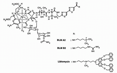
FIGURE 16-1 Structure of bleomycin·Fe(II) complex. The various substitutions on the amino-terminal end of the molecule are shown for bleomycin A2 (BLM A2), for bleomycin B2 (BLM B2; also a component of the clinical preparation), and for one congener, liblomycin.


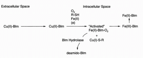
FIGURE 16-2 Schematic representation of bleomycin (Blm) transformation as it moves from the extracellular to the intracellular space. The Cu(II)·Blm complex in the extracellular space is converted to a cytotoxic Fe(II)·Blm·O2 complex in the cell. Inactivation of Blm by Blm hydrolase is also shown.


Mechanism of Action

Early mechanistic studies identified a concentration-dependent loss in DNA integrity with loss of cell viability in the absence of marked decreases in either RNA or protein composition. Thus most investigators accepted direct DNA damage as the most attractive candidate for the cytotoxicity and, consequently, the antitumor activity of bleomycin. Single- and double-strand DNA damage are readily observed in cultured cells and isolated DNA incubated with bleomycin in solution. This breakage is reflected in the chromosomal gaps, deletions, and fragments seen in cytogenetic studies of whole cells incubated with the drug. Although this may simply reflect rapid DNA repair, other biochemical targets, including the various species of RNA and lipid, are attacked by bleomycin and may contribute to its action.15,16


Chemistry of Bleomycin-Mediated DNA Cleavage

The mechanism by which Fe(II)·bleomycin cleaves DNA has been examined using viral, bacterial, mammalian, and synthetic DNAs. Bleomycin is unlike most DNA-damaging agents because it attacks neither the nucleic bases nor the phosphate linkage. In this multistep process, initially an “activated” Fe(II)·bleomycin·O2 complex is formed that is kinetically competent to cleave DNA. The binding of dioxygen to Fe(II)·bleomycin proceeds most rapidly in the presence of DNA, which stabilizes the complex.17 The proposed sequence of events responsible for the production of an activated bleomycin has been deduced from in vitro studies and is briefly outlined in Figure 16-3. Fe(II) combines with apobleomycin, producing an EPR-silent, high-spin Fe(II)·bleomycin complex. With dioxygen, this is rapidly converted to a ternary Fe(II)·bleomycin·O2 species, which can be trapped with isocyanide, CO, or NO or can be activated by a 1e reduction. The e can be supplied by a second Fe(II)·bleomycin·O2 molecule,18 by H2O2, by microsomal enzymes19 and nicotinamide-adenine dinucleotide phosphate (reduced form) (NADPH) organic reductant, or by nuclei and nicotinamide-adenine dinucleotide (reduced form) (NADH).20 Mossbauer studies19 suggest that the activated bleomycin has a half-life of a few minutes at 0°C, so it is likely to be reasonably long lived even at 37°C. In the absence of DNA, the activated species will self-destruct. The association constant of Fe(II)·bleomycin for duplex DNA, however, is approximately 105/M.21 Thus the second step in the DNA cleavage process readily occurs. The interaction of bleomycin with DNA shows nucleotide sequence selectivity22 and most likely occurs at the minor groove. At saturating concentrations of bleomycin, one molecule of drug associates with four or five base pairs of DNA. The binding between bleomycin and DNA appears to be through electrostatic interactions and partial intercalation (insertion between base pairs) of the amino-terminal tripeptide of bleomycin (called the S tripeptide)23,24 (Fig. 16-4). The bithiazole of the S tripeptide bonds to guanine groups in the favored sequence of GpC and GpT.22,23 The terminal dimethylsulfonium of bleomycin A2 and the positively charged terminal amines of other bleomycins also participate in DNA binding, as indicated by proton magnetic resonance studies.22 Binding of either bleomycin or its S tripeptide to DNA produces unwinding of the double-helical structure as the result of intercalation.24 Fe(II)·bleomycin exhibits a strong preference for the B-form of DNA rather than for the Z-form,25 consistent with interactions with the minor groove of DNA.


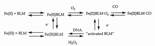
FIGURE 16-3 Model for the activation of cleavage-competent bleomycin (BLM).


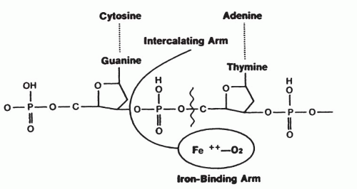
FIGURE 16-4 Intercalation of the bithiazole groups between DNA base pairs, at least one of which contains the GpT of GpC sequence. Also shown is the apposition of the Fe(II)-binding portion of bleomycin to the deoxyribose group, which is cleaved via hydrogen abstraction at the C-4′ of the deoxyribose.

The third step in the action of bleomycin is the generation of single- and double-strand DNA breaks. During the DNA cleavage process, Fe(II)·bleomycin functions catalytically as a ferrous oxidase26 with the oxidation of Fe(II) to Fe(III); regeneration of the active Fe(II) requires endogenous reductants, including cytochrome P-450 reductase and NADPH,27 an enzyme found in the nucleus and nuclear membrane. Under very controlled in vitro conditions, the short-lived oxygenated iron-bleomycin species28 participates in almost four cleavage events per bleomycin molecule. Others have estimated the reduction of dioxygen by bleomycin, as monitored by
measurement of oxygen consumption, with a maximum velocity of 27 mol oxygen consumed per minute per mole of bleomycin.26

The mechanism of DNA cleavage has been defined by the DNA fragments produced after incubation of the substrate with activated bleomycin.16,29,30,31 Incubation of DNA with bleomycin in an aerobic environment results in the scission of the C-3′-C-4′ ribose bond via a Criegee-type rearrangement, which produces three types of product, including a 5′-oligonucleotide terminating at its 3′ end with a phosphoglycolic acid moiety, a 3′-oligonucleotide containing a 5′-phosphate, and a 3′-(thymin-9′-yl)propenal.13 Exposure to Fe(II)·bleomycin produces the release of all four bases (thymine, cytosine, adenine, and guanine)29 (Fig. 16-5, pathway A). Under anaerobic conditions, the free-base release is accompanied by production of an oxidatively damaged sugar in the intact DNA strand, which yields DNA cleavage only under basic conditions, namely, pH 12 (Fig. 16-5, pathway B). No base propenal is released. Which of these pathways predominates in intact cells is not known, although both free bases and base propenal adducts are detected in most cells. The base propenal compounds have intrinsic cytotoxicity and may contribute to further damage to cells.32


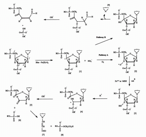
FIGURE 16-5 Scheme for the cleavage of the 3′-4′ deoxyribose bond by the activated bleomycin·Fe(II)·O2 complex. The activated drug complex initially abstracts a hydrogen radical from the 4′ position to produce the unstable intermediate [1] that decomposes in the presence of oxygen (pathway A) to produce the free base propenal [7], leaving a 3′-phosphoglycolate ester [8] and a 5′-phosphate [6] at the free ends of the broken DNA strand. Under conditions of limited oxygen (pathway B), a free base [9] is released, and DNA strand scission occurs only in the presence of alkali (pH 12).

Bleomycin produces both single- and double-strand DNA breaks in a ratio of approximately 10:1. The unexpectedly high frequency of double-strand breaks has been addressed in elegant studies of the effect of bleomycin on hairpin-shaped oligonucleotides that have single-strand gaps corresponding to those produced by bleomycin.33 The highly electronegative 3′-phosphoglycolate and 5′-phosphate
groups remaining at the site of DNA single-strand cleavage may promote access of a second bleomycin molecule to the opposing strand, resulting in a double-strand break.

Analysis of the products of DNA cleavage, using either viral or mammalian DNA, has consistently shown a preferential release of thymine or thymine-propenal, with lesser amounts of the other three bases or their propenal adducts.16,34 The propensity for attack at thymine bases probably results from the previously mentioned preference for partial intercalation of bleomycin between base pairs in which at least one strand contains the sequence 5′-GpT-3′. The specificity for cleavage of DNA at a residue located at the 3′ side of G seems to be absolute.35 A schematic representation of the intercalation and cleavage processes as conceived by Grollman and Takeshita36 is given in Figure 16-4 and summarizes the structural and sequence specificities discussed in this chapter.


Cellular Pharmacology

The cellular uptake of bleomycin is slow, and large concentration gradients are maintained between extracellular and intracellular spaces.37 14C-bleomycin accumulates at the cell membrane of murine tumor cells, with gradual appearance of labeling at the nuclear membrane only after 4 hours of exposure.38 The plasma membrane acts as a barrier for the highly cationic bleomycins, which also have a significant size that limits their diffusion.39 A bleomycin-binding membrane protein, which may participate in its internalization, has a molecular mass of 250 kD and becomes half-maximally saturated with a bleomycin concentration of 5 μmol/L.8 Using a fluorescent mimic of bleomycin or agents that disrupt vacuoles, Mistry et al.40 and Lazo et al.7 concluded that the internalized bleomycin is sequestered in cytoplasmic organelles. The process by which the entrapped bleomycin is released from the vesicles is not known.

Once bleomycin is internalized, it either translocates to the nucleus to effect DNA damage or can be degraded by bleomycin hydrolase, which has been characterized and cloned from human sources.41,42 This homomultimeric enzyme metabolizes and inactivates a broad spectrum of bleomycin analogs. The enzyme cleaves the carboxamide amine from the β-aminoalaninamide, yielding a weakly cytotoxic (<1/100) deaminobleomycin.41 Both the primary amino acid sequence and higher-order structure determined by x-ray crystallography reveal that bleomycin hydrolase is a founding member of what is a growing class of self-compartmentalizing or sequestered intracellular proteases.43,44 Both yeast and human enzymes are homohexamers with a ring or barrel-like structure that have the papain-like active sites situated within a central channel in a manner resembling the organization of the active sites in the 20S proteosome.44 The central channel, which has a strong positive electrostatic potential in the yeast protein, is slightly negative in human bleomycin hydrolase. The yeast enzyme binds to DNA and RNA, but human bleomycin hydrolase lacks this attribute.42,44,45 The C-terminus requires autoprocessing of the terminal amino acid, and the processed enzyme has both aminopeptidase and peptide ligase activities. The kinetic properties of bleomycin hydrolase, such as its pH optimum and salt requirements, are distinct from those of other cysteine proteinases, although the substrate specificity of bleomycin hydrolase is similar to that of cathepsin H. Human bleomycin hydrolase is located on chromosome band 17q11.2 and has one polymorphic site encoding either a valine or isoleucine.46 Bleomycin hydrolase is found in both normal and malignant cells.42,47 That this is the only enzyme responsible for metabolizing bleomycin was documented with bleomycin hydrolase-null or “knockout” mice.48 This inactivating enzyme is present in relatively low concentrations in lung and skin, the two normal tissues most susceptible to bleomycin damage.41,47 Interestingly, pulmonary bleomycin hydrolase levels are highest in animal species or strains resistant to the pulmonary toxicity of bleomycin.41 Mice that lack the functional gene are more sensitive to the toxic effects of bleomycin.48 A polymorphism, A1450G, in the coding region is found in 10% of patients with testicular cancer and is associated with a 20% decrease in survival in patients receiving a regimen containing bleomycin. These findings suggest that the G/G genotype is associated with an increased hydrolytic activity.49

DNA is more sensitive to DNA cleavage at the G2-M and G1 phases of the cell cycle than at S phase, which may reflect differences in chromatin structure.50 The degree of chromatin compactness dramatically influences bleomycin-induced DNA damage.51

Despite the apparent increased toxicity for cells in G2, no agreement exists regarding preferential kill of logarithmically growing cells as compared with plateau-phase cells; indeed, some workers have observed greater fractional cell kill for plateau-phase cells.52 The possibility of enhancing cell kill by maximizing exposure during G2 has led to a trial of bleomycin by continuous infusion, with unimpressive clinical results.

The intracellular lesions caused by bleomycin include chromosomal breaks and deletions and both single-strand and (less frequently) double-strand breaks. In nonmitotic cells, DNA is organized into nucleosomes, or small beads, which are joined by long strands, or linker regions. The primary point of attack seems to be in the linker regions of DNA, between nucleosomes.53 Interestingly, the resulting 180- to 200-base-pair fragments are similar in size to those formed by endonucleases activated during apoptosis.39 Cell kill and DNA strand breakage increase in proportion to the duration of drug exposure for at least 6 hours; this finding again implies a possible advantage for giving bleomycin as a prolonged infusion.

Cells are able to repair bleomycin-induced DNA breaks via a complex array of enzymes and pathways specific for both single-strand and double-strand breaks. A delay in plating cells after bleomycin exposure increases plating efficiency, presumably by allowing time for repair of potentially lethal damage.54 Inhibitors of DNA repair, such as caffeine and 3′-aminobenzamide,55 accentuate DNA strand breakage and cell kill by bleomycin. Indirect evidence suggests that repair processes similar to those required for repair of lesions induced by ionizing radiation play a role in limiting damage due to bleomycin.56 Cells from patients with ataxia-telangiectasia, which arises from an inherited defect in DNA repair, have increased sensitivity to bleomycin,57 as do cells deficient in BRCA1and in other components of repair pathways.58,59


Resistance

Several intracellular factors have been identified as contributors to bleomycin tumor resistance: increased drug inactivation, decreased drug accumulation, and increased repair of DNA damage,
particularly double-strand breaks.58,59 Early studies60 demonstrated increased rates of bleomycin inactivation in two bleomycin-resistant rat hepatoma cell lines. Morris et al.61 demonstrated an increased level of bleomycin hydrolase in cultured human head and neck carcinoma cells with acquired resistance to bleomycin. Metabolic inactivation of bleomycin also can contribute to intrinsic bleomycin resistance in human colon carcinoma cells.62

Increased bleomycin hydrolase activity is not the only mechanism of bleomycin resistance.63 Some cells selected in culture for bleomycin resistance display enhanced DNA repair capacity.64 Because Fe(III)·bleomycin requires reduction to Fe(II)·bleomycin, sulfhydryl groups on proteins and peptides are potential factors in drug resistance. Tumor lines with elevated levels of glutathione, selected for resistance to doxorubicin, are collaterally sensitive to bleomycin.65 The evidence for glutathione enhancement of bleomycin activity is not entirely clear, as buthionine sulfoxamine, a glutathione-depleting agent, enhances tumor sensitivity to bleomycin.66 Increasing the major protein thiol metallothionein produces a small increase in bleomycin sensitivity, consistent with the proposal that this cysteine-rich protein may assist in the removal of Cu(I) from bleomycin.9 Bleomycin is not affected by P-glycoprotein, the product of the multidrug resistance gene.


Clinical Pharmacokinetics

A number of techniques have been developed for assay of bleomycin in biologic fluids, including microbiologic methods,67 HPLC,68 biochemical techniques (degradation of DNA),69 and radioimmunoassay methods,70 which, using bleomycin labeled with iodine-125 or 57Co, may be the most rapid and simple. The antibodies described by Broughton and Strong70 react quantitatively with the component peptides of the clinically used bleomycin formulation. The primary component peptides A2 and B2 give 75% to 100% reactivity compared with the mixture in standard curve determinations. HPLC, using the ion-pairing technique, allows resolution of the component peptides but is more time consuming.

The hallmark of bleomycin pharmacokinetics in patients with normal serum creatinine is a rapid two-phase drug disappearance from plasma; 45%71 to 70%72 of the dose is excreted in the urine within 24 hours. For intravenous bolus doses, the half-lives for plasma disappearance have varied somewhat among the published studies. Alberts et al.73 reported α and β half-lives of 24 minutes and 4 hours, respectively, whereas Crooke et al.74 estimated the β half-life to be approximately 2 hours. Peak plasma concentrations reach 1 to 10 mU/mL for intravenous bolus doses of 15 U/m2.

For patients receiving bleomycin by continuous intravenous infusion, the postinfusion half-life is approximately 3 hours. Intramuscular injection of bleomycin (2 to 10 U/m2) gave peak plasma levels of 0.13 to 0.6 mU/mL, or approximately one tenth the peak level achieved by the intravenous bolus doses.75 The mean half-life after intramuscular injection was 2.5 hours, or approximately the same as that after intravenous injection. Peak serum concentrations were reached approximately 1 hour after injection (Fig. 16-6). Bleomycin pharmacokinetics also have been studied in patients receiving intrapleural or intraperitoneal injections. These routes have proved effective in controlling malignant effusions due to breast, lung, and ovarian cancers.76 Intracavitary bleomycin, in doses of 60 U/m2, gives peak plasma levels of 0.4 to 5.0 mU/mL, with a plasma half-life of 3.4 hours after intrapleural doses and 5.3 hours after intraperitoneal injection.77 Corresponding intracavitary levels are 10- to 22-fold higher than simultaneous plasma concentrations.78 Approximately, 45% of an intracavitary dose is absorbed into the systemic circulation, and 30% is excreted in the urine as immunoreactive material.


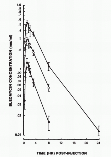
FIGURE 16-6 Pharmacokinetics of bleomycin after intramuscular administration of 2 (•), 5 (m), and 10 (▲) mg of bleomycin per meter square. (From Oken MM, Crooke ST, Elson MK, et al. Pharmacokinetics of bleomycin after IM administration in man. Cancer Treat Rep 1981;65:485.)

As might be expected, bleomycin pharmacokinetics is markedly altered in patients with abnormal renal function, particularly those with creatinine clearance of less than 35 mL/min. Alberts et al.73 noted a terminal half-life of approximately 10 hours in a patient with a slightly elevated creatinine clearance of 1.5 mg/dL, and Crooke et al.71 reported a patient who showed a creatinine clearance of 10.7 mL/min and a β half-life of 21 hours. Others have reported a high frequency of pulmonary toxicity in patients with renal dysfunction secondary to cisplatin treatment.72,79 One report described fatal pulmonary fibrosis that occurred after three doses of 20 U each given to a patient with chronic renal insufficiency (blood urea nitrogen, 48 mg/dL; creatinine, 4.8 mg/dL).80 The available data are too limited to provide accurate guidelines for dosage adjustment in patients with renal failure. One retrospective study identified a glomerular filtration rate of less than 80 mL/min as conferring an increased risk of pulmonary toxicity.81 The prudent course is to decrease dosages by 50% for patients with clearances below 80 mL/min or to give an alternative regimen such as vinblastine, ifosfamide, and cisplatin.82


Clinical Toxicity and Side Effects

The most important toxic actions of bleomycin affect the lungs and skin; usually little evidence of myelosuppression is apparent except in patients with severely compromised bone marrow function due to extensive previous chemotherapy.83 In such patients, myelosuppression is usually mild and is seen primarily with high-dose therapy. Fever occurs during the 48 hours after drug administration in one quarter of patients. Some investigators advocate using a 1-U test dose of bleomycin in patients receiving their initial dose of drug,84 because rare instances of fatal acute allergic reactions have been reported.


Pulmonary Toxicity

Pulmonary toxicity is manifest as a subacute or chronic interstitial pneumonitis complicated in its later stages by progressive interstitial fibrosis, hypoxia, and death.85 Pulmonary toxicity, usually manifested with cough, dyspnea, and bibasilar pulmonary infiltrates on chest radiographs, occurs in 3% to 5% of patients receiving a total dose of less than 450 U bleomycin; it increases significantly to a 10% incidence in those treated with greater cumulative doses.85 Toxicity is also more frequent in patients older than age 70, in those with underlying renal dysfunction or emphysema, and in patients receiving single doses greater than 25 U/m2.86 The use of bleomycin in single doses of more than 30 U should be discouraged because instances of rapid onset of fatal pulmonary fibrosis 7 to 8 weeks after high-dose bleomycin have been reported.87 Previous radiotherapy to the chest predisposes to bleomycin-induced pulmonary toxicity.88 Although the risk of lung toxicity increases with cumulative doses greater than 450 U, severe pulmonary sequelae have been observed at total doses below 100 U. In the standard regimen for treating testicular cancer, bleomycin is given in doses of 30 U/wk for 12 doses, and the incidence of fatal pulmonary toxicity in this low-risk population of young male patients is less than 2%.89


Pathogenesis of Pulmonary Toxicity

The potential for bleomycin A2, A5, A6, or B2 to cause pulmonary toxicity is easily demonstrated by intravenous infusion or by direct instillation of the parent molecule into the trachea of a rodent, where it induces an acute inflammatory response, epithelial apoptosis, an alveolar fibrinoid exudate, and, over a period of 1 to 2 weeks, progressive deposition of collagen.90 The terminal amines of these bleomycins are sufficient, by themselves, to cause the toxicity in rodents, and the toxic potency of the bleomycins is directly correlated with the potency of their individual terminal amines, with the A2 aminopropyl-dimethylsulfonium and the A5 spermidine having greater effect than the B2 agmatine.91 These findings raise the possibility that modification of the terminal amine might allow selection of a less toxic analog for clinical use. Several such analogs have been tested, but clinical superiority has not been demonstrated.

The pathogenesis of bleomycin pulmonary toxicity in rodents serves as a model for understanding pulmonary fibrosis, an end result of a broad range of human diseases induced by drugs, autoimmunity, and infection.90 The primary model has been the intratracheal instillation of bleomycin in mice or hamsters,92 although in clinical drug use the agent is administered parenterally. The drug has direct toxicity to alveolar epithelial cells, causing induction of epithelial apoptosis, intra-alveolar inflammation, cytokine release by alveolar macrophages, fibroblast proliferation, and collagen deposition,92,93 as well as endothelial cell damage in small pulmonary vessels.94 As changes progress from acute inflammation to interstitial fibrosis, pulmonary function deteriorates, as indicated by a decrease in lung compliance, a decrease in carbon monoxide diffusion capacity, and terminal hypoxia.95 Hydroxyproline deposition parallels the increase in collagen and serves as a quantitative measure of the progression of fibrosis in animal models.

A broad array of cytokines, produced by alveolar macrophages and by endothelial cells in response to bleomycin, have been implicated in the molecular pathogenesis of pulmonary fibrosis. These include transforming growth factor β (TGF-β),96,97 tumor necrosis factor α (TNF-α),98,99 interleukin 1β,99 interleukins 2, 3, 4, 5, and 6,100,101 and various chemokines. Bleomycin and TGF-β both stimulate the promoter that controls transcription of a collagen precursor.97 Interleukin 1 augments TGF-β secretion stimulated by bleomycin, whereas TNF-α enhances prostaglandin secretion and fibroblast proliferation.99

Genetic experiments have provided further insight into factors that influence susceptibility to fibrosis99, 100, 101, 102, 103 and into the central role of cytokines in bleomycin lung toxicity. They illustrate the importance of drug inactivation, fibrin deposition, and cytokine action in mediating lung injury. Travis et al. have shown that strains of mice with greatly increased susceptibility to bleomycin toxicity (and simultaneously to radiation toxicity) can be inbred, although the specific genetic defect is still unclear.102,103 Other experiments have shown that specific genetic lesions do predispose to pulmonary fibrosis. Bleomycin hydrolase-knockout mice have significantly greater lung and epidermal toxicity than normal controls.104 Mice lacking plasminogen activator inhibitor 1, a protein that blocks the activation of the major fibrinolytic protease in plasma and in the alveolar space, have decreased susceptibility to bleomycin pulmonary fibrosis,105 as do mice lacking matrilysin, a matrix metalloproteinase.106

Perhaps the most compelling genetic experiments implicate the central role of TGF-β, which is secreted by alveolar macrophages in response to bleomycin.107 TGF-β is secreted in a complex with a latency-associated peptide and is activated by binding of the complex to αvβ

Only gold members can continue reading. Log In or Register to continue

Stay updated, free articles. Join our Telegram channel

May 27, 2016 | Posted by in ONCOLOGY | Comments Off on Bleomycin and Other Antitumor Antibiotics

Full access? Get Clinical Tree

Get Clinical Tree app for offline access