It is not an uncommon scenario for a patient to be referred to an endocrinologist for an adrenal mass that proves to not be an adrenal mass. Surgically resected nonadrenal masses that were initially diagnosed as an adrenal disease represent about 3.5% of all adrenal surgeries. The differential diagnosis includes: retroperitoneal tumors that may be malignant (e.g., kidney tumors, lymphoma, sarcoma), benign vascular and lymph-related anomalies (e.g., cystic lymphangioma, angiomyolipoma), other retroperitoneal tumors (e.g., ganglioneuroma, paraganglioma), and normal anatomic structures that are mistaken for adrenal tissue (e.g., gastric diverticulum). Herein we share several cases to highlight this clinical scenario.
Case Report: Malignant Renal Epithelioid Angiomyolipoma
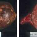
The patient was a 31-year-old woman who presented with daily epigastric discomfort that responded to treatment with a proton pump inhibitor. However, an ultrasound of the abdomen detected an 8-cm right retroperitoneal mass. Computed tomography (CT) and magnetic resonance imaging (MRI) scans of the abdomen showed a 6.5 × 4–cm mixed-density right retroperitoneal mass of either renal, adrenal, or hepatic origin ( Fig. 85.1) . Immunohistochemistry on a CT-guided biopsy was positive for synaptophysin, S-100 epithelial membrane antigen, and vimentin, whereas the chromogranin stain was negative. The pathologist thought that the likely tissue of origin was adrenocortical and unlikely to be renal—the report read: “adrenocortical neoplasm with oncocytic features.” She was referred to endocrinology for evaluation of an adrenal mass. She showed signs or symptoms of adrenocortical or medullary hormone excess. Her blood pressure was normal and she had no change in body weight. The 24-hour urine studies were normal for cortisol, fractionated metanephrine levels, and fractionated catecholamines. She underwent a flank incision with a right radical nephrectomy and right adrenalectomy. Pathology documented a 9.2 × 8 × 3–cm malignant renal epithelioid angiomyolipoma—a malignant mesenchymal neoplasm, characterized by proliferating epithelioid cells. The adrenal gland was anatomically normal. Epithelioid renal angiomyolipoma is a rare malignant variant of angiomyolipoma. There was no evidence for recurrent disease on periodic abdominal cross-sectional imaging completed over the subsequent 14 years.

Case Report: Retroperitoneal Lymphangioma
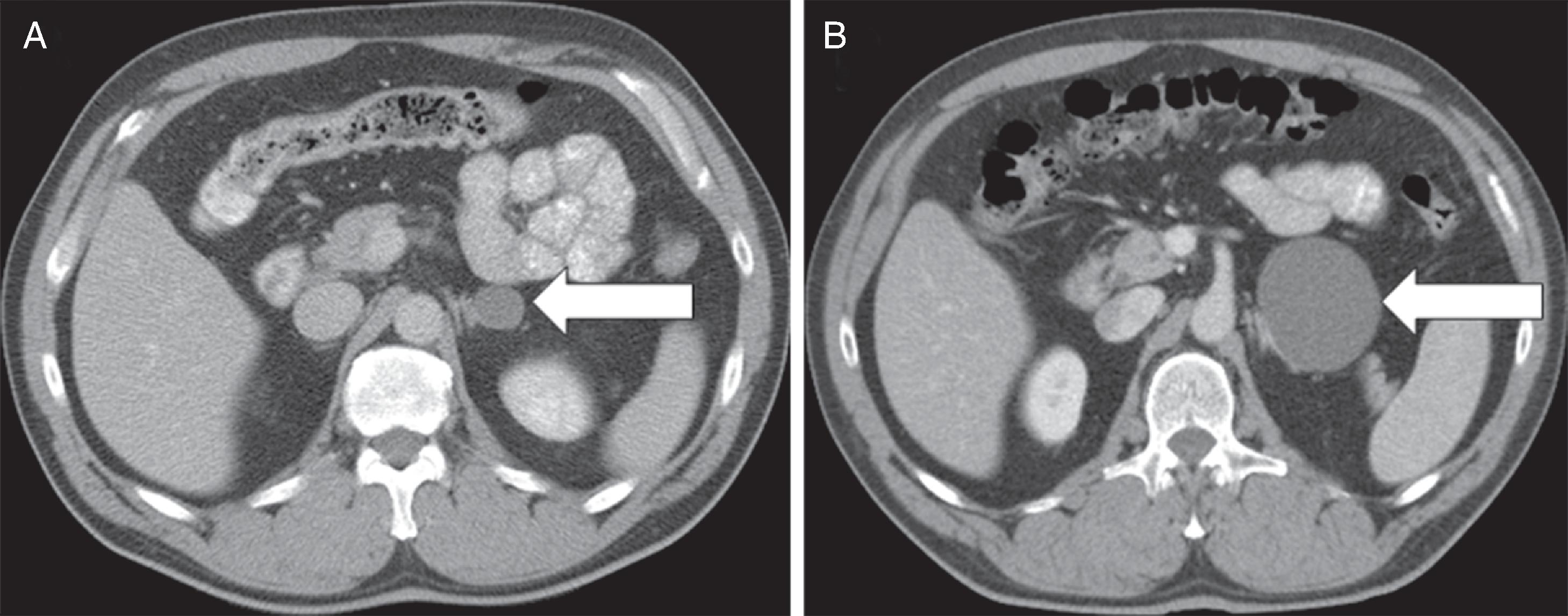
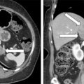
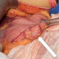
The patient was a 58-year-old man who had a slowly growing incidentally discovered cystic left retroperitoneal mass ( Fig. 85.2 ). Seven years previously, it measured 2.3 × 1.8 cm in diameter. It measured 6.7 × 6.6 cm on the most recent CT scan (see Fig. 85.2 ). The mass was located along the lateral wing of the left adrenal gland. It was clearly distinct from the left kidney, pancreas, and surrounding vascular structures. There was no apparent solid component or an enhancing ring to the cystic mass. The patient had no signs or symptoms of pheochromocytoma. He had been troubled by a left flank discomfort over the past year. He graded the pain at 6–7 out of 10 in severity and he felt it every day. Plasma fractionated metanephrine levels were normal. In view of his flank discomfort and the slow enlargement over 7 years, surgical resection was advised. At laparoscopic surgery, the cystic mass abutted the adrenal gland but did not involve the adrenal gland. At pathology the mass proved to be a lymphangioma. An abdominal CT scan completed 6 months postoperatively showed no evidence of persistent lymphangioma. His flank pain resolved after surgery. Lymphangioma is a rare benign proliferation of lymph vessels, which results in fluid-filled cysts as a result of the blockage of lymphatic flow. Surgical resection should be considered if there is associated pain or documented enlargement.

