Protein C, Protein S, Thrombomodulin, and the Endothelial Protein C Receptor Pathways


Protein C, Protein S, Thrombomodulin, and the Endothelial Protein C Receptor Pathways


Laurent O. Mosnier

John H. Griffin


THE PROTEIN C PATHWAYS—OVERVIEW

Control of coagulation reactions is essential for normal hemostasis. As part of the tangled web of host defense systems that respond to vascular injury, the blood coagulation factors act in concert with the endothelium and blood cells, especially platelets, to generate a protective fibrin-platelet clot, forming a hemostatic plug. Pathologic thrombosis occurs inter alia when the protective clot extends beyond its beneficial size, when a clot occurs inappropriately at sites of vascular disease, or when a clot embolizes to other sites in the circulatory bed. For normal hemostasis, both procoagulant and anticoagulant factors must interact with the vascular components and cell surfaces, including the vessel wall and platelets. Moreover, the actions of the fibrinolytic system must be integrated with coagulation reactions for timely formation and dissolution of blood clots. The protein C pathways provide diverse important functions to maintain a regulated balance between hemostasis and host defense systems in response to vascular injury. On one hand, the anticoagulant protein C pathway is designed to regulate coagulation, maintain the fluidity of blood within the vasculature, and prevent thrombosis. On the other hand, the cytoprotective protein C pathway provides anti-inflammatory and cytoprotective activities and it acts to prevent vascular leakage. This chapter summarizes current knowledge of the protein C pathway components and their activities, with particular emphasis on the recently elucidated, novel direct cellular effects due to activated protein C (APC) and its cofactors, protein S, thrombomodulin (TM), and endothelial protein C receptor (EPCR). Several reviews are available on the biochemistry and clinical impact of the anticoagulant and cytoprotective protein C pathway.1,2,3,4,5,6,7,8,9,10,11,12,13,14

Thrombin is central to the protein C pathways. Thrombin that is generated by the blood coagulation pathways (see Chapters 8, 9, and 14) binds with high affinity to the cell-surface receptor, TM, and converts the protein C zymogen that is bound to EPCR to APC. For its anticoagulant activity, APC acts in concert with cofactors, including protein S, to inactivate factors Va and VIIIa, the major cofactors for thrombin generation. Cytoprotective APC activities that include antiapoptotic activity, anti-inflammatory activity, regulation of gene expression, and stabilization of endothelial barrier protection can be initiated when APC targets two key receptors, protease-activated receptor-1 (PAR-1) and EPCR.6,15,16,17,18,19,20 Target cells for APC’s cytoprotective activities are not limited to the endothelium but also include epithelial cells, various lymphocytes, dendritic cells, and neurons, and additional receptors may also play key roles.

Many questions arise related to APC’s effects on cells, such as (a) what are the signaling pathways and/or molecular mechanisms underlying APC’s cytoprotective effects on each type of cells; (b) what are the target(s) for APC’s effects on cells that allow for APC’s many pleiotropic properties; (c) to what extent are molecular mechanisms for APC’s antiapoptotic activity, anti-inflammatory activity, regulation of gene expression, and endothelial or epithelial barrier protection shared, overlapping, or distinct; and, perhaps most critically for mechanistic insights, (d) what are the relative contributions of APC anticoagulant activity and APC’s cytoprotective activities for the observed beneficial effects, including mortality reduction, in various in vivo injury models? Partial answers to these questions are provided in this chapter. Because of their critical roles in the body’s host defense system, APC and the protein C pathway components are ideal targets for translational medicine research, and they provide opportunities for therapeutic treatment of complex and challenging medical disorders, including thrombosis, severe sepsis, and stroke among others.


IDENTIFICATION OF THE PROTEIN C PATHWAY COMPONENTS

Although protein C was originally identified as “autoprothrombin-IIa” more than 50 years ago, it was subsequently rediscovered by Stenflo and named “protein C” because of its elution in the third peak (“peak C”) on an ion-exchange chromatogram.21,22 The identification of TM, an endothelial cell cofactor for thrombin-catalyzed activation of protein C, provided a rationale for how a kinetically inefficient enzyme, such as thrombin, could be a physiologically significant activator of protein C.23,24 The clinical importance of the protein C pathway was based on discovery of deficiencies in protein C or in protein S linked to thrombosis and, for severe or total deficiency, to inflammation.25,26,27,28,29 Efforts to elucidate the molecular basis of the anti-inflammatory effects of APC were sparked by the identification of the EPCR.30 The finding of the PROWESS trial in which recombinant APC (Xigris) reduced 28-day all-cause mortality in adult severe sepsis patients was a watershed event.31 However, 10 years after that trial, in late 2011 APC’s mortality reduction efficacy was not reproduced in the PROWESS-SHOCK trial, raising multiple questions about each trial and their comparisons. Nonetheless, many studies between 2001 and 2011 were driven by the PROWESS trial’s success, including major breakthroughs in understanding APC cellular effects involving the direct action of APC on cells that induce signaling through activation
of PAR-1 when bound to EPCR and possibly other receptors.16 Additional dimensions to studies on the protein C pathway include preclinical studies showing beneficial effects of second-generation recombinant APC mutants in sepsis and ischemic stroke, the generation of protein C- or protein S-deficient mice, and the findings that protein S can exert direct receptor-mediated effects on cells.16,17,18,19,20,21,22,23,25,27,28,29,30,31,32,33,34,35,36,37,38,39,40,41,42,43,44,45,46,47


Protein C

Protein C was discovered in search for vitamin K-dependent coagulation factors that are not detected by clotting assays.21 The protein C gene (PROC) is homologous to the genes for factors VII, IX, and X and is comprised of nine exons and eight introns, located on chromosome 2q13-14 (Table 19.1).48,49,50,51 After removal of the prepro leader peptide, mature protein C contains 419 amino acids (Mr ˜ 62 kDa) (figure 19.1) and is synthesized mainly in the liver. Protein C in plasma circulates at 70 nM (4 µg/mL) with a half-life of approximately 8 hours.25 A significant posttranslational modification involves the cleavage of the zymogen between residues 157 and 158 by a furin-like enzyme, followed by the loss of the Lys156-Arg157 dipeptide connecting the light chain (residues 1 to 155; Mr ˜ 25 kDa) to the heavy chain (residues 158 to 419; Mr ˜ 41 kDa). As a result, 85% to 90% of human protein C is a two-chain form, linked by a single disulfide bond between Cys141 and Cys277. Other posttranslational modifications involve β-hydroxylation at Asp71, N-linked glycosylation at residues 97, 248, 313, and 329, and γ-carboxylation at the first nine Glu residues in the γ-carboxyglutamic acid (Gla) domain. The three subforms of the heavy chain, designated alpha (Mr ˜ 41 kDa), beta (Mr ˜ 37 kDa), and gamma (Mr ˜ 32 kDa), represent tri-, di-, and monoglycosylated heavy chain due to partial glycosylation of Asn residues 329 and 248.52 Ca2+-binding sites that are located in the Gla domain are required for binding to phospholipids and EPCR, and such sites are also present in the first epidermal growth factor (EGF) domain and the protease domain.53,54,55 The characteristic structural elements of protein C include an amino-terminal Gla domain (residues 1 to 37), an aromatic stack (residues 38 to 45), two EGF-like regions (EGF-1, residues 46 to 92 and EGF-2, residues 93 to 136), an N-terminal activation peptide (residues 158 to 169) on the heavy chain, and the serine protease domain (residues 170 to 419) (figure 19.1).
Thrombin cleavage of the zymogen at Arg169 removes the activation peptide and generates APC, a trypsin-like serine protease with a typical serine protease active site triad (His211, Asp257, and Ser360). Endogenous circulating levels of plasma APC are low (<40 pM), and inactivation of APC in plasma is driven by serine protease inhibitors (SERPINs) that contribute to a remarkably long circulation half-life of APC in man of approximately 20 to 25 minutes. Most important inhibitors of APC in plasma are protein C inhibitor (PCI) and α1-antitrypsin and, to a lesser extent, α2-macroglobulin and α2-antiplasmin.


Table 19.1 Characteristics of the protein C pathway components


Protein


Gene


Locus


cDNA


Residues


Theoretical Mass (kDa)


Observed Mass (kDa)


Plasma Concentration (nM)


ε1%; 1 cm


Coagulation Factors


Protein C


PROC


2p13-14


NM_000312


419


47


62


70


14.5


Protein S


PROS1


3q11.2


NM_000313


635


71


77


320a


9.5


FV


F5


1q23


NM_000130


2,196


249


330


25


8.9


FVIII


F8


Xq28


NM_000132


2,196


265


330


0.7


11.9


C4b-binding proteinb


C4BPA


1q32


NM_000715


549


70


500-570b


260


14.1


C4BPB


1q32


NM_000716c


235


45


Cell Receptors


TM


THBD


20p12-cen


NM_000361


557


60


75 (105)d


0.05e


9.2f


EPCR


PROCR


20q11.2


NM_006404


221


25


46


2.5g


11.3f


PAR-1


F2R


5q13


NM_001992h


400


47


65


n/a


ND


ApoER2


LRP8


1p34


NM_004631i


899


102


180


ND


24f


a The total protein S concentration (320 nM) includes free protein S (130 nM) and protein S in complex with C4BP (190 nM).

b C4BP contains two polypeptide chains in varying proportions and circulates in different isoforms, alpha-7-beta-1 being the most common. About 17% (45 nM) of C4BP contains only alpha-chains and lacks the beta-chain that binds protein S. The α6, α6β1, and α7 isoforms have molecular weights of approximately 500, 530, and 570 kDa.

c C4BP beta-chain variants 1 (NM_000716), 3 (NM_001017365), and 5 (NM_001017367) encode for the longer isoform 1 but vary in the 5′-untranslated region, whereas variants 2 (NM_001017364) and 4 (NM_001017366) encode for isoform 2 with a shorter N-terminus due to an in-frame alternate splice site.

d The TM apparent molecular weight is dependent on the presence of the GAG attachment.

e Soluble TM is believed to be the product of proteolysis of TM on the cell surface and comprises different molecular forms (Mr 35-65 kDa) that lack the transmembrane sequence. The concentration of soluble TM in normal individuals is approximately 2 to 5 ng/mL but can increase many-fold under pathologic conditions. Increased soluble TM levels are regarded to be a sign of endothelial cell activation.

f Values are given for the soluble extracellular domain of the receptor. Soluble ApoER2 is truncated after EGF-like domain B but includes all seven ligand-binding domains.

g Soluble EPCR is derived from proteolysis of EPCR on the cell surface and lacks the transmembrane domain (Mr ˜ 43 kDa).

h The reference sequence was adjusted in 2008 between revision 2 (NM_001992.2) and 3 (NM_001992.3), resulting in two amino acid changes compared to the sequence originally published for PAR-1.284

i Several cell-type specific ApoER2 isoforms are reported. Transcript variant 1 (NM_004631) contains all 19 exons, transcript variant 2 (NM_033300) lacks exons 5 and 6 encoding the ligand-binding domain (LBD) 4-6 and 7. Transcript variant 3 (NM_017522; e.g., found in endothelial cells [HUVEC]) lacks exon 5 (LBD4-6), exon 15 (O-linked sugar), and exon 18 (part of the intracellular domain). Transcript variant 4 (NM_001018054) is similar to full-length transcript variant 1 except for the lack of exon 18. In addition, U937 cells were found to express an additional transcript that lacks exons 5 and 18 but that contains exon 15.43


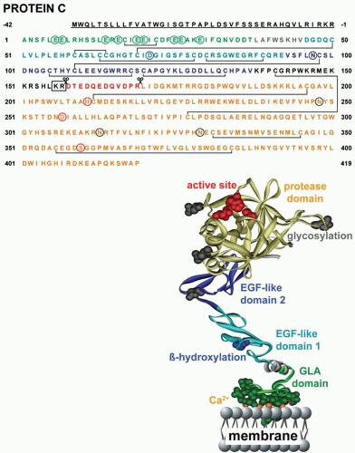
FIGURE 19.1 Amino acid sequence of protein C and ribbon polypeptide scheme of APC. Amino acids are numbered from the amino-terminus of the mature protein with the signal peptide sequence underlined. Specific domains are color coded as indicated in the ribbon cartoon. Green circles depict γ-carboxylation, blue circles depict β-hydroxylation, and black circles represent sites of N-linked glycosylation. Sites of proteolytic cleavage during posttranslational processing and protein C activation are indicated by the scissors. The dipeptide that is proteolytically removed during the posttranslational processing of most protein C molecules in the liver is identified by the box. The serine, aspartic acid, and histidine residues that constitute the active site triad are identified by red circles.


Protein S

Protein S, named after its birth city, Seattle, is a vitamin K-dependent plasma glycoprotein and is best known for its function as a nonenzymatic cofactor to APC in the inactivation of factors Va and VIIIa. The relatively large gene for protein S (PROS1) is located on chromosome 3 (3q11.2) and contains 15 exons (Table 19.1). Chromosome 3 also contains an inactive pseudogene for protein S (PROSP at location 3p21-cen). After removal of the prepro leader peptide, mature protein S contains 635 amino acids (Mr ˜ 77 kDa) (figure 19.2) and is predominantly synthesized in
the liver but is also made by endothelium and other cells. Protein S circulates in plasma at a total concentration of 320 nM, and approximately 60% of the protein S is noncovalently complexed with C4b binding protein (C4BP) such that free protein S is 130 nM in plasma.56 C4BP is an octopus-like shaped glycoprotein of approximately 500 to 570 kDa and consists of six or seven disulfide-linked, identical alpha-chains and one smaller beta-chain (see Table 19.1).57 C4BP regulates protein S functions by binding via the beta-chain.58 Protein S binds to the C4BP beta-chain in a 1:1 stoichiometric complex with high affinity (Kd ˜ 0.1 to 0.6 nM).59 Although C4BP is an acute-phase response protein, only alpha-chains are increased causing the levels of free protein S levels to remain stable during an acute-phase response.60


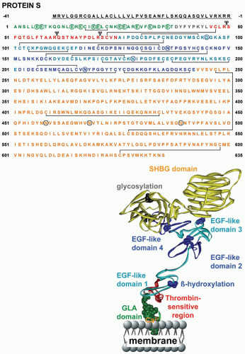
FIGURE 19.2 Amino acid sequence and ribbon polypeptide scheme of protein S. Amino acids are numbered from the amino-terminus of the mature protein with the signal peptide sequence underlined. Specific domains are color coded as indicated in the ribbon cartoon. Green circles depict γ-carboxylation, blue circles depict sites of potential β-hydroxylation, and black circles represent sites of N-linked glycosylation. Sites of potential proteolytic cleavage are indicated by the scissors.

Mature protein S has a defined structural organization consisting of five distinct domains, including an N-terminal Gla domain (residues 1 to 37) and aromatic stack (residues 38 to 45), an unique 29-amino acid sequence known as the thrombin-sensitive region (TSR; residues 46 to 74), a string of four EGF-like domains (EGF-1 [residues 75 to 115], EGF-2 [residues 116 to 159], EGF-3 [residues 160 to 201], and EGF-4 [residues 202 to 242]), and finally a large 393-amino acid domain on the C-terminus referred to as the sex hormone-binding globulin-like domain (residues 243 to 635) that is composed of two laminin G-type domains (figure 19.2).


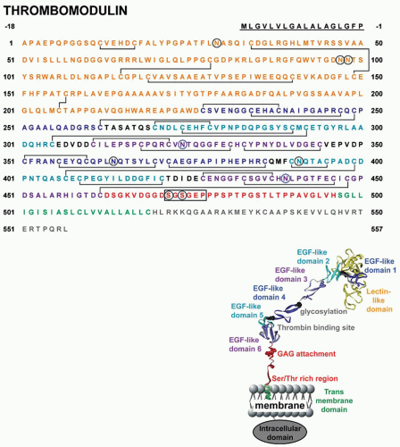
FIGURE 19.3 Amino acid sequence and ribbon polypeptide scheme of TM. Amino acids are numbered from the amino-terminus of the mature protein with the signal peptide sequence underlined. Specific domains are color coded as indicated in the ribbon cartoon. Blue circles depict sites of potential β-hydroxylation, black circles represent sites of N-linked glycosylation. The consensus sequence for the GAG attachment is boxed.


Thrombomodulin

The endothelial cell receptor TM (CD141) was discovered and named for its ability to modulate the protease specificity of thrombin (figure 19.3).24,61 The gene for TM (THBD) is on chromosome 20 (20p12-cen) and is intron-less (Table 19.1).62,63 Based on its cDNA sequence, TM consists of 575 amino acids in
the form of a 60.3-kDa single-chain transmembrane protein.63 Posttranslational O-linked and N-linked glycosylation accounts for approximately 20% of the apparent molecular weight of TM, and other modifications include covalently bound chondroitin sulfate moieties.64

Functionally, five regions of the mature TM can be identified (figure 19.3)63: An N-terminal lectin domain (residues 1 to 226), a repeat of six EGF-like domains (EGF-1 [residues 227 to 262], EGF-2 [residues 270 to 305], EGF-3 [residues 311 to 344], EGF-4 [residues 351 to 386], EGF-5 [residues 390 to 421], and EGF-6 [residues 427 to 462]), a serine- and threonine-rich region (residues 463 to 496), a transmembrane region (residues 497 to 519), and finally a C-terminal intracellular tail (residues 520 to 557). The EGF-like modules 5 and 6 mediate thrombin binding, whereas EGF-like module 4 is required for interactions with protein C. The major site for the glycosaminoglycan (GAG) attachment is within the chondroitin consensus sequence of Ser472-Gly-Ser474-Gly-Glu-Pro at Ser472 or Ser474. GAGcontaining TM has somewhat different properties than TM devoid of a GAG attachment as the presence of GAG increases the affinity for thrombin approximately 10-fold, reduces thrombin’s ability to clot fibrin and to activate platelets, and modulates the Ca2+-dependent profile of protein C activation. Furthermore, the GAG on TM can bind a second thrombin molecule and facilitate inactivation of thrombin by the SERPINs, PCI, and antithrombin (AT).65 Besides promoting protein C activation by thrombin, TM also supports activation of a plasma carboxypeptidase, known as thrombin-activatable fibrinolysis inhibitor (TAFI). Activated TAFI is not only a potent attenuator of fibrinolysis but also an effective inactivator of a variety of proinflammatory peptides such as the activated complement components C3a and C5a, bradykinin, thrombin-cleaved osteopontin, and other peptides.66 For a more detailed discussion of TAFI, see Chapters 20 and 21.


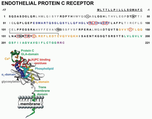
FIGURE 19.4 Amino acid sequence and ribbon polypeptide scheme of the EPCR. Amino acids are numbered from the amino-terminus of the mature protein with the signal peptide sequence underlined. Specific domains are color coded as indicated in the ribbon cartoon. Black circles represent sites of N-linked glycosylation. EPCR residues implicated in binding of APC are indicated in red.54

Low levels of soluble TM, generated by shedding from the endothelium due to proteolysis, circulate in plasma and are generally considered to be a marker of endothelial cell damage.61 The functional significance of circulating TM is unknown, although variations in its plasma level arise in different clinical conditions.


Endothelial Protein C Receptor

The EPCR (CD20167) was discovered in a search for the endothelial cell receptor responsible for binding of APC.30 The gene for EPCR (PROCR) is on chromosome 20 (20q11.2) (Table 19.1).68 EPCR is homologous to the CD1/MHC superfamily. MHC class I molecules are type 1 integral membrane proteins and consist of three extracellular domains, alpha-1, alpha-2, and alpha-3. The latter domain typically associates noncovalently with β2-microglobulin. EPCR lacks the alpha-3 domain and thus does not associate with β2-microglobulin; rather, in EPCR, the transmembrane domain is connected directly to the alpha-2 domain (figure 19.4).68 The alpha-1 and alpha-2 domains form a binding groove by each providing an alpha-helix that
is located along opposing edges of a planar platform generated by an eight-stranded antiparallel beta-sheet. The crystal structure of soluble EPCR revealed that a phospholipid, most likely phosphatidylcholine, was bound in this groove between the two alpha-helices.69,70 EPCR’s short cytoplasmic tail (Arg-Arg-Cys-CO2H) suggests that induction of direct cell signaling is unlikely and that palmitoylation of the C-terminal Cys residue may help localize EPCR to certain lipid rafts or caveolae and facilitate modulation of cell-signaling pathways that depend on EPCR.71,72

In addition to binding protein C and APC with similar affinity (KD ˜ 60 nM), EPCR also binds factor VII and factor VIIa to the same end of an EPCR alpha-helix that binds to the ligand’s Gla domain (figure 19.4). Although EPCR is named after its expression on endothelial cells, EPCR is also present on epithelial cells, monocytes, macrophages, neutrophils, eosinophils, natural killer cells, and in mice on specific bone marrowderived dendritic cells.30,73,74,75,76,77,78,79

EPCR is susceptible to shedding of the EPCR ectodomain due to proteolysis in the region of residues 193 to 200, just above the transmembrane domain, by tumor necrosis factor-alpha (TNF-α)-converting enzyme/ADAM17 (TACE). Several proinflammatory cytokines, for example, TNF-α and interleukin-1β, induce soluble EPCR formation and increase soluble EPCR levels in mice exposed to endotoxemia; soluble EPCR is increased in plasmas of patients with a prothrombotic and proinflammatory tendency.80,81,82,83,84


THE ANTICOAGULANT PROTEIN C PATHWAY

The protein C system is a dual function cofactor-dependent system that exerts direct cytoprotective activities on cells as well as its well-studied anticoagulant activity anticoagulant. By targeting factors Va and VIIIa, major procoagulant cofactors for thrombin generation, APC regulates coagulation and prevents thrombosis. The physiologic importance of thrombosis prevention by the anticoagulant protein C system is evident from clinical observations of thrombotic complications in individuals with deficiencies of protein C and protein S.


Anticoagulant Protein C Pathway Defects and Thrombosis

Homozygous and compound heterozygous severe protein C deficiency typically presents itself as neonatal purpura fulminans, a rapidly progressing hemorrhagic necrosis of the skin due to microvascular thrombosis, inflammation, and disseminated intravascular coagulation (DIC).85 Infants with severe protein C deficiency suffer massive thrombotic complications, and if they survive due to maintenance replacement therapy, they tend to present with mental retardation and/or visual impairment.85,86,87 Replacement therapy with fresh-frozen plasma or with purified plasma protein C concentrate (CEPROTIN) is beneficial.88,89,90 The incidence of clinically significant protein C deficiency carries a significantly increased risk for venous thrombosis (see Chapters 80, 81 and 82).91,92,93 Hundreds of mutations have been identified (see protein C mutation databases92,94,95), and the basis for hereditary protein C defects can most often be rationalized based on the structure of protein C.96,97,98 Type I mutations, characterized by reductions of both antigen and activity, typically involve amino acid mutations that affect protein folding, secretion, or stability. In contrast, most type II mutations, characterized by reduction of anticoagulant activity but not antigen, involve residues that cause dysfunctional molecules either by affecting thermodynamic stability or by proteinprotein interactions important for expression of anticoagulant activity. Acquired protein C deficiency occurs upon initiation of vitamin K antagonists (coumarin/warfarin) therapy due to the relatively short half-life of protein C (T1/2 ˜ 8 hours) compared to most procoagulant vitamin K-dependent factors (T1/2 ˜ 20 to 48 hours); this potential problem is addressed by using heparin during initiation of therapy, or even protein C concentrate for special circumstances.99,100

Severe (homozygous or compound heterozygous) protein S deficiency, although extremely rare, manifests with purpura fulminans in the neonatal period similar to severe protein C deficiency.101,102,103 Hereditary protein S heterozygous deficiency conveys a significantly increased risk of thrombosis.29,93,104,105,106 Due to the binding of protein S to C4b-binding protein in plasma, testing to establish protein S deficiency is challenging.105 Deficiencies are classified as follows: type I, reduction of both free and total protein S antigen; type II or qualitative deficiency, functional protein S defect with a low functional activity but normal antigen level; and type III, with a low level of free protein S level but a normal level of total protein S antigen.29,102,104,105,106,107,108,109 Mutations are found in the majority of patients and families with hereditary protein S deficiency (see Chapters 80, 81 and 82 and protein S mutation databases).105,110 Two polymorphisms causing deficiencies are known as protein S Heerlen (Ser460Pro), a type III defect among subjects of European ancestry, and protein S Tokushima (K196E), a type II defect.111,112,113,114,115,116 Protein S Tokushima is a common risk factor for venous thrombosis among Japanese and is found in approximately 10% of venous thrombosis patients.105,111,117,118,119 It is likely that the protein S Tokushima mutation of K196E (residue 155 in mature protein S) arose from a founder effect in Japan and is a balanced polymorphism, like factor V Leiden or prothrombin 20210A that carries a beneficial effect (e.g., prevention of excess fatal bleeding) and a mild risk factor for venous thrombosis. In contrast, the Heerlen polymorphism that alters glycosylation is not as obvious a risk factor. Acquired protein S deficiency is common during pregnancy and also may arise during the use of certain oral contraceptives, or after initiation of oral anticoagulant treatment.120,121,122,123,124

APC resistance conveys an increased risk for venous thrombosis and is defined as an abnormally reduced anticoagulant response of a plasma sample to APC. It can be caused by many potential abnormalities in the protein C anticoagulant pathway but among whites a mutation in factor V, which arose in a European ancestor involving an APC-cleavage site (Arg506Gln, factor V Leiden), is the most common cause for hereditary thrombosis.38,39,125 Other known APC cleavage site mutations in factor V that contribute to an increased thrombotic risk are Arg306Thr (factor V Cambridge) and Arg306Gly (factor V Hong Kong).126,127,128,129,130,131 APC resistance with no identifiable genetic or acquired abnormalities is well described in patients with venous and arterial thrombosis, and further studies are needed to identify the causes of APC resistance in such patients.132,133,134,135

TM genetic mutations may be associated with increased risk of arterial thrombosis and myocardial infarction, whereas there is less supportive data for association with risk for venous thrombosis.61,136,137,138,139,140,141,142 Mutations in TM have also been linked to atypical hemolytic-uremic syndrome patients; this syndrome
is strongly linked to excessive complement activation.143 The lectin-like domain of TM (figure 19.3) can contribute to inhibition of complement activation and provide direct anti-inflammatory activity, although the molecular basis for this remains somewhat unclear.144,145,146,147

The potential association of EPCR defects with venous and arterial thrombosis remains a bit controversial and seems difficult to prove (see Chapters 81 and 82).148,149,150,151,152,153,154,155 To date, four EPCR gene haplotypes have been characterized containing certain polymorphisms.68 Interestingly, the H1 haplotype was associated with a reduced risk of venous thromboembolism and diminished risk of thrombosis in carriers of the factor V Leiden mutation.156,157 In contrast, the H3 haplotype of EPCR that is characterized by a Ser219Gly mutation in EPCR causes enhanced EPCR shedding from the membrane, resulting in increased levels of soluble EPCR in the circulation, which might be associated with an increased risk of venous thrombosis.81,156,158,159,160


Protein C Activation

Activation of zymogen protein C by thrombin is due to cleavage at the Arg169-Leu170 peptide bond (figure 19.1) in a reaction that is accelerated by TM and EPCR on the surface of the endothelial cells (figure 19.5).1,6,7,23,161,162 Binding of thrombin to TM shields thrombin’s procoagulant exosite I and unveils its anticoagulant properties.7,163,164,165 Subsequent activation of protein C by the thrombin-TM complex is facilitated by localization of protein C on the endothelial cell surface by binding to EPCR.166,167 Available data suggest that EPCR and TM must be in close proximity on cell surfaces, though this has yet to be experimentally demonstrated. In blood vessels, TM is generally considered more abundant in the microcirculation while EPR is relatively more abundant in large vessels than in small vessels.1,168,169


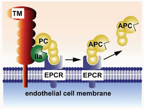
FIGURE 19.5 Protein C activation on endothelium. Physiologic activation of protein C (PC) by the thrombin (IIa)-thrombomodulin (TM) complex occurs on the surface of endothelial cell membranes. The most efficient activation of protein C occurs when protein C is bound to the endothelial receptor (EPCR). Since protein C and APC have a similar affinity for EPCR, after activation APC can either dissociate from EPCR to exert anticoagulant activity or remain bound to EPCR where it might express multiple direct cellular activities. (This scheme was originally published in Blood. Mosnier et al. The cytoprotective protein C pathway. Blood 2007;109(8):3161-3172. © the American Society of Hematology.)

APC is constantly generated in the resting state because APC is present at approximately 40 pM in all healthy human subjects’ plasma. The concentration of zymogen protein C in plasma is an important determinant for circulating APC levels, reflecting the kinetics of the protein C activation on cultured cells in vitro.166,167,170,171,172,173,174,175,176,177 An inverse correlation between circulating fibrinopeptide A and APC levels suggests that APC is a significant in vivo regulator of basal thrombin activity.178,179,180 Impaired activation of protein C occurs in patients with inflammation and sepsis.181 Protein C levels in patients with severe sepsis often are markedly decreased, due to a combination of protein C consumption, dysfunctional protein C synthesis in the liver, and/or reduced levels of TM or EPCR on the endothelium.


APC Anticoagulant Activity

Anticoagulant activity of APC involves proteolytic inactivation of factors Va and VIIIa (figure 19.6).182,183 Multiple cofactors, including protein S, negatively charged phospholipids, neutral glycosphingolipids, and high-density lipoprotein (HDL), are known to enhance APC’s inactivation of factor Va and/or factor VIIIa.184,185,186,187,188

Only gold members can continue reading. Log In or Register to continue

Stay updated, free articles. Join our Telegram channel

Jun 21, 2016 | Posted by in HEMATOLOGY | Comments Off on Protein C, Protein S, Thrombomodulin, and the Endothelial Protein C Receptor Pathways

Full access? Get Clinical Tree

Get Clinical Tree app for offline access