Early detection and improvement in adjuvant systemic therapy has resulted in a decrease in breast cancer mortality. However, each year 500,000 women still die of breast cancer worldwide. It is clear that more work is needed to identify which breast cancers have a high propensity to metastasize, and to guide clinicians in selecting the most appropriate therapies.
Prognostic factors reflect the biology of the cancer and are defined as those factors associated with outcome without consideration of treatment. Predictive factors reflect the tumor sensitivity or resistance to a particular treatment, and are defined as those factors that predict which patients are likely to respond to a specific therapy. The relative worth of predictive factors will obviously differ with varying treatments. While some factors are primarily prognostic, some tumor markers are both prognostic and predictive. The classic example is the overexpression of HER-2/neu, which is associated with a worse outcome (prognostic), but also associated with response to treatment with trastuzumab (predictive).
The relative benefit of a predictive or prognostic factor can be classified as weak, moderate, or strong. For prognostic factors, the relative strength is related to the difference in the likelihood of an adverse event (recurrence, death) between a patient with the prognostic factor and one who is negative. An example of a strong prognostic factor is lymph node status, as a node-positive patient is 2 to 3 times more likely to have an event than a lymph node-negative patient. An example of a weak prognostic factor is estrogen receptor (ER) expression, as patients with ER-positive cancers have only slightly better outcomes than ER-negative patients. Predictive factors can also be classified as weak, moderate, and strong based on the likelihood that a patient with the factor will respond to treatment compared to that of a patient without the factor. While ER expression is a weak prognostic factor, it is a strong predictive factor for hormonal therapy, as ER-positive patients may realize a 50% reduction in the risk of recurrence with hormonal therapy, while ER-negative patients obtain almost no benefit from hormonal therapy.
When discussing prognostic and predictive factors, it is crucial to separate the statistical significance of a factor from the clinical significance. As an independent factor, many measurable factors may correlate with outcome. However, when examined in the context of other prognostic or predictive factors (multifactorial analysis), they may lose statistical significance. So while there may be biological significance to the factor it may provide little help to the clinician in estimating the likelihood of death or the benefit of therapy. Moreover, even if a factor retains statistical significance on multifactorial analysis, this still does not guarantee clinical significance. If the absence or presence of the factor is relatively rare among breast cancer patients, its clinical utility is severely limited. One example is tumor grade. While grade III (poorly differentiated) patients may have worse outcomes than grade I (well-differentiated) patients, the great majority of patients are either grade III or II (moderately differentiated), greatly limiting the clinical utility of grade as a prognostic factor.
More importantly, the measurement of that factor must be technically reliable and reproducible. The more variation there is in its interpretation, the less useful it is. Again, grade is a good example, as the subjective labeling of tumors as grade I, II, or III allows for significant variation among pathologists. Recent changes to the grading system have attempted to increase the objectivity and decrease variation, but differences in interpretation can never be fully eliminated. Even for “standard” tumor markers, such as ER expression or HER-2/neu overexpression, there can be variability in the assays used for measurement. Researchers and clinicians can use different assays to measure the factor (immunohistochemistry [IHC], Western blotting, or enzyme-linked immunosorbent assay [ELISA] for protein expression; Northern blotting or reverse transcriptase-polymerase chain reaction [RT-PCR] for RNA expression; or florescence in situ hybridization [FISH] for DNA amplification) and different reagents for the assay (such as different antibodies for IHC). Even minor technical differences such as the concentration of a reagent or the method by which the specimen is procured and processed can impact the results of the test. Finally, whenever visual assays are used for the final interpretation, intra- and interobserver variability will always be a concern. For example, several studies have demonstrated significant discordance for HER-2/neu overexpression between laboratories.1,2
It is important to realize that true estimations of prognosis are dependent upon weighing the relative impact of all known prognostic factors. One extremely useful method of determining the risk of recurrence and of death based on multiple prognostic factors is the use of computer programs. The most popular is the Web-based Adjuvant Online. The prognostic information was created based on prognosis estimates derived mainly from the Surveillance, Epidemiology, and End Results (SEER) data, which are then adjusted using a Bayesian method on the relative risks conferred and the prevalence of positive test results. After entering in patient and tumor data, a 10-year risk of recurrence and 10-year risk of death are calculated. In addition, estimates of the benefit of hormonal therapy and chemotherapy, based on the Oxford Overviews and other sources, are also presented. This information and program have been extremely useful in facilitating physician–patient communication regarding outcome and the benefits of adjuvant therapy.
One of the first described and still most important prognostic factors in breast cancer is the size of the primary tumor. Many studies have demonstrated a linear correlation between the diameter of the primary tumor and both the presence of lymph node metastases and clinical outcome. Among node-negative patients, tumor size is the single most important prognostic factor.3
The reasons that tumor size is so strongly associated with outcome are multifactorial. Part of this is explained by the increased presence of metastases in the regional lymph nodes with increasing tumors size. However, tumor size is still an independent prognostic factor; small tumors associated with positive nodes have a better prognosis than large tumors with positive nodes. Larger tumor size appears to be associated with increased occult blood vascular dissemination. It must always be remembered, however, that there may be a detection bias at play. More aggressive tumors are typically found when they are large given the intervals at which we screen, while slow-growing tumors have an inherent favorable prognosis and are found at a small size.4
For the most precise prognostic information, tumor size should be obtained from the pathologic specimen. The correlation with the clinical assessment (physical examination, radiographic studies) may be inaccurate.
Although reporting of pathologic size has not yet been standardized, numerous studies have demonstrated that it is the size of the invasive component that is associated with outcome. There are some relevant practical considerations in reporting tumor size; especially the determination of microinvasion, multifocal tumors, and tumors removed in more than one specimen.
Microinvasion is defined by the current AJCC Cancer Staging Manual (sixth edition) as “the extension of tumor cells beyond the basement membrane into the adjacent tissues with no focus more than 0.1 cm in greatest dimension.” When multiple foci of microinvasion are present, it is the size of only the largest focus that is used, not the sum of the microinvasive foci. Most studies have shown that the prognosis of ductal carcinoma in situ (DCIS) with microinvasion is similar to the prognosis of DCIS alone.5,6
When more than one focus of invasive carcinoma is present, the largest focus is used for staging purposes. Similarly, when invasive carcinomas are removed in more than one specimen, pathologists measure the size of the largest tumor. In difficult cases, radiologic correlation can be very helpful in determining tumor size.7 A few studies have demonstrated that invasive carcinomas with multiple foci of invasion have higher rates of lymph node metastasis and that the number of lymph node metastasis is predicted best by the aggregate size of the invasive foci.8,9 Even though at this time size determination is based on the size of the largest focus, emerging data may result in revisions to our current staging system in the future.
The most powerful prognostic factor in breast cancer remains the presence of disease in the regional lymph nodes. Repeated studies show an inferior outcome when disease is identified in the lymph nodes and the more nodes involved, the worse the outcome.
Breast cancer can metastasize to both the axillary lymph nodes and the internal mammary (IM) lymph nodes (as well as the supraclavicular nodal basins, although this is less common). Metastases to either the IM or axillary nodes are associated with a poor outcome compared to lymph node-negative patients. However the presence of regional disease in both the IM and axillary lymph nodes portends a worse prognosis than disease in either basin alone.10-14 The majority of these data come from the era of the extended radical mastectomy. At that time, it was thought that including an IM lymph node dissection with the radical mastectomy might improve outcomes. This was not the case, and support for the extended radical mastectomy has faded. However, the importance of staging the IM nodes has reemerged with the introduction of radioactive colloid for intraoperative lymphatic mapping. Depending on the method of injection, drainage to the IM nodes is often identified on lymphoscintigraphy. Whether the prognostic information and potential therapeutic benefit of excising SLN in the IM chain justifies the morbidity of the procedure is an area of ongoing debate.
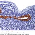
The SLN biopsy was introduced in the 1990s. As pathologists were faced with fewer lymph nodes to evaluate, we began using multiple levels and/or IHC in the routine evaluation of SLN.
Before we can discuss the impact of SLN biopsy on prognosis we need to define two commonly used terms: micrometastasis and isolated tumor cells. Micrometastases are foci of cancer in a lymph node that are greater than 0.2 mm but with none greater than 2.0 mm. In the most recent AJCC staging system for breast cancer, these are designated as pN1mi. Isolated tumor cells are defined as single cells or small clusters of cells smaller than 0.2 mm. These are staged as pN0. As compared with micrometastases, which often show evidence of proliferation or stromal reaction, isolated tumor cells or small cell clusters usually do not show such evidence of malignant activity.
Although more definitive evidence is needed, available literature suggests that micrometastasis not only are associated with a risk of additional disease in the non-SLN (justifying complete axillary lymph node dissection) but also provide prognostic information.15,16 Until recently, there have been no convincing data suggesting that patients with isolated tumor cells in SNL have a worse prognosis.7 However, a recent study found that isolated tumor cells or micrometastasis in the SLN were associated with a reduced 5-year disease-free survival compared to node negative patients, and that disease-free survival was improved with adjuvant therapy.16B Additional data from prospective studies of SLN biopsy (NSABP B-32, ACOSOG Z-10) are pending.
There are practical with applying the strict size definitions of the AJCC that arise in daily pathology practice. Those issues include methods to measure very small metastasis, how to assess the diffuse single-cell pattern of metastasis of invasive lobular carcinomas, and the presence of multiple clusters of tumor cells.
To measure accurately the size of isolated tumor cells, pathologists need to employ a calibrated ocular micrometer, or estimate from field sizes.
Invasive lobular carcinomas have long been recognized to metastasize in a diffuse, sometimes single-cell pattern. In this pattern, there can be thousands of cells, but no cell cluster larger than 0.02 mm. Sometimes the identification of rare metastatic cancer cells is extremely hard on routine hematoxylin and eosin staining, and the use of cytokeratin stains is recommended. The designation of isolated tumor cells and N0 classification seems inappropriate for such cases, and several pathologists have advocated classifying these cases as N1, based on the number of tumor cells. This will likely be addressed in the upcoming seventh edition of the AJCC.
Another difficulty that pathologists face is the presence of multiple clusters of tumor cells in a lymph node. In this instance, if 2 or more clusters are separated by normal lymph node tissue, the distance between the clusters should not be included in the measurement. Sometimes a lymph node may contain over 5 clusters smaller than 0.2 cm. Is this best classified as a micrometastasis or a bonafide metastasis? Some of these issues need clarification in the very near future. We recommend that pathologists communicate with the surgeons and clinicians when these cases arise to be considered in clinical decision-making.
Both histologic grade and type are routinely used to make clinical recommendations.
The most common method of determining histologic grade is the Nottingham combined histologic grade (the Elston-Ellis modification of the Scarff-Bloom-Richardson grading system). This system is based on scoring 3 factors: tubule formation, degree of pleomorphism, and mitotic count. While this modification is more objective than previous methods of determining grade, there are still issues of variability in tissue fixation and observer interpretations.
Histologic grade correlates with survival independent of lymph node status and tumor size.17 Additionally, some studies have demonstrated that histologic grade can be prognostic among certain subsets of breast cancer patients. Among patients with tumors smaller than 1.0 cm (and primarily node negative), Stierer et al18 found that high histologic grade was a significant predictor of recurrence and death. Winchester et al19 found histologic grade to be a significant predictor of disease-free survival among aneuploid tumors. Grade was the most powerful prognostic factor in a multiple regression analysis of 654 reported patients by Sundquist et al.20
It is important to note that in addition of being a powerful prognostic factor, histologic grade may have predictive value. Adjuvant chemotherapy may produce a greater benefit in high-grade tumors than in low-grade tumors, particularly among node-negative patients.21-23 For this reason, histologic grade is commonly used in adjuvant therapy decision-making.
Certain histologic types of invasive breast carcinoma including tubular, mucinous, cribriform, adenoid cystic, and medullary are associated with favorable outcome. Pathologists need to apply strict criteria in order to make these diagnoses. If strict diagnostic criteria are used, the 20-year disease-free survival of these favorable histologic tumor types measuring up to 3.0 cm is similar to that of invasive ductal carcinomas of 1.0 cm or smaller.24 The College of American Pathologists (CAP) recommends using the World Health Organization classification for the determination of histologic type.25


