Stage
Clinic
CCDS
I. Prodromal phase
Red/white spot; telangiectasia
Structureless; low-echo space
Blurred vision; swelling
No signs of pathological vessels
II. Initial phase
Loss of typical skin structure
Hyposonoric center
Increasing thickness and induration
Hypervascularization beginning at edges
III. Proliferation phase
Bright red cutaneous infiltration; flat spreading
Increasing intratumoral hyperperfusion
Subcutaneous growth of thickness;
Central vessel density
Infiltration of surroundings possibly even at organ borders
Nutrition of tumor vessels
Possible early primary central ulceration
Drainage veins with arterial flow profile
IV. Maturation phase
Pale and livid color
Declining central vessel density
Possible central exulceration
Increasing ectatic drainage veins
Decreasing growth
Declining arterialization of drainage veins
Possible late ulceration over drainage veins
Central increasing hypersonoric aspect
V. Regression phase
Hypopigmentation; wrinkled skin/telangiectasis
Circumscribed hypersonoric area
Surrounding subcutaneous drainage veins
Loss of typical tissue structure
Subcutaneous palpable induration
Nearly no central tumor vessels
Residuals supplying tumor arteries
Residuals of ectatic drainage veins
Spontaneous Course
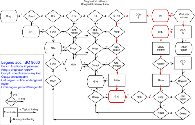
Because hemangiomas may involute spontaneously, waiting for spontaneous regression remains a viable therapeutic option. But the “wait-and-see” principle is always wrong (Table 10.2). If it means that treatment arrives too late, then “see and wait” as a control is correct, because at the first sign of progression of complication an action can be taken. Therefore, in case of small, uncomplicated infantile hemangiomas in non-problematic areas (extremities, body) without any tendency to proliferate, especially in cutaneous hemangiomas, one can “see and wait.” If delayed growth cannot be excluded, frequent controls are required. Clinical checkups alone may not be sufficient, as subcutaneous infantile hemangiomas may remain unnoticed as they grow deeply and are then only recognized after complications result. This is why a periodic duplex scan control is mandatory. For hemangiomas in the quiescent or regression phase, a “see-and-wait” attitude should normally be recommended. However, if complications are expected from ulcerations, treatment is also required for these forms. As therapy may cause adverse systemic or cutaneous side effects, particularly scarring, sometimes intervention has been reserved for patients with significant complications. Therefore, it is difficult to choose a therapy that eliminates hemangiomas before the development of complications and without systemic side effects [2]. For this reason, the following infantile hemangiomas must be considered as “problem hemangiomas,” for which active treatment is mandatory (Table 10.3):

Table 10.2
Grade 1 means uncomplicated hemangioma with no risks; Grade 2a hemangiomas with low risk factors need controls; Grade 2b with risk factors needs close controls and laser therapy if any progress is observed; Grade 3 has a laser indication due to risks of complication; and Grade 4 requires additional systemic therapy
Grade | Definition | Procedure |
|---|---|---|
G1 | Uncritical region/organ | No treatment |
No action | Signs of/or final regression | No control |
G2a Control | Uncritical region/organ | Control; if no progress or sign of regression, follow G1 |
No expectation of progress | If with slight progress follow G2b | |
G2b Close control | Although no progress expected but critical region/organ | If with slight progress and no complication, close control |
If with major progress or slight progress but with complication, follow G3a | ||
Uncritical region/organ but progress expected | Follow G3a | |
G3a Laser treatment | Progress, in case of further progress complication expected | If progress or no progress but remaining hemangioma causes functional impairment; start laser therapy to prevent complication |
No further progress, but critical region/organ complication expected | If regression starts, follow G2b | |
If complication occurs, follow G3b | ||
G3b Add. systemic treatment | Critical region/organ complication has occurred with early regression/reduction necessary | If primary complication due to critical localization and/or biological aggressivity occurs, perform additional laser therapy systemic therapy |
Monotherapy not successful | If stabilization achieved, follow G3a |
Table 10.3
Therapeutic algorithm for infantile hemangiomas and congenital hemangioendotheliomas


