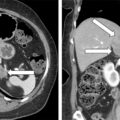
Androgen hypersecretion in a premenopausal woman may be due to an ovarian tumor, an adrenal tumor, polycystic ovarian syndrome, or nonclassic congenital adrenal hyperplasia. The expansiveness of the diagnostic evaluation of a woman with androgen excess is dependent on the clinical situation and the degree of androgen hypersecretion. For example, adrenal gland imaging is not needed when the signs and symptoms related to androgen excess are chronic and mild and serum testosterone concentration is mildly increased in a woman with a typical history of polycystic ovarian syndrome. However, with more abrupt onset of marked signs and symptoms and more marked increases in serum testosterone concentrations, it is important to assess the patient with a full androgen profile and imaging studies of the ovaries and adrenal glands. When an adrenal tumor with a suspicious imaging phenotype is identified in a woman with androgen excess, it should be determined if other adrenocortical hormones are also being autonomously secreted. Herein we present a case of a patient with a nearly pure testosterone-secreting adrenocortical carcinoma (ACC).
Case Report
The patient was a 44-year-old woman seen for progressive hirsutism over the prior 2 years. Her self-graded Ferriman-Gallwey score was 25 (normal, <8; maximum = 36). She also had marked androgenic scalp hair loss. The patient had lifelong regular menstrual cycles until 5 months ago, when they became irregular. She has also had treatment-resistant hypertension diagnosed 3 years ago. An abdominal computed tomography (CT) scan, obtained as part of her secondary hypertension evaluation, detected a 2.6 × 2.9 × 2.7–cm left adrenal mass with suspicious imaging characteristics ( Fig. 90.1 ). Although she was overweight, her body weight had been stable. She had no signs or symptoms of Cushing syndrome. There was no history of hypokalemia. Her medications included amlodipine, 5 mg daily; hydrochlorothiazide, 25 mg daily; and spironolactone, 100 mg daily. On physical examination, her body mass index was 36 kg/m 2 , blood pressure 176/104 mmHg, and heart rate 89 beats per minute. She had marked hirsutism and male pattern balding, but she was not virilized.

INVESTIGATIONS
Laboratory test results are shown in Table 90.1 . Marked testosterone hypersecretion was documented. Although the midnight salivary cortisol concentration and the 24-hour urinary cortisol excretion were normal, the serum corticotropin concentration was low-normal and a 2-mg overnight dexamethasone suppression test documented glucocorticoid secretory autonomy. These data were consistent with dominant testosterone hypersecretion and mild autonomous cortisol cosecretion. Although the adrenal mass was relatively small, the imaging phenotype on CT scan and the plurihormonal pattern on laboratory testing favored an ACC.

