Primary Gastric Lymphoma


Primary Gastric Lymphoma


Frank X. Zhao

Sanford A. Stass


INTRODUCTION

The gastrointestinal tract (GI) is the most common site of extranodal malignant lymphomas, accounting for 30% to 50% of cases.1 Within the GI organs, the stomach is the most commonly involved followed by the ileocecal region, small and large bowel. Since primary nodal lymphoma can also involve secondarily the stomach, it may be difficult to define primary gastric lymphoma (PGL). Gastric non-Hodgkin lymphomas (NHLs) have been defined as lymphomas that uniquely involve the stomach or for which the involvement represents >75% of total tumor volume.2 More strictly, PGL may be defined as the stomach being the only organ involved by lymphoma. Since there were no consensus criteria for the origin of PGL in the past, some cases previously reported in the literature may have included lymphomas of nongastric origin.


PATHOGENESIS OF PRIMARY GASTRIC LYMPHOMA

The pathogenesis of extronodal marginal zone B-cell lymphoma of mucosa-associated lymphoid tissue (MALT), also called MALT lymphoma, serves as a classical mechanism for gastric lymphomas. This type of lymphoma originates from MALT, which represents the accumulation of extranodal lymphoid tissue (characterized by follicular hyperplasia) in the setting of chronic inflammation.3 Although there is normally no organized lymphoid tissue in the stomach, MALT forms in response to chronic inflammation or bacterial infection.4 Helicobacter pylori has been confirmed as the pathogen responsible for some chronic gastritis and PGL.5 If chronic stimulation persists, H. pylori-specific T cells will promote the proliferation of polyclonal B cells in the MALT. Over time one clone may become dominant,6 by either activation of an oncogene (such as BCL10) or inactivation of a tumor suppressor gene (such as p53), and gain an advantage in proliferation over the other B-cell clones. This dominant B-cell clone eventually develops into a MALT lymphoma.


EPIDEMIOLOGY OF PRIMARY GASTRIC LYMPHOMA

PGL comprises approximately 2% of all NHLs. A British study of 153 PGL reported an annual incidence of 1.2% of all gastric malignancies, with most (97%) being NHL.7 A more recent Danish study revealed an incidence of approximately 7% for PGL.2 Significant geographic disparities have been noted. Comparing the incidence of PGL in northern Italy to UK communities, Doglioni et al. reported 13 times more cases of PGL in the Italian community (66 vs. 5 per 100,000 per 5 years).8 Notably, a much higher incidence of gastric carcinoma and H. pylori chronic gastritis was also found in the same Italian community,8 supporting the role of H. pylori in the pathogenesis of PGL as well as gastric carcinoma.


FIGURE 10-1 Endoscopic appearance of MALT lymphoma. MALT lymphoma can have protean presentation from thickened gastric folds and polypoid projection (like in this example) or be flat or ulcerated.


MACROSCOPIC MORPHOLOGY OF PRIMARY GASTRIC LYMPHOMA

The appearance of PGL at endoscopy or on gross examination can be diverse and similar to other malignant tumors of the stomach (Fig. 10-1).They may present as a polypoid nodular growth, as a large fungating mass, or thickened mucosal folds or perforating ulceration. PGL may involve a portion of the stomach, but may also be diffuse or multifocal. Therefore, malignant lymphoma should always be on the list of differential diagnoses of gastric cancers.


CLASSIFICATION OF PRIMARY GASTRIC LYMPHOMA

Currently, the WHO classification is used for PGL. Except for the unique gastric MALT lymphoma, almost all the nodal counterparts of lymphomas can be found in the stomach. Like lymphomas in other organs, PGLs are largely divided into Hodgkin and NHLs. Primary gastric Hodgkin lymphomas are extremely rare. Based on the histology, primary gastric NHLs can be seen as either small cell type or large cell type. Immunophenotyping further divides the lymphomas into B-cell and T-cell types. The distribution of PGL histologic types differs among several studies.9, 10 and 11 However, the most cited are the data of a 2005 German multicenter study (GIT NHL 02/96) of 747 patients11 that indicated that the most common PGL is diffuse large B-cell lymphoma (DLBCL), accounting for more than half of all the 398 PGLs (Table 10-1).


GASTRIC MALT LYMPHOMA

MALT lymphoma is the most common PGL in Western countries.9 The stomach is also the most common organ involved by MALT lymphoma (Table 10-2). The concept of MALT lymphoma was first proposed by Isaacson and Wright in 1983.12 Its connection with H. pylori infection was later demonstrated by Wotherspoon et al.13 Since the progression of chronic gastritis to MALT lymphoma is a continuous process, morphological overlap exists between gastritis and early MALT lymphoma.13 Although there is no consensus on the morphologic criteria for differentiating
gastritis and MALT lymphoma, Wotherspoon et al.
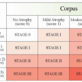
14 have proposed a scoring system for this purpose (
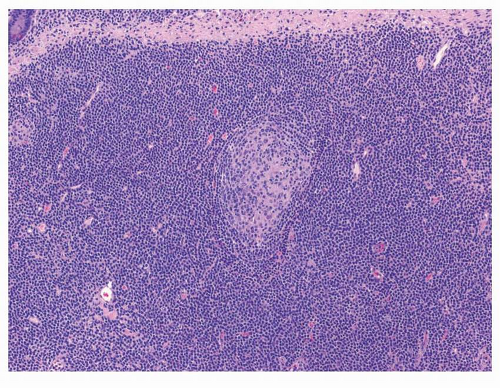
Table 10-3). Generally speaking, expansion of the marginal zone of the hyperplastic lymphoid follicles (Fig. 10-2), dense lymphoid infiltrate composed of predominantly small B cells (
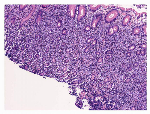
Fig. 10-3), increase in plasmacytoid lymphocytes, dropping out of gastric glands (
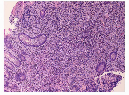
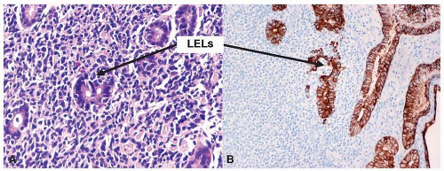
Fig. 10-4), and conspicuous lymphoepithelial lesions (LELs) (Fig. 10-5) are all morphological features that favor MALT lymphoma. However, these features also can be seen in rare cases of severe gastritis. When in doubt, molecular study to detect VDJ rearrangement should be performed to rule out a B-cell clonal process.
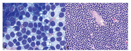
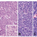
15


Table 10-1 Histological types of the localized PGL


Histological Types


Frequency (%)


Diffuse large B-cell lymphoma


59.5


With small cell component (18.1%)


Without small cell component (81.9%)


MALT lymphoma


37.9


T-cell lymphoma


1.3


Mantle cell lymphoma


0.8


Follicular lymphoma


0.5


Source: From Koch P, Probst A, Berdel WE, et al. Treatment results in localized primary gastric lymphoma: data of patients registered within the German multicenter study (GIT NHL 02/96). J Clin Oncol. 2005;23:7050-7059.


Microscopically, MALT lymphoma is morphologically heterogenous. The characteristic lymphoma cells are small to medium in size and have a bland appearance with relatively increased cytoplasm, slightly irregular nuclei, moderately dispersed chromatin, and inconspicuous nucleoli, resembling centrocytes. The relative abundant, pale cytoplasm allows the cells to exhibit a “monocytoid” morphology16 (
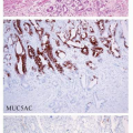
Fig. 10-6). The neoplastic cells can also infiltrate the gastric glands and form clusters within the glandular epithelium, fostering “lymphoepithelial lesions” (Fig. 10-5A). The characteristic LELs can be highlighted by staining for pancytokeratin (Fig. 10-5B).


Table 10-2 Organ distribution of MALT lymphoma


Lymphoma (Other Names)


Organ


Frequency (%)


Primary gastric MALT lymphoma


Stomach


70


Extranodal marginal zone B-cell lymphoma of bronchus-associated lymphoid tissue (BALT lymphoma)


Lung


14


Ocular MALT lymphoma


Ocular adnexa (conjunctiva; eye socket; lacrimal glands)


12


Thyroid MALT lymphoma


Thyroid


4


Immunoproliferative small intestinal disease


Small intestine


1


Table 10-3 Scoring system for the diagnosis of MALT lymphoma


Grade


Description


Histological Features


0


Normal


Scattered plasma cells in lamina propria. No lymphoid follicles.


1


Chronic active gastritis


Small clusters of lymphocytes in lamina propria. No lymphoid follicles. No LELs


2


Chronic active gastritis with florid lymphoid follicle formation


Prominent lymphoid follicles with surrounding mantle zone and plasma cells. No LELs.


3


Suspicious lymphoid infiltrate in lamina propria, probably reactive


Lymphoid follicles surrounded by small lymphocytes that infiltrate diffusely in lamina propria and occasionally into epithelium


4


Suspicious lymphoid infiltrate in lamina propria, probably lymphoma


Lymphoid follicles surrounded by CCL that infiltrate diffusely in lamina propria and into epithelium in small groups


5


Low-grade B-cell lymphoma of MALT


Presence of dense diffuse infiltrate of CCL cells in lamina propria with prominent LELs


CCL, centrocyte-like; LEL, lymphoepithelial lesion.


Source : From Wotherspoon AC, Doglioni C, Diss TC, et al. Regression of primary low-grade B-cell gastric lymphoma of mucosa-associated lymphoid tissue type after eradication of Helicobacter pylori. Lancet. 1993;342:575-577.


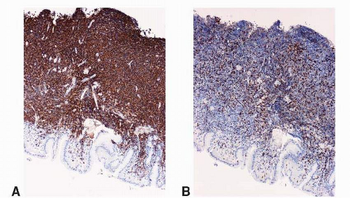
The cells are almost always positive for CD19, CD20 (Fig. 10-7), CD22, and CD79a and show surface immunoglobulin light chain kappa or lambda restriction. They are frequently positive for CD43. Typically negative for CD5 and CD10, an immunophenotype differs from those of most other small B-cell lymphomas (chronic lymphocytic leukemia/small lymphocytic lymphoma, follicular lymphoma, and mantle cell lymphoma [MCL]). The neoplastic B cells often are accompanied by plasmacytic differentiation as well as monoclonal plasma cells. Although primary lymphoplasmacytic lymphoma in the stomach is much rarer than MALT lymphoma, it has been reported.17 Because of the overlapping features, differentiating between these two diseases is extremely difficult. In addition to Waldenstrom macroglobulinemia, which is often
associated with lymphoplasmacytic lymphoma, cytogenetic analysis is helpful in differentiating these two lymphomas.18


FIGURE 10-2 Histology of MALT lymphoma (medium power). Marginal zone expansion of a monotonous population of small bland lymphoid cells is one of the characteristic histology.


FIGURE 10-3 MALT lymphoma (low power). Gastric glands with dense lymphoid infiltration of small lymphoid cells in the lamina propria with occasional dropping out of gastric glands.


FIGURE 10-4 MALT lymphoma (low power). Dropping out of atrophic gastric glands with intestinal metaplasia in an otherwise lamina propria crowded with Lymphoma cells.


FIGURE 10-5 MALT lymphoma (high power). A: On the touch prep, the lymphoma cells are bland with small amount of basophilic cytoplasm, round or occasionally cleaved nuclei, clumped chromatin and inconspicuous nucleoli. Mitotic figures are not present. B: A monotonous population of monocytoid lymphoid cells with relatively more abundant cytoplasm provide ample clear space between the nuclei of the adjacent lymphoma cells.


FIGURE 10-6 LELs of MALT lymphoma (high power). A: Characteristic LELs in the gastric glands (arrows). B: LELs can be easily demonstrated by immunostaining for pancytokeratin.


FIGURE 10-7 Immunophenotype of MALT lymphoma (low power). Immunohistochemistry for CD20 (A) and CD3 (B) reveals that the majority of the infiltrate cells are positive for CD20.

Only gold members can continue reading. Log In or Register to continue

Stay updated, free articles. Join our Telegram channel

May 22, 2016 | Posted by in ONCOLOGY | Comments Off on Primary Gastric Lymphoma

Full access? Get Clinical Tree

Get Clinical Tree app for offline access