Gastric Carcinoma: Classifications and Morphologic Variants


Gastric Carcinoma: Classifications and Morphologic Variants


Do Youn Park

Hye Seung Han

Gregory Y. Lauwers


INTRODUCTION

Several chapters of this book cover the epidemiologic and biologic characteristics of gastric cancer. Others discuss the staging of gastric adenocarcinoma in detail, including its classification into early and advanced gastric cancers. This chapter focuses on the classification of gastric adenocarcinoma, specifically as it relates to anatomic location and especially with regard to cancers of the proximal stomach (i.e., cardia); genetic predisposition; and, most particularly, the various morphologies of gastric cancer.


CARCINOMA OF THE PROXIMAL STOMACH

Worldwide, most gastric adenocarcinomas are diagnosed in the antropyloric region and preferentially on the lesser curvature. Nevertheless, the incidence of proximal stomach cancers has increased, particularly in geographic areas of low risk for gastric cancer.1 However, this phenomenon has not been noted uniformly worldwide.2


Anatomic Controversies

These previous considerations notwithstanding, there is confusion with regard to proximal gastric adenocarcinoma.

First, there is no consensus on the origin of the cardiac-type mucosa that lines the most proximal stomach. This debate is beyond the scope of this chapter, but suffice it to say that this mucosa is variably considered as an original, native lining of the stomach as it connects to the esophagus or as developing (i) secondary to columnar metaplasia of the distal esophageal squamous mucosa due to gastroesophageal reflux and (ii) due to atrophy of the oxyntic mucosa immediately distal to the normal cardia mucosa from Helicobacter pylori infection.3, 4 and 5

Just as there is debate about the origin of the cardia, there is no clear anatomic landmark, either. Pragmatically, the term “cancer of the proximal stomach” (cardia cancer) is applied to adenocarcinomas involving primarily the very first centimeters of the stomach near the gastroesophageal junction (GEJ). A complicating issue is that esophageal tumors may grow downward and involve the cardia. Thus, these tumors frequently overlap, and it is challenging to clarify whether a given tumor is an esophageal adenocarcinoma, or a GEJ or cardial tumor. In an attempt to clarify the issue, Siewert and Stein6 classified tumors of the GEJ region into three types based on the estimated location of the tumor center. Type 1 tumors were classified as true Barrett adenocarcinomas of the distal esophagus, and type 3 tumors represented subcardial gastric tumors. Type 2 tumors were those centered
at the GEJ. However, there does not seem to be appropriate justification for separating GEJ tumors from adenocarcinomas of the distal esophagus, especially from a pathophysiologic standpoint. More recently, the International Gastric Cancer Association has endorsed another scheme for classifying these tumors.7 Type I tumors are defined as those in the distal esophagus, type II are tumors of the true gastric cardia, and type III are those in the gastric mucosa of the subcardia (Fig. 5-1). Finally, the recently published 7th edition of the TNM classification by the American Joint Committee on Cancer proposes a novel classification scheme. Cancers whose epicenter is in the lower thoracic esophagus or EGJ, or within the proximal 5 cm of the stomach (i.e., cardia) and extending into the EGJ or esophagus are to be stage grouped similar to adenocarcinoma of the esophagus, while cancers with an epicenter in the stomach >5 cm distal to the EGJ, as well as those within 5 cm of the EGJ but not extending into the EGJ or esophagus, are to be classified and staged as gastric cancer.8


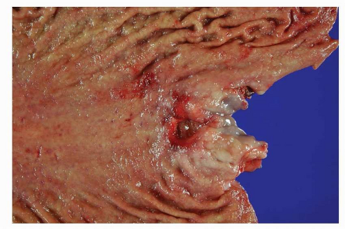
FIGURE 5-1 Adenocarcinoma of proximal stomach. The ulcerated tumor is located just below the GEJ. (Courtesy of center for Gastric Cancer, National Cancer Center, Korea.)


Epidemiologic Characteristics and Risk Factors of Adenocarcinoma of the Proximal Stomach

In geographic areas of low incidence of gastric cancers, proximal stomach tumors are characterized by a higher M/F ratio than distal tumors, and in the United States, they are more common in the white population than in African Americans.9 Furthermore, a proportion of adenocarcinomas arising in the proximal stomach may have an etiology and pathogenesis similar to esophageal adenocarcinoma, that is, an association with a history of reflux symptoms. However, this association is much weaker than that between reflux symptoms and esophageal adenocarcinoma. High body mass index, smoking, and alcohol intake have not been universally accepted as risk factors of the proximal stomach cancer.2,10,11 Furthermore, the association between chronic H. pylori infection and cancer of the proximal stomach is complex. The prevalence of H. pylori infection in patients with cancer of the proximal stomach was the same as in the control population, suggesting that it is not involved in the pathogenesis. However, a trend toward a positive association between H. pylori infection and cancers of the proximal stomach is reported in China.12

Overall, it appears that cancer of the proximal stomach may be connected to at least two disparate etiologies. This provides an explanation for the differing association between H. pylori and cardia cancer in different regions of the world. In regions of the Western world, where H. pylori atrophic gastritis is uncommon and gastroesophageal reflux disease common, the majority of these cancers are of esophageal type. In contrast, in China, where H. pylori atrophic gastritis is common and reflux disease less prevalent, the majority of cancers of the proximal stomach are of the H. pylori atrophic gastritis type and thus resemble distal gastric cancers.


Morphology and Phenotype of Proximal Gastric Adenocarcinoma

Cancers of the proximal stomach share the same morphologic patterns as distal tumors. However, the prevalence of adenocarcinoma has been noted to be higher than for distal tumors at early and
late stages.13 With regard to phenotypic differentiation, differentiated proximal gastric cancers are also more likely to have a gastric immunophenotype. It has been shown that the incidence of human gastric mucin (HGM) expression is significantly higher in proximal tumors at an early stage, whereas in advanced cancers, MUC2 expression is lower than in distal tumors.


HEREDITARY GASTRIC CANCER SYNDROMES

About 10% to 15% of gastric cancers can be qualified as familial.14 Several syndromes and genes are involved, and usually, several organs are at risk of developing various malignancies (Table 5-1).


Hereditary Diffuse Gastric Cancer

Hereditary diffuse gastric cancer (HDGC) patients typically present with diffuse type gastric cancer with signet ring cells and, at late stage, linitis plastica. In addition to a high susceptibility of developing diffuse gastric carcinoma, an increased risk for lobular breast carcinoma is reported.15 Germline mutations in the E-cadherin gene (CDH1) account for 30% to 40% of HDGC cases.16, 17 and 18 The penetrance of the gene varies between 70% and 80%, and the average age for the diagnosis of cancer is 37 years.18 The lifetime risk of developing a gastric cancer is about 67% in men and 83% in women.19

Analysis of prophylactic gastrectomy, the sole preventive treatment, has led to the detection of in situ signet-ring cell carcinomas (SRCCs) and the pagetoid spread of signet ring cells between the foveolar epithelium and basement membrane (Fig. 5-2),20 as well as the presence of multiple foci of stage T1a SRCC confined to the superficial lamina propria and without nodal
metastases.20,21 These lesions are not readily identified by endoscopic examination. In most cases, the neoplastic foci spare the antral mucosa, and there is a marked increase in density and size within the transition zone between the antrum and the body21 There is wide variation in the number of foci both within and between HDGC kindred, from an average over 100 to <20.20,22, 23 and 24 No correlation between patient age and number of foci has been observed. Phenotypic differentiation and proliferation studies support that the neoplasms are developing from the upper isthmus of the neck region of the gastric gland.25


Table 5-1 Hereditary Gastric Cancer Syndromes


Hereditary Syndromes


Genes


Tumor Types


HDGC syndrome


CDH1


Lobular Breast Cancer


Colorectal Cancer


Hereditary nonpolyposis colorectal cancer syndrome


MLH1, MSH2,


MSH6, PMS2


Colorectal Cancer


Endometrial Cancer


Urinary Tract Cancer


Ovarian Cancer


Peutz-Jeghers syndrome


STK11


Colon Cancer


Ovarian Cancer


Cervical Cancer


Breast Cancer


Pancreatic Cancer


Juvenile polyposis syndrome


SMAD4, BMPR1A


Colon Cancer


FAP


APC


Colon Cancer


Brain Tumors


LFS


Tp53


Osteosarcoma


Soft Tissue Sarcoma


Breast Cancer


Brain tumors


Leukemia


Adrenal Cortical Carcinoma


Lung Cancer


Colorectal cancer


Lymphoma


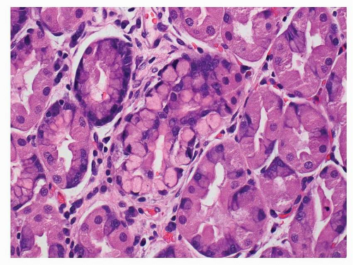
FIGURE 5-2 In situ signet-ring cell carcinoma in a patient with hereditary gastric cancer syndrome. The neoplastic signet ring cells are located within the confines of the pit’s basement membrane.


Hereditary Nonpolyposis Colorectal Cancer Syndrome

Gastric carcinoma, usually of the intestinal type, accounts for 5% to 11% of all carcinomas in these patients. The lifetime risk is 10% for patients of Western ancestry and up to 30% for patients of Asian ancestry26, 27, 28 and 29 An RER phenotype is present in 65% of these cases.


Familial Adenomatous Polyposis Coli

Familial adenomatous polyposis (FAP) patients frequently develop multiple gastric polyps, with fundic gland polyps more common than adenomas. The former have been proven to be neoplastic in nature, with frequent somatic mutations of the APC gene.30 However, the development of carcinoma in fundic gland polyps remains extremely rare.31 Adenomas, which also arise in these patients, are much less common, but at risk for transformation.


Li-Fraumeni Syndrome

Li-Fraumeni syndrome (LFS) is an autosomal dominant hereditary cancer syndrome associated with germline mutations in the tp53 tumor suppressor gene. Gastrointestinal tract tumors are relatively infrequent in this syndrome, accounting for < 10% of all neoplasms that develop; however, among these patients, gastric carcinomas occur more frequently than colon cancers, representing over 50% of the cases.32


Peutz-Jeghers Syndrome

Most patients who meet the clinical criteria present with germline mutations of the serine/threonine protein kinase STK11, found on chromosome 19p. Those who are afflicted with this syndrome develop characteristic polyp and have an increased risk of gastric cancer.33


HISTOMORPHOLOGIC CLASSIFICATION

Just as gastric cancers represent a biologically and genetically heterogeneous group of tumors, their histology is characterized by marked heterogeneity at the architectural and cellular level, reflected, for example, by a combination of foveolar, intestinal, and endocrine-type cells.34

Population-based differences may be seen as well. For example, following the Lauren classification, in high-risk regions of the world, intestinal-type adenocarcinoma is more common than the diffuse type that is relatively more common in low-risk areas. A shift in the proportion of subtypes has also been noted, with the incidence of gland-forming adenocarcinoma decreasing mainly in young patients, and an increasing rate of diffuse-type carcinoma localized to the proximal stomach. Finally, in young individuals, a higher proportion of tumors are of the diffuse type that affects females more often than males.35,36


Table 5-2 Gastric Adenocarcinoma Classification Systems


WHO (2010)


CARNEIRO (1997)


GOSEKI (1992)


MING (1977)


LAUREN (1965)


  • Papillary adenocarcinoma


  • Tubular adenocarcinoma


  • Mucinous adenocarcinoma


  • Glandular carcinoma


  • Well-differentiated tubules Intracellular mucin poor


  • Well-differentiated tubules Intracellular mucin rich


  • Pushing


  • Intestinal type


  • Signet-ring cell carcinoma and other poorly cohesive carcinomas


  • Isolated-cell type carcinoma


  • Solid carcinoma


  • Poorly differentiated tubules Intracellular mucin poor


  • Poorly differentiated tubules Intracellular mucin rich


  • Infiltrating


  • Diffuse type


  • Mixed carcinoma


  • Mixed carcinoma


  • Indeterminate type


  • Adenosquamous carcinoma


  • Squamous cell carcinoma


  • Hepatoid adenocarcinoma


  • Carcinoma with lymphoid stroma


  • Choriocarcinoma


  • Carcinosarcoma


  • Parietal cell carcinoma


  • Malignant rhabdoid tumour


  • Mucoepidermoid carcinoma


  • Paneth cell carcinoma


  • Undifferentiated carcinoma


  • Mixed adeno-neuroendocrine carcinoma


  • Endodermal sinus tumour


  • Embryonal carcinoma


  • Pure gastric yolk sac tumour


  • Oncocytic adenocarcinoma


  • Adenosquamous carcinoma


  • Squamous carcinoma


  • Choriocarcinoma


  • Embryonal carcinoma


  • Hepatoid carcinoma


  • Parietal cell carcinoma


  • Others


WHO Classification

This descriptive classification recognizes four main patterns: tubular, papillary, mucinous, and poorly cohesive (including signet ring cell type), with other variants. Most gastric cancers show significant variability, and the predominant pattern is used to represent the final descriptive diagnosis (Table 5-2).


Tubular Adenocarcinoma

This type of cancer, which tends to form polypoid or fungating masses, is composed of distended and/or anatomizing branching tubules of various sizes (Fig. 5-3A-D). Mucus and cellular
and inflammatory debris can be noted intraluminally. Individual neoplastic cells can be either columnar or cuboidal, or with various degrees of atypia and mitoses. In some well-differentiated cases, the neoplasm can mimic intestinal metaplasia perfectly37 A poorly differentiated variant is sometimes called solid carcinoma (Fig. 5-4). Tubular adenocarcinoma is the most common histologic type of early gastric cancers, representing 52% of the cases.


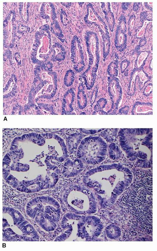
FIGURE 5-3 A: Tubular adenocarcinoma, enteric type. This tumor is formed by irregular-shaped glands lined by hyperchromatic pencillate nuclei. B: Tubular adenocarcinoma, not otherwise specified. This tumor is composed of irregular-sized and -shaped tubules.

Only gold members can continue reading. Log In or Register to continue

Stay updated, free articles. Join our Telegram channel

May 22, 2016 | Posted by in ONCOLOGY | Comments Off on Gastric Carcinoma: Classifications and Morphologic Variants

Full access? Get Clinical Tree

Get Clinical Tree app for offline access