Chronic Gastritis and Gastric Precancerous Conditions


Chronic Gastritis and Gastric Precancerous Conditions


Massimo Rugge

Donato Nitti

Gregory Y. Lauwers

David Y. Graham


INTRODUCTION

This chapter describes the basic clinical context, natural history, and histological phenotypes of gastric precancerous conditions (particularly chronic gastritis); it also addresses the clinicobiological relationship between these diseases and the natural history of gastric cancer. Information is also provided to support the biological rationale for follow-up and preventive therapeutic strategies.

Within gastric mucosa, as in other regions of the gastrointestinal tract, full-blown cancer is the final event of a multistep pathway, usually triggered by (multifactorial) inflammation (Table 20-1).1,2 This link between inflammation and cancer is mediated by a progressive accumulation of genotypic changes associated with dedifferentiation of the native epithelial phenotype. The cascade of biological events results in a new, neoplastic cell capable of infiltrating the surrounding stroma and metastasizing. It has recently been demonstrated that either resident or bone marrow-derived stem cells may be involved in the onset of cancer.3, 4 and 5 This oncogenic pathway is defined as “environmental” because the etiologic agents responsible for inflammatory diseases are of environmental origin, as opposed to the pathway resulting mainly from (host-related) genetic syndromes.

Less than 10% of stomach cancers are hereditary.6 While most of the genetic factors involved in familial gastric cancers are unknown, specific mutations have been well characterized as being responsible for a minor subset of gastric epithelial malignancies. Families harboring germline truncating E-cadherin mutations (with an autosomal dominant pattern of inheritance) have a high prevalence of diffuse-type gastric cancer.7,8 Other hereditary conditions predisposing to gastric cancer include familial adenomatous polyposis (FAP), hereditary nonpolyposis colorectal cancer (HNPCC), and the Li-Fraumeni and Peutz-Jeghers syndromes.6

The natural history of sporadic gastric cancer involves a multistage oncogenic cascade. This chapter will specifically address the “inflammatory step” along this biological pathway.1

In the stomach, a distinction has been established between precancerous conditions and precancerous lesions. Precancerous conditions are clinicopathological situations due to hereditary syndromes and environmental etiologies that are associated with an increased risk of cancer, while precancerous lesions are histological abnormalities in the cell substrate that can lead to cancer.9


Table 20-1 Long-standing inflammatory conditions potentially leading to cancer in the gastrointestinal tract


Anatomical Site


Etiology of Disease


Inflammatory Disease > Malignancy


Esophagus


Gastroesophageal reflux


Barrett mucosa > adenocarcinoma


Stomach


Gastritis (H. pylori associated) Immunomediated


Atrophic gastritis > adenocarcinoma


Small bowel


Immunomediated


Celiac enteritis > lymphomas


Small and large bowel


Immunomediated


Crohn disease > adenocarcinoma


Large bowel


Immunomediated


Inflammatory bowel disease > adenocarcinoma


Liver


Viral, metabolic, toxic


Cirrhosis > hepatocellular cancer


Pancreas


Toxic, immunomediated


Chronic pancreatitis > adenocarcinoma


NONHEREDITARY PRECANCEROUS CONDITIONS

The risk of gastric cancer increases significantly for patients with (a) gastritis, (b) gastric ulcer disease, and (c) gastric mucosa hyperplasia (be it diffuse or focal/polypoid). None of these conditions are primarily hereditary, though a genetic predisposition has been claimed for some, and gene polymorphisms leading to a stronger inflammatory response may increase the risk of onset.10


Gastritis

Gastritis is defined as histologically proven inflammation of the gastric mucosa. Worldwide, the primary cause of gastritis is Helicobacter pylori infection. Approximately 50% of the world’s population is infected, the prevalence being higher in developing and disadvantaged countries, since the prevalence of H. pylori is inversely related to socioeconomic status.11

The etiology of gastritis is established from clinical examination, serology (pepsinogens and antibodies against infectious agents and/or autoantigens), endoscopy (which should include biopsy), and histology. From a clinical standpoint, gastritis may be acute or chronic, though no clear cutoff in duration of disease has been established to distinguish them. In practice, the best way to separate them is by histological examination of gastric mucosal biopsies.

There are robust criteria for classifying gastritis by etiology, and strong correlations have been established between etiology and clinical course. Table 20-2 lists the most common agents responsible for inflammatory conditions of the gastric mucosa and also indicates those most frequently associated with a prolonged (chronic) clinical course.


Gastritis: Basic Morphology

Histology identifies two main histological variants of the disease, that is, nonatrophic and atrophic gastritis.12,13

Nonatrophic gastritis is basically characterized by the presence of inflammatory cells within the lamina propria (lymphocytes, monocytes, polymorphs, and granulocytes), with no loss of the gland units normally present. However, inflammation may coexist with visible alterations, such as columnar epithelia hyperplasia, fibrosis of the lamina propria, and smooth muscle hyperplasia.

Atrophic gastritis is characterized by loss of appropriate glandular units.12 The distinction between atrophic and nonatrophic gastritis is important because there is a well-established clinicobiological relationship between gastric atrophy and adenocarcinoma. The loss of appropriate
glands may coexist with the histological (inflammatory and/or hyperplastic) changes that also occur in nonatrophic gastritis.


Table 20-2 Etiological classification of gastritis


Etiological Category


Agents


Specific Etiology


Clinical Presentation


Notes (Prevalence: High***, Low**, Very Low*)


Infectious Agents


Virus


Cytomegalovirus


Acute


Nonatrophic**


Herpes virus


Acute


Nonatrophic**


Bacteria


H. pylori


Acute or chronic


Nonatrophic and atrophic, type B***


Mycobacterium tuberculosis


Acute (?)


Nonatrophic*


Mycobacterium avium complex


Acute(?)


Nonatrophic*


Mycobacterium diphtheriae


Acute


Nonatrophic*


Actinomyces


Acute


Nonatrophic*


Spirochaeta


Acute


Nonatrophic*


Fungi


Candida


Acute


Nonatrophic**


Histoplasma


Acute


Nonatrophic*


Phycomycosis


Acute


Nonatrophic*


Parasites


Cryptosporidium


Acute


Nonatrophic*


Strongyloides


Acute


Nonatrophic*


Anisakis


Acute


Nonatrophic*


Ascaris lumbricoides


Acute


Nonatrophic*


Chemical Aaents (with Low inflammatory Trait)


Environment (dietary and drug related)


Dietary factors


Chronic


Nonatrophic and atrophic***


Drugs: NSAIDs, ticlopidine


Acute or chronic


Nonatrophic; type C***


Alcohol


Acute


Nonatrophic; type C**


Cocaine


Acute


Nonatrophic; type C**


Bile (reflux)


Acute or chronic


Nonatrophic; type C***


Physical Agents


Radiation


Acute or chronic


Nonatrophic and atrophic*


Immunomediated


Various pathogenesis (mostly unknown)


Autoimmune


Chronic


Atrophic (corpus); type A**


Drugs (ticlopidine)


Acute


?Gluten


Chronic


Lymphocytic gastritis**


Food sensitivity


Acute or chronic


Eosinophilic gastritis**


H. pylori (autoimmune component)


Chronic


Nonatrophic and atrophic


GVHD


Acute or chronic


Nonatrophic and atrophic*


Idiopathic


Crohn disease


Chronic (?)


Nonatrophic/focal atrophy**


Sarcoidosis


Chronic (?)


Nonatrophic or focal atrophy*


Wegener granulomatosis


Chronic (?)


Nonatrophic or focal atrophy*


Collagenous gastritis


Chronic


Nonatrophic*


Idiopathic “isolated” gastritis


Acute or chronic


Nonatrophic and atrophic


The Inflammatory Infiltrate

The inflammatory infiltrate mainly consists of lymphocytes, plasma cells, histiocytes, and granulocytes within the lamina propria and, less commonly, within the single glandular units. Lymphocytes may be either dispersed or packed in follicular lymphoid structures; lymphoid follicles are usually seen in H. pylori infections. Visual analog scales (VASs) have been developed to improve interobserver consistency in scoring the mononuclear infiltrate.14

The term lymphocytic gastritis is applied when a prominent lymphocyte infiltrate is detected within the glandular gastric epithelia. Different cutoff values have been suggested for assessing lymphocytic gastritis, ranging from about 20 to 25 inflammatory cells per 100 epithelial cells. Lymphocytic gastritis is suggestive of, but not diagnostic for, an immunomediated component in the inflammatory disease. An association has also been suggested between lymphocytic gastritis and celiac disease.15, 16, 17 and 18 A more severe (nodular) lymphocytic intraglandular infiltrate that destroys or partially replaces the glandular structures (i.e., “lymphoepithelial lesion”) is almost pathognomonic of primary gastric B-cell lymphoma, which is almost always associated with H. pylori infection.

Active inflammation of the gastric mucosa is defined as the presence of neutrophils within the lamina propria and/or in the glandular lumen. Activity is scored on a VAS according to the intensity of the neutrophilic infiltrate. Inflammatory activity suggests H. pylori infection, the presence of which in cases of chronic inflammation should suggest the presence of an active infection; otherwise, the activity score is clinically of no prognostic value. Active inflammation also is seen in association with any mucosal erosion, as in NSAID-related gastritis, for instance (see below). Intramucosal eosinophilic infiltrate is prominent in eosinophilic gastritis, though the cutoff for its histological assessment (the number of eosinophils per high-power microscopic field) has not been firmly established. The etiopathogenesis and clinical significance of this histological category remain to be clarified.17


Accessory Lesions

These include mucosal erosions, fibrosis of the lamina propria, smooth muscle hyperplasia, and columnar cell hyperplasia.

Mucosal erosions are commonly seen at endoscopy and histology; they can be due to infectious (H. pylori) or chemical (NSAIDs, drugs, bile) agents. Gastric epithelia adjacent to the erosion may undergo reactive hyperplastic changes (see below).

Expansion of the collagen tissue of the lamina propria (fibrosis), coupled with loss of glandular units, is defined as mucosal atrophy. Fibrosis of the lamina propria also may be focal, as seen in scars from prior peptic ulcers.

Hyperplasia of the muscularis mucosae may result from long-term proton pump inhibitor therapy; smooth muscle fascicle hyperplasia may push the glandular coils apart, expanding the interglandular spaces and giving rise to a pseudoatrophic pattern.

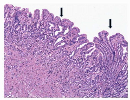
All inflammatory conditions of the gastric mucosa are associated with some degree of regenerative epithelial changes (regenerative hyperplasia), and this is generally seen associated topographically with erosions and peptic ulcers.19 Chemicals (NSAIDs, bile reflux) or infectious stimuli increase the cell turnover by expanding the proliferative compartment of the glands (neck region), resulting in hyperplastic foveolae (Fig. 20-1). Atypical regeneration of the glandular neck may be difficult to differentiate from dysplastic lesions (lesions “indefinite for noninvasive neoplasia”).17,20

Changes occurring in the oxyntic epithelia as a result of proton pump inhibitor (PPI) are sometimes considered hyperplastic changes, but they may simply represent a remodeling of the epithelial structure due to cytoskeletal rearrangements.21


FIGURE 20-1 Foveolar hyperplasia in antral mucosa (hyperplastic foveolae indicated by arrows) (hematoxylin & eosin [H&E], original magnification 25×).

May 22, 2016 | Posted by in ONCOLOGY | Comments Off on Chronic Gastritis and Gastric Precancerous Conditions

Full access? Get Clinical Tree

Get Clinical Tree app for offline access