Plasminogen Activation and Regulation of Fibrinolysis


Plasminogen Activation and Regulation of Fibrinolysis


Nicola J. Mutch

Nuala A. Booth


Hemostasis requires mechanisms both to stop bleeding by formation of a hemostatic plug and to dissolve this plug following wound repair, thereby allowing normal blood flow through the vessel. The latter function relies on local activation of plasminogen (PLG) to plasmin, which dissolves fibrin. The system is finely tuned, to allow healing of a vascular lesion without compromising the early stability of the hemostatic plug and to limit fibrinolytic activity to the injured area. There is a dynamic equilibrium between coagulant and fibrinolytic activities and, within these systems, a further balance between proteolytic and inhibitory proteins. Excessive local or systemic fibrinolytic activity can result in bleeding, often occurring after a delay, as the weakened plug is dissolved. Conversely, an inadequate fibrinolytic response may retard lysis of a thrombus and contribute to its extension.

Plasmin is the central enzyme responsible for thrombus degradation. It is a trypsin-like serine protease generated by activation of the zymogen, PLG. The central players governing the system are plasminogen activators (PAs) and inhibitors of both the activation stage and plasmin activity. Additional players include fibrin and cell surfaces and further proteins that interact with the PLG activation system. This chapter first introduces the key components and then considers how they interact to regulate the activation of PLG and the degradation of fibrin.


PROTEASES AND SUBSTRATES

Fibrinolytic proteases include plasmin, the enzyme that degrades fibrin, and two major PAs that occur in the circulating blood, tissue-type plasminogen activator (tPA) and urinary-type plasminogen activator (uPA), also called urokinase (UK). Like the serine proteases of blood coagulation, they generally exist as a zymogen, a single-chain form of the molecule that is activated by cleavage of a single peptide bond. All the active serine proteases cleave arginine and/or lysine bonds and originated from a simple trypsin-like ancestor protease.1,2 In the case of the fibrinolytic proteases, the products are all in a two-chain form held together by one or more disulfide bonds. An exception to this rule is tPA, which is an active protease in its single-chain form; tPA can be considered to be at one end of the scale of zymogenicity, with PLG as a totally inactive example at the other.3

The typical structure of these proteases (FIGURE 20.1) is that they have an N-terminal A chain containing several independently folded modules or domains, which have particular binding properties, and a C-terminal B chain, which contains the protease domain. The kringle domain—named after a Danish pastry—is approximately 80 residues long and is held together by three internal disulfide bonds. The finger domains are homologous to type I and type II fibronectin regions. Type I finger domains are approximately 50 residues long, contain two internal disulfide bonds, and may play a role in fibrin binding. The epidermal growth factor (EGF) domains, which are homologous with a module in the EGF precursor, contain 53 residues and three internal disulfide bonds. In several proteins, these domains bind to specific cell-surface receptors or binding proteins. The protease domain (about 250 residues, including four disulfide bonds) contains the catalytic triad, Ser195, His57, and Asp102 (chymotrypsin numbering). The protease domain is well conserved, with particularly high-sequence identity in the vicinity of the three active site residues and at the N-terminus.

The individual components of the fibrinolytic system are now discussed in more detail, beginning with the central enzyme, plasmin, and its action on fibrin.


PLASMIN DEGRADATION OF FIBRIN(OGEN)

Fibrin is the substrate most relevant to this chapter but many studies of cleavage sites used fibrinogen, a soluble protein that is more amenable to study (reviewed by4). Plasmin cuts primarily at lysyl and arginyl bonds. Plasmin initially cleaves the α chain of fibrinogen to release the αC fragments, thereby producing fragment X (˜260 kDa; FIGURE 20.2). Further cleavage of fragment X in the α, β, and γ chains along one-coiled coil, connecting the central E and terminal D domains of fragment X, splits the molecule asymmetrically generating fragment Y (˜160 kDa) and fragment D (˜100 kDa).5 Plasmin then cleaves fragment Y into two additional products, a second fragment D and fragment E (˜60 kDa), which contains the amino-terminal portion of all six polypeptide chains. Fragment X can be subsequently clotted by thrombin while fragments Y and D have antipolymerizing effects.

Plasmin digestion of non-cross-linked fibrin is identical to that of fibrinogen, supporting the hypothesis that fibrinogen does not undergo major structural reorganization during polymerization.6 Cross-linked fibrin is degraded more slowly by plasmin and produces different fibrin degradation products (FDPs). Initially a fragment, termed D-dimer, consisting of two fragment D moieties from adjacent fibrin monomers covalently bound between their γ-chain remnants is generated (FIGURE 20.2). Fragment E exists free and in a noncovalent complex with D-dimer (DD/E complex). A variety of large degradation products are released from cross-linked fibrin. These complexes represent a noncovalently bound two-stranded, halfstaggered fibrin protofibril. The DD/E fragment is the smallest complex, with larger forms representing longitudinal strings of DD/E held together by uncleaved coiled coils.5 These larger FDPs are able to associate, in a size-dependent manner, both with themselves and with the clot.6


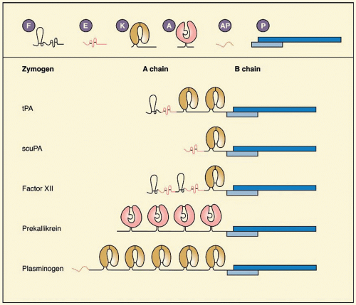
FIGURE 20.1 Structural elements of several fibrinolytic serine proteases. A, apple domain (˜80 residues); AP, activation peptide; E, epidermal growth factor domain (˜32 residues); F, fibronectin finger domain (˜40 residues); K, kringle (˜90 residues); P, protease domain (B chain; ˜250 residues); scuPA, single-chain urokinase; tPA, tissue-type plasminogen activator.


PLASMIN(OGEN)

Human PLG is the zymogen form of plasmin (EC 3.4.21.7). PLG is a single-chain glycoprotein of 92 kDa, consisting of 791 amino acid residues and approximately 2% carbohydrate (Table 20.1). In the human adult, the plasma concentration of PLG is approximately 200 mg/L, or 2 µM (Table 20.1) and is generally rather stable, but there is an increase in the acute-phase response.7 The circulating half-life (t1/2) of native PLG, which has a glutamic acid residue at its N-terminus (Glu-PLG), is approximately 2.2 days; the partially cleaved Lys-PLG (see subsequent section, “Activation of Plasminogen”) has a much shorter t1/2 of 0.8 days.8 The principal production site for PLG is the liver, but PLG is present in the extravascular space of most tissues. Some tissues, such as eosinophils, kidney, cornea, brain, and adrenal medulla, may be capable of synthesizing it.9,10,11,12


Expression and Structure

The PLG gene is 52.5 kb and is located on chromosome 6q26-q27,13 close to two genes for apolipoprotein (a) and for the PLG-related genes A and B.14,15 Two sequence elements of CTGGGA common to acute-phase reactant genes are at position 76 to 81 and -553 to -558.16 Three interleukin-6-responsive elements are at positions -830, -518, and +117 in the 5′-flanking region.17 Two main regions conferring liver specificity of expression to the PLG gene were found in the cis-acting element, AAAAATA, at -2,194 to -2,188 and +48 to +61.18 Several other putative regulatory transcription elements are present in the 5′-flanking region (reviewed in Ref. 17). On the 3′-flanking region, the location of the consensus polyadenylation sequence AATAAA and of potential CAYTG sequence indicates the presence of three polyadenylation sites.16

PLG consists of an N-terminal activation peptide, five kringles, and the protease domain with the catalytic triad His603, Asp646, and Ser74116 (FIGURE 20.3). The kringles are well conserved between species19 and consist of some 80 residues, the structure being shaped by a characteristic 1-6, 2-4, 3-5 disulfide bond pattern using the conserved Cys residues in positions 1, 22, 51, 63, 75, and 80 (kringle 5 numbering). K2 and K3 are covalently linked by a further disulfide bond.20 Functionally, the kringles give PLG the ability to bind to exposed lysyl residues in fibrin,21,22 α2antiplasmin (α2AP),23 tetranectin,24 histidine-rich glycoprotein (HRG),25 collagen,26 high molecular weight (HMW) and low molecular weight (LMW) kininogen,27 thrombospondin,28 and cell-surface receptors or binding proteins for PLG (discussed
in more detail below). The affinity of the kringles for lysine is exploited in affinity chromatography.29 The kringles also bind to ω-aminocarboxylic acids, such as 6-aminohexanoic acid (εACA, 6-AHA), p-aminomethyl benzoic acid (p-AMBA), and trans-4-amino-methyl-cyclohexane-carboxylic acid (trans-AMCA, also known as tranexamic acid). These are used experimentally and clinically to inhibit the functional activity of plasmin by competing with the binding of plasmin(ogen) to fibrin and cell surfaces.


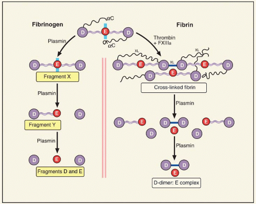
FIGURE 20.2 Degradation of fibrin(ogen) by plasmin. The principal structure of fibrinogen (top) is a three domain globular protein with extending αC domains. Through the action of plasmin the degradation fragments D, E, α-helical coiled coils that connect them, and the Aα chains that extend from the carboxy-terminus of the D-domains. Fibrinogen is degraded asymmetrically (left panel). Intermediate degradation product, fragment X, consists of all three domains connected by coiled coils, but lacks the Aα chains and the Bβ1-42 sequence. Fragment Y is composed of the central E-domain connected by a coiled coil to the D-domain. Cross-linked fibrin (right panel), occurs through the enzymatic actions of thrombin and activated factor XIII (FXIIIa). Thrombin cleaves fibrinopeptides A and B from the E-domain, and FXIIIa cross-links (XL) fibrin longitudinally between D-domains and within the α-chain extensions (see Chapter 16). Plasmic cleavage of the two-stranded protofibrils initially removes the cross-linked α chains, then the coiled coils to liberate a series of FDPs, the smallest of which is DD/E. Larger complexes, such as DY/YD, are also liberated from cross-linked fibrin, but these are subsequently degraded to the DD/E moiety.

Human PLG kringles have different affinities and specificities for ω-amino acids, such as εACA. Kringle 1 exhibits the tightest binding (Kd 9 µM),30,31 followed by kringle 430,32,33 and kringle 5,32,33 with kringle 2 interacting only weakly and kringle 3 not at all.34 Kringle structures have been solved both in solution and in crystals and show the general feature of a hydrophobic groove with acidic and basic residues that are ideally spaced for docking of zwitterions such as lysine or εACA, the opposite charges of which are approximately 7 Å apart. The lack of binding by kringle 3 is explained by the positive charge at position 57, which in the other kringles is negatively charged.35

Experimentally, kringle 4 can be isolated from PLG by digestion with elastase, typically yielding three major components: a fragment containing kringles 1 to 3, kringle 4, and kringle 5 attached to the protease domain.20 The latter fragment, also called mini-PLG, can be activated to plasmin by PAs,36,37,38 as can microplasmin, which has only the last 31 amino acid residues of the A chain and a complete B chain.38 Kringle 1 can be obtained from kringles 1 to 3 by further digestion with other enzymes, such as Staphylococcus aureus V8 protease.39 Other fragments of PLG have effects on tumor growth and metastasis; these fragments are known as angiostatins. Their activity depends crucially on their disulfide pattern.40


Activation of Plasminogen

All known PAs cleave the Arg561-Val562 bond in PLG (FIGURE 20.4), generating a two-chain plasmin molecule in which the A and B chains remain attached by two disulfide bonds. PLG exists in different conformations, affected by its N-terminus and also by the presence of lysine analogues. A particularly important consideration is whether PLG is free in solution or bound to a surface, either fibrin or cells, as discussed more fully in the section on “Balance of Plasminogen
Activation and Regulation of Fibrinolysis” At this stage of our discussion, it can be assumed that the binding is primarily dependent on exposed lysine residues, and that the binding to fibrin and to cell receptors for PLG are generally equivalent.


Table 20.1 Properties of proteins of the fibrinolytic system


Factor


Mr (kDa)


Residues


Concentration (mg/L)


Molar Concentration


t1/2


Function


PLG


92


791


200


2 µM


2.2 d


Zymogen


tPA


68


530


0.005


70 pM


4 min


Protease


uPA


54


411


0.002


40 pM


7 min


Zymogen


PAI-1


52


379


0.02


400 pM


8 min


Inhibitor


PAI-2


46/70


393


<0.005


<70 pM


Inhibitor


Factor XII


80


596


30


0.375 µM


2-3 d


Zymogen


PK


88


619


40


0.45 µM


7-10 d


Zymogen


HK


110


626


70


0.60 µM


9 h


Cofactor


Vitronectin


78


459


350


4.5 µM


Cofactor


C1-inhibitor


105


478


180


1.7 µM


70 h


Inhibitor


α2-Antiplasmin


70


452


70


1 µM


3 d


Inhibitor


α2-Macroglobulin


725


1,451


2,500


3 µM


Inhibitor


HRG


75


507


100


1.5 µM


3 d


Inhibitor


Tetranectin, monomera,b


22.5


181


10


0.5 mM


3 d


Cofactor


Apolipoprotein(a)c


300-800


<7


Modulator


TAFI (pro-CpU)


60


401


5


75 nM


10 mind


Zymogen, Inhibitor


uPAR


55


313


Receptor


Annexin IIa,e


38


338


Receptor


LRP (α2-MR)


600


4,525


Receptor, inhibitor


α-Enolase


54


433


Receptor


Mannose receptor


175


1,437


Receptor


a Mr of nonglycosylated form as listed in Swiss protein base ExPASy.

b Heavy chain of homotrimer.

c Many isoforms due to 13 up to 37 type 4 kringle motifs.

d Activated form.

e Heavy chain of heterotetramer (formed by two heavy and two 10-kDa light chains).


tPA, tissue-type plasminogen activator; uPA, urinary-type plasminogen activator; PAI-1, plasminogen activator inhibitor 1; PAI-2, plasminogen activator inhibitor 2; HMW, high molecular weight; C1-inhibitor, complement component 1 esterase inhibitor; TAFI, thrombin-activatable fibrinolysis inhibitor; uPAR, urinary-type plasminogen activator receptor; LRP, low-density lipoprotein receptor-related protein; MR, macroglobulin receptor.


Cleavage of Glu-PLG generates trace amounts of plasmin cleaved at the N-terminus at one or several of the following bonds: Arg68-Met69, Lys77-Lys78, or Lys78-Val79.41,42 The final product obtained is termed Lys-plasmin (FIGURE 20.4). These two conformations differ in several features, including efficiency of activation and binding to fibrin. Small-angle neutron scattering revealed that Glu-PLG has a more closed form than Lys-PLG,43 making it less readily activated. In the closed form, the five kringle domains and the catalytic domain interact to produce an ellipsoid shape. The open form of Lys-PLG, in contrast, has increased flexibility, which facilitates the binding of PAs some 10-fold.44,45,46 Binding of εACA to Glu-PLG elicits a similar, but probably not identical change in conformation.43 Lys 50 of Glu-PLG binds to kringle lysine binding site (LBS),47,48 making them less available for binding to C-terminal lysine in fibrin, PLG receptors, and other proteins, so that Lys-PLG binds with higher affinity to fibrin than does Glu-PLG. The susceptibility of Glu-PLG to activation is dependent on additional factors, such as the presence of anions, which in the human plasma are mainly chloride ions, and of the divalent cations Ca2+, Mg2+, and Mn2+. These ions counteract the effect of εACA and stabilize PLG in the Glu-form.46,49


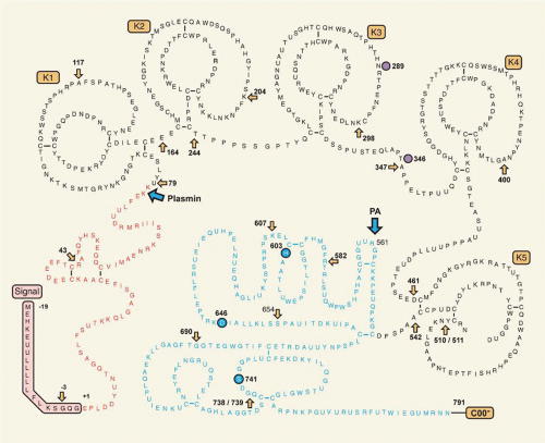
FIGURE 20.3 Sequence of the human PLG molecule. Positions of the 18 introns are indicated by yellow arrows at (for type 1 and 2 introns) and between (for type 0 introns) residues. The 19-residue signal peptide (shown in the pink box with negative numbers) is cleaved by the signal peptidase at the Gly-Glu peptide bond. Conversion of PLG to plasmin by PAs occurs after the peptide bond Arg561-Val562 (shown by a blue arrow). Plasmin cleaves primarily at Lys77-Lys78 bond (shown by a blue arrow) to release the activation peptide (red lettering) generating Lys-plasmin(ogen). K1-K5 kringles are located in the A chain (black lettering). The B chain remains attached to the A chain (blue lettering) after cleavage at 561-562 by PAs via the disulphide bonds Cys548-Cys666 and Cys558-Cys566. Carbohydrate attachment sites (Asn289 and Thr346) are shown as purple circles. The residues that comprise the classical serine protease Ser-His-Asp catalytic triad are circled in blue. (Redrawn based on Petersen TE, Martzen MR, Ichinose A, et al. Characterization of the gene for human plasminogen, a key proenzyme in the fibrinolytic system. J Biol Chem 1990;265:6104.)


Variants of Plasminogen

The PLG molecule is subject to considerable variation in the normal population. In addition to the limited proteolysis and activation processes already discussed, the two important sources of variation are glycosylation and genetic polymorphism.


Glycosylation

Human Glu-PLG occurs in two variants that differ in glycosylation. PLG variant 1 is diglycosylated, with N-linked carbohydrate moiety of the mannose type with 10 to 11 monosaccharide units on Asn289, and an O-linked moiety of three to four residues on Thr346 (FIGURE 20.3).50 PLG 2, which accounts for a little more than half the circulating PLG, contains only the carbohydrate group at Thr346.51 Both of these forms exhibit additional heterogeneity with respect to their sialic acid content, generating six additional isoforms with pIs between 6.1 and 6.6.41,42,52,53 All 12 molecular forms are present in plasma from single donors.8

Activation of PLG 1 in solution is faster than PLG 2, but this difference is not evident on fibrin. PLG 1 switches more readily to the open conformation in the presence of fibrin or tranexamic acid than PLG 2.54 PLG 2 is more rapidly cleared than PLG 1 and bound five times faster to a de-endothelialized
vessel surface.55,56,57 Additional glycosylation sites at Ser24958 and Ser33959 have been described; a further serine residue, Ser578, can be phosphorylated.60


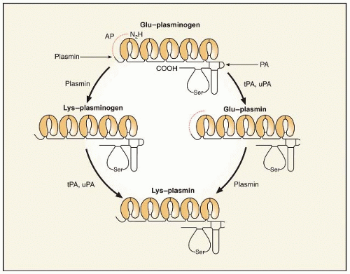
FIGURE 20.4 Activation of native Glu-PLG. PA cleaves at Arg561-Val562, separating the B (light, protease, or catalytic) and the A (heavy, kringle) chains. Glu-PLG and Glu-plasmin forms both contain the amino-terminal activation peptide from Gln1 to Lys76 (shown in red). Plasmin has the potential to cleave this activation peptide (left side), producing Lys-PLG, an intermediate form that interacts with fibrin more efficiently and is readily cleaved by the PAs (tPA and uPA). Plasmin can also cleave the activation peptide from Glu-plasmin, generating Lys-plasmin (right side). The five kringle structures of the A chain are involved in binding of PLG to both fibrin and cell receptors. The catalytic center contains the typical Ser-His-Asp residues and is the major site of interaction with α2antiplasmin.


Polymorphism and Deficiency

Isoelectric focusing of neuraminidase-treated plasma (which removes sialic acid) revealed several bands of activatable PLG, representing the genetic polymorphism of PLG alleles. The two most common variants found in all races are called PLG A (A for acidic pI) and PLG B (B for basic pI)61 with Asp and Asn at position 453, respectively.62 Variants with intermediate pI are designated as PLG M. Variants with more acidic pI than PLG A are assigned a numerical suffix that increases with decreasing pI. Similarly, variants with more basic pI than PLG B receive a numerical suffix that increases with increasing pI. Alleles are designated with an asterisk (e.g., PLG*A3), and silent (null) PLG alleles are designated PLG*Q0 (Q0 standing for quantitatively zero or nonexpressed). The frequency of the allele PLG*A in the Japanese population is considerably higher (95%) than in the Western population (70.1%).63

Recurrent thrombosis is associated with a deficiency of PLG antigen and activity (type I), or a normal level of antigen but reduced activity (type II, dysplasminogenemia). In most instances, a thromboembolic event or ligneous conjunctivitis prompt family studies. Cases in which the molecular defect has been identified are listed in Table 20.262,63,64,65,66,67,68,69,70,71,72,73,74,75,76; updates can be found on the OMIM (Online Mendelian Inheritance in Man) Web site. The data do not prove conclusively that the heterozygous presence of one mutant PLG gene results in an increased incidence of thromboembolism.77,78 Homozygous deficiencies, however, are clearly associated with ligneous conjunctivitis, caused primarily by Arg216 to His or Trp597 to stop mutations,67 and replacement therapy with PLG is effective.69 It is not clear why some mutations cause ligneous conjunctivitis and why very low functional PLG can be tolerated in some individuals, without thrombosis occurring.67

PLG-deficient mice also develop ligneous conjunctivitis.79 These mice are predisposed to severe thrombosis and deposit intravascular and extravascular fibrin in many tissues.80,81 PLG-deficient mice die prematurely and exhibit retarded growth and frequent rectal prolapse, but ovulation, embryonic development, and reproduction are normal.82 Wound healing and tissue remodeling are impaired,83,84 and response to an inflammatory stimulus is diminished.85 After electric injury to the femoral artery, wound healing, removal of necrotic debris, leukocyte infiltration, smooth muscle cell immigration, and arterial neointima formation were greatly delayed in Plg-/- mice,86 suggesting that PLG deficiency might reduce development of atherosclerosis. However, Plg-/- mice crossed with apolipoprotein E-deficient mice develop early atherosclerotic lesions.87 Plg-/- mice crossed with fibrinogen-/- mice were restored to normality in many of these cases, demonstrating the causative role of intravascular and extravascular fibrin deposition.88,89


PLASMINOGEN ACTIVATORS

There are two physiologic PAs, tPA (tissue-type) and uPA (urinary-type, UK), named after the original sources of purified proteins. A further activation system, which is dependent on factor
XII (FXII), prekallikrein (PK), and high molecular weight kininogen (HK), is known as the contact PA pathway. There are also PAs from bacteria and the vampire bat, which are briefly included here. The two human proteases, tPA and uPA, are also implicated respectively in neurobiology (for review see Ref. 90) and tumor biology (for review see Refs. 91 and 92). These aspects are not considered further in this chapter, which focuses on PAs in hemostasis.


Table 20.2 Congenital PLG mutations


Residue


Mutation


Type


Other Names


Clinical Manifestations


References


9


Thr to Asn


I


69


19


Lys to Glu


I


Ligneous conjunctivitis


69


128


Lys to Pro


I


Ligneous conjunctivitis


64,69


133


Cys to stop


I


67


134


Arg to Lys


I


64


212


Lys deletion


I


Ligneous conjunctivitis


69


216


Arg to His


I


Ligneous conjunctivitis


68


355


Val to Phe


II


Nagoya


Thrombophilia


70


374


Val to Phe


Nagoya-I


Thrombophilia


70


453


Asp to Asn


64


460


Glu to stop


I


71


513


Arg to His


I


Ligneous conjunctivitis


69


572


Ser to Pro


I


Thrombophilia (secretion defect)


71,72


597


Trp to Cys


I


Ligneous conjunctivitis


68,69


597


Trp to stop


I


Ligneous conjunctivitis


67,68


601


Ala to Thr


II


Tochigi, Osaska II


Thrombophilia


70,75


620


Ala to Thr


I


Nagoya-II, Tochigi, Kagoshima


Thrombophilia


70


675


Ala to Thr


I


Thrombophilia?


72,74


676


Asp to Asn


II


Osaka-II, III


None


75,76


693


Gly to Arg


II


64


732


Gly to Arg


II


Kanagawa-I


77


8,771


Ala to Gly


I


Ligneous conjunctivitis


78


TISSUE-TYPE PLASMINOGEN ACTIVATOR

tPA, a serine protease of 68 kDa (EC 3.4.21.68), previously known as vascular PA or extrinsic activator, is a glycoprotein of 527 residues (FIGURE 20.1). It exerts its effect primarily in the vascular system, because it is produced and secreted by endothelial cells93; many other cells in culture also synthesize tPA. In normal plasma, the antigen concentration of tPA is approximately 5 µg/L, which corresponds to approximately 70 pM (Table 20.1),94 but most is in complex with its primary inhibitor, plasminogen activator inhibitor 1 (PAI-1).95,96


Expression and Structure

The gene for tPA is located on bands p12-p11 on chromosome 897; it contains four exons98 and comprises 32.7 kb. The mature protein exists in two forms, the N-terminus being Gly or Ser in Bowes melanoma cell tPA,99 but predominantly Ser in recombinant tPA,100 and the numbering in this chapter is based on Ser at position one. tPA expression in cultured cells can be stimulated through multiple intracellular signaling pathways. Vasoactive substances, such as thrombin and histamine, increase tPA synthesis in human umbilical vein endothelial cell (HUVEC), acting through their G-coupled receptors and the protein kinase C pathway.101,102,103 Steroid hormones104,105 and retinoids can increase synthesis of tPA.106,107 The expression of tPA has been reviewed108 and will not be covered in detail here. It is important to note that not all endothelial cells synthesize tPA in vivo and that expression is restricted to small vessels.109,110

The structure of tPA (FIGURE 20.1) shows a finger and EGF domain, and two kringle domains in the A chain, whereas the protease domain resides in the B chain. The finger domain extends from residues 6 to 43. It contains a binding site for fibrin, as mutants lacking kringle domains are still capable of binding fibrin.111 Consistent with this, a degraded form of tPA that had lost the N-terminal 12 kDa bound less well to fibrin than wild-type tPA.112 The binding of the finger domain is independent of LBS, in that it cannot be blocked by εACA.113 Structurally, the finger domain is very similar to the seventh type 1 repeat of human fibronectin.114


The EGF domain (residues 44 to 92) is structurally similar to other EGF structures, as shown by nuclear magnetic resonance (NMR).115 There is some evidence from deletion mutagenesis that the EGF domain binds to the mannose receptor and is involved in tPA clearance.116 Kringle 2 has affinity for lysine, ω-amino acids, and fibrin, whereas no function has yet been ascribed to kringle 1.111 The affinity for the binding of εACA (a model for C-terminal lysine residues) and of N-acetyllysine methyl ester (a model for intrachain lysine residues) is approximately equal, suggesting that unlike PLG, tPA does not have a preference for C-terminal lysine residues binding.113 Intact tPA and a variant consisting only of kringle 2 and the protease domains were found to bind to the cyanogen bromide (CNBr) fibrinogen fragment FCB-2, which also binds PLG and acts as a stimulator of tPA-catalyzed PLG activation.111 In both cases, binding was completely inhibited by εACA, pointing to the involvement of LBS in this interaction.111

The structure-function relations among kringle 2 and ω-amino acids and lysine have been studied in detail, using NMR,117 microcalorimetry,118 crystallography,119 and site-directed mutagenesis.118,120,121 The crystal structure of kringle 2 resembles that of PLG kringle 4; there are, however, differences in the lysine-binding pocket. The core of kringle 2 is formed by a hydrophobic cluster of three tryptophan residues in positions 25, 63, and 74, surrounded by aromatic and hydrophobic side chains that form, at the surface of the kringle, a hydrophobic grove. Ligand binding appears to rely mostly on the integrity of Trp63 and Trp74, and an aromatic residue at position 76, which is normally Tyr.122 Mutation of the critical amino acids Lys33, Asp55, Asp57, or Trp72 markedly diminished binding to lysine-Sepharose, εACA, or both.123

The protease domain of tPA has the typical catalytic triad His322, Asp371, and Ser478. The 2.3-Å crystal structure of the protease domain revealed strong structural similarity with other trypsin-like serine proteases, thrombin in particular.124 The active site cleft is shaped and narrowed by four surface loops. The loop around Arg299 exhibits five additional residues, Arg298-Arg-Ser-Pro-Gly302, compared with chymotrypsin. It projects out of the molecular surface as a β-hairpin and is of fundamental importance for the interaction with PAI-1.3 The 60-loop around Arg327 is similar to but shorter than the corresponding loop in thrombin. Further loops are found around Ser381 and Gly465. The fully solvent-exposed hydrophobic region, comprising amino acids 420 to 423 of tPA, which forms a surface loop near one edge of the active site of tPA, is an important secondary site for the interaction of tPA with PLG in the absence of fibrin.

tPA is secreted as a single-chain molecule (sctPA) that is converted to the two-chain form (tctPA) by plasmin by cleavage at Arg275-Ile276. Unlike most serine proteases the single-chain form is not a zymogen but a protease that, in the presence of fibrin, is nearly as active as the two-chain form.125 Mutagenesis studies have revealed that tPA lacks a specific zymogen triad Asp194, His40, and Ser32 (chymotrypsin numbering).3,126 The constructed double mutant Phe305His and Ala292Ser was more zymogenic.126


Release

The major source of tPA in the circulation under basal conditions is thought to be the endothelial cell, from which it is constitutively released.127 A secondary pool of tPA is present in specialized storage vesicles and is released in response to specific extracellular stimuli.128,129,130,131 High concentrations of tPA are contained within these storage vesicles that occur in various cells, such as endothelial, neuroendocrine, and adrenal chromaffin cells.129,131 In rats, the tissue stores of tPA were calculated to be sufficient to maintain a steady-state plasma level of tPA for 2 days in the absence of protein synthesis.132 Compounds that triggered release within a few minutes include bradykinin, histamine, eledoisin, acetylcholine, β-adrenergic agents, platelet-activating factor, endothelin, calcium ionophore A-23187, and acidosis.133,134 These compounds induce calcium influx into the endothelial cell and activate G-protein-coupled receptors.135 Some interventions that cause elevated tPA act through decreased clearance, including exercise and α-adrenergic agents.136 In all situations in which epinephrine levels are increased, such as stress, anxiety, and exercise, tPA antigen levels increase.96,137

Acute release of tPA is established from early studies on venous occlusion.138,139,140 Young patients with a history of recurrent venous thromboembolism exhibited an abnormal response to venous occlusion and this could be attributed either to subnormal tPA release or, more often, to elevated PAI-1.95,139,140 In isolated cases with severe forms of von Willebrand disease, a complete lack of response to venous stasis and the infusion of 1-deamino-8-D-arginine vasopressin (DDAVP) was found.141 These patients may have a functional or structural defect in their endothelial cells. It should be noted, however, that release of von Willebrand factor (vWF) and tPA occur by different mechanisms.142 Lower PA activity is found in leg veins than in proximal veins and may be one etiologic factor for development of deep venous thrombosis. DDAVP was widely used in the 1980s to study the release potential for tPA in patients with idiopathic or recurrent thrombosis.143,144,145 Comparison of the effects of DDAVP and sodium nitroprusside showed that nitroprusside produced a greater increase of forearm blood flow but no increase of tPA; however DDAVP stimulated tPA release from the vascular bed.146 The increase of circulating tPA levels after intra-arterial infusion of acetylcholine and methacholine was shown to be mediated by muscarinic receptors.146,147,148 Bradykinin and substance P both induced tPA release after being infused into the forearm of volunteers,149,150 and the releasable pool of tPA, rather than the availability of PAI-1 to inhibit it, was the key factor.151


Enzymatic Properties of Tissue-Type Plasminogen Activator

tPA is unusually specific among the serine proteases. Its major substrate is PLG, in which it cleaves the Arg561-Val562 bond but is a relatively inefficient activator of PLG in the absence of its cofactor, fibrin.152 Binding of sctPA and tPA to fibrin is roughly comparable.153 The Kd for binding of tPA to fibrin clots in the absence of PLG ranges from 140 to 400 nM.125,153,154 In the presence of PLG, the affinity of tPA to fibrin increases approximately 20-fold (Kd 20 nM).155 These observations are explained by formation of a ternary complex between tPA, PLG, and fibrin or binding of tPA to PLG, which subsequently, upon binding to fibrin, takes on an open conformation. The isolated A chain of tPA has been shown to bind to Glu- and mini-PLG with a Kd of 100 nM.156

There is a wide range of kinetic parameters reported for the activation of PLG by tPA due to many potential experimental variables, such as different protein preparations, substrate
concentrations, and nature of the fibrin stimulator. In the absence of fibrin, KM values for the activation of Glu-PLG by tPA range from 9 µM to slightly more than 100 µM.125,157,158 In general, KM is three to four times lower with tPA than with sctPA for the activation of Glu-PLG in the absence of fibrin. In the presence of fibrin, this difference is less apparent with KM values typically two orders of magnitude smaller, with only moderate increases of kcat. Values for KM in the presence of fibrin range from 0.16 to 1.1 µM PLG, and kcat values from 0.1 to 1.1 per second.125,158 Several authors found nonlinear enzyme kinetics,156,157,159 and there appear to be two phases in the activation of Glu-PLG by tPA in the presence of fibrin, with an initial KM of 1.05 µM and kcat of 0.15 per second. Later in the process, as new high-affinity binding sites for PLG and tPA are exposed in partially digested fibrin,160,161,162,163,164 KM decreases to 0.07 µM, whereas the kcat is unchanged.159 The key message is that PLG is not readily activated by tPA except in the presence of fibrin, because it is only in this situation that the KM for the reaction is consistent with the circulating PLG concentration of 2 µM.

Distinct sites in the fibrin molecule have been identified as accelerating PLG activation; notably, they are not available for binding in fibrinogen but become exposed in fibrin.165 The D-region of fibrin binds both PLG and tPA in a region that encompasses the residues Aα148-160. Both these associations are of low affinity and in the circulation the Aα148-160 site would bind only PLG, which is present in much higher concentrations than tPA. Another binding in the D-region encompasses γ311-336 and γ337-379, which are linked by a disulfide bond.166 Antibodies have revealed that there is a tPA binding site that includes γ312-324.167 Higher affinity sites for binding tPA and PLG are in the C-terminus of the α chain, within the region Aα392-610.168 The conformational changes that occur on fibrinogen cleavage and fibrin assembly169 reveal new sites for binding of tPA and PLG, and for enhancement of PLG activation.

Mutations have been engineered in tPA to change its characteristics, with the aim of making it an even more effective therapeutic agent. The most striking of these is the series of mutations to make tPA resistant to PAI-1. Madison et al.170 identified the importance of interactions between the positively charged tPA sequence 298 to 302 and the negatively charged PAI-1 sequence 350 to 355. Mutagenesis of Arg298, 299, and 304 resulted in a mutant that was inhibited 120,000 times less rapidly by PAI-1 than wild-type tPA.171 These observations led, in part, to the development of the new tPA mutant tenecteplase (TNK-tPA), in which the sequence 296 to 299 (Lys-His-Arg-Arg) is replaced by four alanines.172 This variant also exhibits slower clearance, by virtue of its changed glycosylation, and its enhancement of PLG activation is more selective for fibrin over fibrinogen and the fibrin degradation product DD/E.173


URINARY-TYPE PLASMINOGEN ACTIVATOR

The urinary-type PA (EC 3.4.2.73; urokinase, uPA) is found in urine at 40 to 80 µg/L,174 and is synthesized by several cell types, particularly cells with a fibroblast-like morphology, but also by epithelial cells175 and monocytes and macrophages.176,177 uPA activates PLG by the cleavage of Arg561-Val562, as does tPA, but importantly does not require fibrin as a cofactor. This characteristic has generated the view that tPA functions in fibrinolysis in the vasculature, whereas uPA’s primary role is in processes such as degradation of extracellular matrix and cell migration, with consequences for wound healing, inflammation, embryogenesis, and invasion of tumor cells and metastasis.178,179 These clear roles of the uPA system outside of fibrinolysis do not argue against its importance in fibrin degradation, as discussed in subsequent text.


Expression and Structure

The human uPA gene is located on chromosome 10q24 and is 6.4 kb in length.180 It contains 11 exons, and the intron-exon organization of the uPA gene closely resembles the tPA gene. The regulation of its expression has been reviewed in detail elsewhere108 and is not included here. uPA is produced as a 54-kDa, single-chain glycoprotein, 411 residues long (Table 20.1) and containing three domains: an EGF domain, a kringle, and a protease domain (FIGURE 20.1). The EGF domain interacts with urokinase plasminogen activator receptor (uPAR) found on many cells, as discussed in subsequent text. The kringle domain has no affinity for fibrin but has been reported to stabilize the interaction of uPA with uPAR.181 The protease domain contains the catalytic triad, His204, Asp255, and Ser356, and has approximately 40% sequence identity with tPA.98 uPA is normally glycosylated at Asn302, whereas a covalently attached single monosaccharide, fucose, to Thr18,182 appears to affect the mitogenicity of uPA.183 There are two phosphorylation sites on Ser138 and Ser303. The phosphorylated form diminishes uPA’s interaction with cells and PAI-1.184

Activation of single-chain urinary plasminogen activator (scuPA, also called proUK) to the active enzyme, uPA (UK), is by cleavage of Lys158-Ile159.185 Many enzymes can cleave this bond but the most important to hemostasis are plasmin,186,187 factor XIIa, and kallikrein.188 In PLG-deficient mice activation of scuPA is ascribed to kallikrein.189 Cleavage close to the activation site (Lys158-Ile159) generates an inactive form of uPA. Thrombin cleaves Arg156-Phe157 in scuPA188,190,191 and the inactive product can be subsequently activated through release of the N-terminal dipeptide of its B chain by cathepsin C or, albeit slowly, by plasmin.185,192 Granulocyte elastase and cathepsin G cleave the Ile159-Ile160 peptide bond to yield an inactive product.193 uPA has two active forms: HMW-UK and LMW-UK.194 These are, respectively, full-length active uPA and a processed form, cleaved at Lys135-Lys136 to yield the amino-terminal fragment (ATF), which interacts with uPAR, leaving 21 residues of the A chain and an intact B chain.


Enzymatic Properties of Urinary-Type Plasminogen Activator

The question of whether scuPA is a true zymogen was debated for several years, a controversy that was fuelled by the problem of trace plasmin or uPA contamination, with reciprocal activation of scuPA and PLG.195 The issue has been resolved by studies on mutant forms of the proteins and by examining scuPA in the absence of PLG. For instance, the Lys158Glu mutant of scuPA, which cannot be cleaved by plasmin into uPA, still converted PLG to plasmin.196 The consensus is that scuPA has approximately 0.5% of the activity of uPA.197,198,199,200 Therefore, scuPA lies on the spectrum between PLG, a true zymogen, and tPA, which is almost fully active in its single-chain form. Lys300 and Asp355 are key structural elements of scuPA’s enzymatic
activity. Lys300, situated in the flexible loop region 297 to 313, forms a weak interaction with Asp355 and pulls the adjacent active site Ser356 into the position found in the fully active protease. Site-directed mutagenesis of Lys300 to Ala201 or of Asp355 to Asn202 resulted in a 40-fold and 270-fold reduction in activity, respectively, compared to wild-type scuPA. Altering the flexibility of the 297-313 loop changes the intrinsic catalytic activity of scuPA.203


Urinary-Type Plasminogen Activator Receptor

A cellular receptor for uPA (uPAR; CD87) was first isolated on human monocytes204 but is expressed on several cell types. The gene for uPAR (gene code PLAUR) is on chromosome 19 and consists of seven exons and six introns extending over 23 kb.205,206 Regulation of its expression has been reviewed108 and is not included here. The 1.4-kb mRNA encodes a signal peptide of 22 and a protein of 313 residues, with a glycosylphosphatidyl inositol (GPI) anchor (FIGURE 20.5). The N-terminus of the receptor consists of three homologous domains; region 1 interacts with uPA and regions 2 and 3 enhance binding.207

Only gold members can continue reading. Log In or Register to continue

Stay updated, free articles. Join our Telegram channel

Jun 21, 2016 | Posted by in HEMATOLOGY | Comments Off on Plasminogen Activation and Regulation of Fibrinolysis

Full access? Get Clinical Tree

Get Clinical Tree app for offline access