Crohn’s related risk factors for small bowel adenocarcinoma
Long duration of CD
Area of CD inflammation
Jejunal CD
Strictures
Fistula
Bypassed segment
CD medications
Young age
Male gender
Tobacco-alcohol-diet
The most common clinical presentation of SBA associated with CD is obstruction with nausea, vomiting, and abdominal pain. Other presentations are hemorrhage, fistula, and perforation [3–4]. These symptoms are commonly seen in CD’s exacerbation, and it is almost always difficult to differentiate small bowel adenocarcinoma from those of the CD’s activation. Majority of the patients with SBA associated with CD are diagnosed at the time of operation or postoperatively and only less than 5% cases are diagnosed preoperatively [3]. Two indicators of the malignancy in patients with CD are exacerbation of the symptoms after long periods of the quiescence and small bowel obstruction refractory to medical treatments [4]. Therefore, it is important to consider surgical assessment of patients with long-standing symptomatic CD who failed to respond to conservative treatments.
The age of diagnosis of SBA associated with CD is 45–55 years [3, 4]. Even though de novo SBA are seen all along the small intestine, SBA associated with CD is almost located within the inflamed areas of the ileum [4–6].
Diagnosis of SBA associated with CD is often challenging. Imaging techniques may miss lesions smaller than 0.5 cm and not be able to distinguish the affected areas from severe CD.
SBA staging can be 47% accurate with computed tomography (CT); however sensitivity is very low in the presence of CD [7]. CT exposes patients to radiation, and magnetic resonance (MR) imaging is not, but MRI takes much longer time and cost is expensive [8]. Enteroclysis is invasive and training is required for exact diagnosis of SBA associated with CD [9]. Positron emission tomography (PET) is used within limits in the diagnosis of SBA associated with CD due to chronic inflammation [10]. Multiphasic dynamic studies may have the potential to improve the diagnostic capacity of multidetector CT for SBA [11].
SBA associated with CD are mainly adenocarcinoma, very rare signet ring cell carcinoma [12].
The prognosis of SBA associated with CD has been reported to be poorer than the de novo small bowel carcinoma [13].
To date, there is no any report regarding the incidence and prognosis as well as management of peritoneal metastasis (PM) from SBA associated with CD. Here, we report our case of a patient with PM from SBA associated with CD who had a limited survival.
1.3 Case Report
A 51-year-old male patient with a history of Crohn’s disease for 30 years developed intestinal obstruction in June 2014. Through a midline incision, he underwent resection of the terminal ileum. Pathological investigations revealed adenocarcinoma of the ileum developed from CD. He underwent a strictureplasty and small bowel resections 10 years ago due to CD. In July 2014, he developed an intestinal obstruction and returned back to the operating room for right hemicolectomy and segmental small bowel resection. The patient had five cycles of adjuvant systemic chemotherapy with FOLFOX, and he developed peritoneal metastasis after systemic chemotherapy. Then, six cycles of FOLFIRI + bevacizumab were given. His PET scan shows the peritoneal metastasis due to seeding from his previous operations.
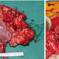
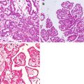
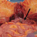
Positron emission tomography (PET), computerized tomography (CT), PET-CT, and computerized axial tomography (CAT) scans of the patient with PM from SBA associated CD are given in Figs. 1.1, 1.2, 1.3, 1.4, 1.5, and 1.6. The patient developed partial intestinal obstruction in December 2015. The patient was presented to the American Society of Peritoneal Surface Malignancy multidisciplinary team via the Internet. Only a limited outcome has been introduced from all over the world. They all concluded that the management of PM from SBA associated with CD is poor. His perioperative findings revealed that he had a recurrence in the ileo-transversostomy site and with extensive peritoneal metastasis (Figs. 1.7, 1.8, and 1.9). His perioperative PCI score was 30. He underwent a cytoreductive surgery and terminal ileostomy and hyperthermic intraoperative intraperitoneal chemotherapy. The small bowel was around 120 cm and had multiple metastatic nodules after the cytoreduction. Small bowel mesenteric nodules are removed. He had a CC-3 resection and HIPEC. His postoperative period was uneventful. He was discharged on postoperative day 12. He had only 3 months free from symptoms. Then, best palliative care was initiated and he died in the end of April 2016, 22 months after his first diagnosis.
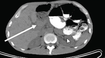
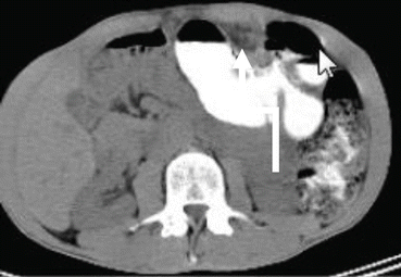
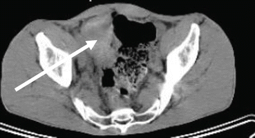
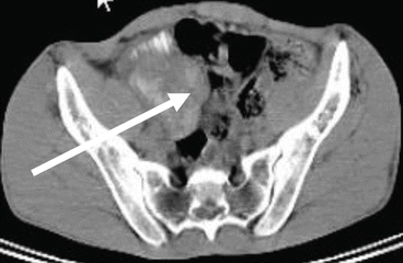
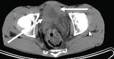
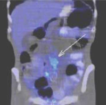

Fig. 1.1
The mass in the anterior abdominal wall in axial CT scan. Arrow shows the intra-abdominal metastatic mass in patients with SBA associated with CD

Fig. 1.2
The mass in the anterior abdominal wall in axial CT scan. Arrow indicates the metastatic tumor in patients with SBA associated with CD

Fig. 1.3
The mass in the lower anterior abdominal wall in axial CT view. Arrow indicates the metastatic abdominal mass in patients with small bowel adenocarcinoma (SBA) associated with Crohn’s disease (CD)

Fig. 1.4
The mass in the lower anterior abdominal wall in axial CT view. Arrow indicates the metastatic abdominal mass in patients with small bowel adenocarcinoma (SBA) associated with Crohn’s disease (CD)

Fig. 1.5
The masses in the lower anterior abdominal wall in axial CT view. Arrows indicate the metastatic abdominal mass in patients with SBA associated with CD

Fig. 1.6


A PET-CT scan of the peritoneal metastasis of the patients with small bowel adenocarcinoma associated with Crohn’s disease. Arrow indicates the peritoneal nodules
Stay updated, free articles. Join our Telegram channel

Full access? Get Clinical Tree


