Perinatal Infections


Perinatal Infections


VIRUSES


Rubella

Rubella (also known as German measles) is usually a mild viral illness with fever, postauricular or suboccipital lymphadenopathy, arthralgia, and a transient erythematous rash. It has been gratifying that since 1966, the incidence rate of reported rubella cases has fallen dramatically from 28 cases per 100,000 population to approximately 1 case per 100,000 in 1982. Yet, there was a resurgence of rubella and congenital rubella syndrome (CRS) from 1989 to 1991, with outbreaks continuing to occur. By 1992 and 1993, however, the reported number of cases of rubella was the lowest ever. Further, in 1996, there were only 238 cases of rubella reported in the United States and only 4 cases of CRS.


Epidemiology

Wild rubella virus is spread by droplets or direct contact with infected persons or articles contaminated with nasopharyngeal secretions. Although it is considered primarily a disease of childhood, in the past few years, more than half the cases have occurred in patients older than 9 years. About 65% of reported cases of rubella now occur in individuals of reproductive age. By reproductive age, about 75% to 85% of the population has had rubella, and about half of the cases are subclinical infections. Once wild virus infection occurs (even if subclinical), immunity is lifelong.

Before vaccination became available in 1969, rubella occurred in 6- to 9-year cycles, but the last major epidemic was in 1964. Since 1982, the rate in the United States has been less than 1 case per 100,000 population; but outbreaks occur, particularly among members of religious communities that traditionally refuse vaccination, among young adults in specific racial/ethnic groups (for example, Hispanics and Asian/Pacific Islanders) who frequently have not been vaccinated, and among recent immigrants from countries, such as Brazil and Mexico, where vaccination is not routine.

In early pregnancy, primary maternal rubella may lead to involvement of the embryo or fetus. As shown in Table 15.1, the risk of CRS after maternal infection in the first trimester is as high as 85% when infants have been followed for up to 40 years. The risk of any defect decreases to approximately 50% if infection occurs during the 9th through 12th week of gestation, and the risk of CRS is low when infection occurs after the 20th week of gestation. In 1996, the Centers for Disease Control and Prevention (CDC) revised the case definitions for rubella and CRS. These are available from CDC or on the website for this textbook.

Cataracts, patent ductus arteriosus, and deafness are the most common abnormalities. When these children have been followed for a few years, additional disorders such as diabetes have occurred more frequently than expected. CRS has not been eliminated from the United States, but progress is being made. In 1969, when the vaccine was licensed, there were 62 cases of CRS in the United States. The number of cases decreased generally over the next two decades, so by the late 1980s, only one to three cases were reported per year. Then paralleling the rise in rubella overall, there was a dramatic surge in CRS cases between 1990 and 1991 to 25 and 31 cases, respectively. In 1991, two thirds of the cases occurred in infants born to Amish mothers in Pennsylvania. By 1992, there were only five cases in the United States, and in 1993—for the first time ever—there were no cases of CRS among infants born in the United States.


Diagnosis

With rubella infection, the virus can be isolated from the bloodstream and throat 7 to 10 days after exposure. Shedding of virus from the throat continues for about 1 week. The rash, which typically starts in the face, commonly develops 16 to 18 days after exposure. The diagnosis of rubella should never be based solely on clinical criteria. To confirm rubella infection, various antibody tests are available. Hemagglutination inhibition (HI) antibody, an immunoglobulin (Ig)G class antibody, has been most commonly used for screening, but several newer antibody tests have replaced the HI because of lower cost and/or greater sensitivity. The most commonly used tests are enzyme immunoassay (EIA) tests, but others include latex agglutination, fluorescence immunoassay, passive hemagglutination, hemolysis in gel, and virus neutralization. Several kits have been approved by the Food and Drug Administration (FDA), but in comparative testing, not all kits are equivalent. The most important aspect of a diagnostic kit for rubella screening tests is a high specificity (i.e., a low false-positive rate), as false-positive test results could result in failure to identify susceptible women. Further, many clinical laboratories now report rubella status as simply present or not present (corresponding to immune or nonimmune). Any antibody level is considered evidence of immunity. Thus, when acute rubella is suspected, the laboratory must be consulted and paired serum obtained so that quantitative antibody levels can be measured.

Also an IgG antibody, complement-fixing (CF) antibody appears later than HI antibody and may be useful in some diagnostic situations, such as in patients with high HI levels or patients first seen 1 to 5 weeks after exposure. The CF test must be requested because it is not performed routinely. Rubella-specific IgM antibodies appear early and last for only
a few weeks. This test may be helpful in some diagnostic situations but is available in few laboratories. Although a positive rubella IgM titer indicates recent primary rubella infection, its absence does not necessarily exclude it, as in some patients, IgM may disappear in less than 4 to 5 weeks. When a patient is first seen with an elevated titer, it is not always possible to determine whether there has been acute primary rubella infection, even though these other tests are properly used. According to the CDC, persons generally can be presumed to be immune to rubella if they have documented vaccination with at least one dose of measles, mumps, and rubella (MMR) vaccine or other live rubella-containing vaccines, when administered on or after the first birthday, or if they have laboratory evidence of rubella immunity, or if they were born before 1957 (except women who could become pregnant). Birth before 1957 is not acceptable evidence for rubella immunity for women who could become pregnant because it provides only presumptive evidence of rubella immunity and does not guarantee that a person is immune. If a person has an “equivocal” serologic test result, that individual should be considered susceptible to rubella unless they have evidence of adequate vaccination or a subsequent serologic test result indicating rubella immunity. Postinfection immunity to rubella appears to be long lasting and is probably lifelong.


TABLE 15.1 ▪ RISK OF CONGENITAL RUBELLA AFTER MATERNAL INFECTION BY GESTATIONAL AGE AT INFECTION


Gestational Age (wk)


Estimated Risk (%)


<8


85


9-12


52


>20


Rare


Reproduced with permission from the Centers for Disease Control and Prevention. Measles, mumps, and rubella-vaccine use and strategies for elimination of measles, rubella, and congenital rubella syndrome and control of mumps: recommendations of the Advisory Committee on Immunization Practices (ACIP). MMWR Morb Mortal Wkly Rep 1998;47:1-57.


Counseling and Management

Pregnant women with confirmed rubella infection must have proper counseling about the risks and types of congenital anomalies. Culturing amniotic fluid for rubella virus is not advised, because this does not reliably distinguish the infected fetus from the uninfected one among pregnancies at risk.

The Advisory Committee to the CDC notes that Ig given after exposure prevents neither infection nor viremia, although it may alter symptoms. Further, infants with congenital rubella have been born to women who received Ig shortly after exposure. Thus, the committee does not recommend Ig for routine use for postexposure prophylaxis. Yet, it suggests that Ig might be useful in susceptible, exposed pregnant women for whom pregnancy termination would not be acceptable. In this case, Ig might offer some protection against fetal infection.


Prevention

For the last 30 years, the rubella virus vaccine has been RA27/3, which contains live attenuated virus. RA27/3 is administered subcutaneously. After vaccination, approximately 95% of susceptible individuals develop HI antibodies, which provide longterm (probably lifelong) protection. Among adults who did not show a positive HI titer after vaccination, nearly all show detectable antibody when a more sensitive test is used. According to the CDC, any detectable rubella antibody or a history of rubella vaccination is presumptive evidence of immunity.

Those vaccinated may shed the attenuated virus from the nasopharynx for a few weeks, but there is no evidence that the vaccine virus can be transmitted. Thus, there appears to be no risk to susceptible pregnant women who contact recently vaccinated children or adults.

Rubella-susceptible women of reproductive age should be considered candidates for immunization, but it is recommended that pregnancy be avoided for 90 days. The immediate postpartum period is often suggested as an excellent time for immunization. Vaccinated women may breastfeed without fear of adverse effects to the newborn, and Rh-negative women may receive Rho(D) immune globulin, if indicated, as well as the rubella virus vaccine.

It has been suggested that to eradicate CRS, one must make an increased effort to ensure that patients are either immune to rubella or vaccinated as part of routine medical and gynecologic care. For reproductive-age women, recommendations have included determining rubella immunity or vaccinating women in family planning clinics, during any hospitalization, at the time of premarital serology testing, or at college entry examinations. Rubella outbreaks in hospitals have led to the recommendation to screen for rubella immunity and vaccinate susceptible hospital employees who may have contact with pregnant women.

Some rubella-susceptible pregnant women have received the RA27/3 rubella virus vaccine within 3 months after the time of conception. With the RA27/3 vaccine, vaccine virus is isolated less frequently (approximately 3%) from the products of conception. Even when the virus is isolated in the products, none of these cases resulted in any anomalies consistent with CRS. The risk of teratogenicity from the RA27/3 vaccine virus is considerably lower than that with the wild virus and has been estimated by the CDC to be 1.2%.

Side effects of the vaccine include arthralgias, but true arthritis occurred in less than 1%. In susceptible adult women, joint symptoms are more frequent and tend to be more severe than in children, but adults have not usually had to disrupt work. Other complaints such as pain or paresthesias have been rare.

Besides pregnancy, contraindications to vaccination are febrile illnesses and immunosuppression. Precautions are necessary in few individuals with neomycin allergy. Breastfeeding is not a contraindication to vaccination. Even though vaccine virus can be transmitted via breast milk to the infant, the infant remains asymptomatic. The eradication of CRS and rubella is feasible worldwide because of the high efficacy of the RA27/3 rubella vaccine and the low cost of vaccine outside of the United States, making it affordable except in the poorest countries. However, universal vaccination of infants without associated vaccination of adults is not likely to be successful. Efforts need to be made to strengthen postpartum vaccination of susceptible women and women at all encounters in the healthcare system.


Prenatal Diagnosis of Congenital Rubella

Investigators have reported a variety of techniques to diagnose fetal infection after primary rubella infection in early pregnancy. These have included testing of amniotic fluid, fetal blood, and placental tissue for virus isolation and rubella antigens, rubella IgM and RNA. Overall, these approaches have had low sensitivity. A more sensitive test has been reported to be a reverse-transcription-nested PCR.


Cytomegalovirus

Cytomegalovirus (CMV), a DNA virus of the herpesvirus group, causes cytomegalic inclusion disease. Characteristic large cells with prominent intranuclear inclusion bodies have been identified with this disease since the early 20th century, but the virus was not isolated until 1956 (Fig. 15.1).

Although this “owl’s eye” appearance is pathognomonic for CMV infection, these cells are relatively rare in infected individuals. Initially, CMV was considered to be rare because only the classic clinically severe form of the disease was appreciated. Subsequent studies using viral cultures have identified the frequent presence of “silent” CMV in which no clinical manifestations are present. There are approximately 40,000 infants born annually in the United States with congenital infection. CMV is now recognized as the most common cause of intrauterine infection, and congenital CMV infection has been reported to occur in 0.5% to 2.5% of all births in the United States. An additional 3% to 5% of newborns acquire CMV during the perinatal period.

Absence of detectable infection at birth may not be innocuous. The persistent and progressive nature of these inapparent congenital infections may result in central nervous system (CNS) pathology and neurologic sequelae, which represent the major impact of CMV infection.


Epidemiology

Approximately 40% of women in the United States and Europe are susceptible to CMV by the time they reach reproductive age, and the highest rate of seroconversion occurs between the ages of 15 and 35 years. Socioeconomic status is a major determinant of susceptibility; only 15% of lower income women are susceptible, compared with 45% of higher income women. The rate of seroconversion in women in the reproductive age range is approximately 2% annually in higher socioeconomic groups, compared with 6% among lower socioeconomic groups.

In a study from Alabama and Texas, 30% to 50% of pregnant women were susceptible to CMV. Primary CMV infection, as evidenced by seroconversion, occurred in 1.6% to 2.2% of pregnancies. Although CMV infection is widespread, it produces serious illness only in fetuses, immunodeficient individuals, and patients receiving immunosuppressive therapy. Most adult infections are subclinical, and the remainder (approximately 10%) cause a mononucleosis-like illness.


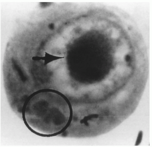
FIGURE 15.1 Intranuclear (arrow) and cytoplasmic inclusion (circle) in a pathognomonic “owl’s eye” cell of cytomegalic inclusion disease.

CMV is a ubiquitous organism that has developed a remarkably successful form of parasitizing humans. It is persistently excreted and is communicable for long periods. Infants infected congenitally excrete CMV on an average of 4 years. Those acquiring CMV at the time of birth excrete the virus for 2 years. Many seropositive young adults shed CMV intermittently.

Asymptomatic CMV infection and excretion is common during pregnancy. The cervix is involved in 3% to 18% of cases, urinary tract in 3% to 9%, breast milk in up to 27%, and the pharynx in 1% to 2%. Overall, CMV can be cultured (cervix and urine) in 2% to 28% of pregnant women. The incidence of CMV infection is highest in low-income, young, primiparous, lower educational status, and unmarried women. These asymptomatic infections occur mainly in seropositive women whose antibody status does not change despite viral shedding. Pregnancy may either increase a woman’s susceptibility to CMV infection or reactivate latent infection.

Congenital CMV infection is acquired in utero, usually as the result of transplacental transmission of CMV. However, ascending infection from the cervix may also occur. Neonates with congenital CMV are culture positive for the virus at birth. Most commonly, CMV is excreted in urine. On average, 1% of all newborns excrete CMV at birth and are congenitally infected. In addition to those fetuses, an additional 3% to 5% of liveborn infants acquire CMV peripartum, presumably as a result of exposure to infected cervical secretions, ingestion of infected milk, or exposure to infected transfused blood. If a maternal genital CMV infection is present at the time of birth, 30% to 50% of the neonates will acquire the virus. Perinatal CMV is the term applied to infants whose initial urine culture at birth is negative but who have subsequent positive cultures several days to months after delivery.

The infections transmitted in utero are the major concern, because they affect infant development. Perinatally acquired CMV infection does not result in serious complications or sequelae except in very-low birth weight neonates (less than 1,200 g). Congenital infections may occur after either primary or recurrent maternal infection. Recurrent maternal CMV is an important cause of intrauterine transmission of CMV. Most intrauterine infections occur in immune, rather than in susceptible, women. In fact, the birth of an infant with congenital CMV infection does not ensure that a subsequent fetus will not become infected in utero. Congenital CMV has been reported in consecutive pregnancies up to 3 years apart. In a prospective study of seroimmune women, the rate of congenital CMV infection was 1.9% among 541 infants born to seropositive women.

However, primary CMV infection acquired during pregnancy poses a significantly more severe risk to the infant than does recurrent infection. Symptomatic congenital CMV infection occurs mainly with primary maternal infection, and sequelae or complications were significantly more frequent in the primary group (35% vs. 7%) (Table 15.2). Only infants born to mothers with primary CMV infection had symptomatic CMV infection at birth (18% vs. 0%). Sequelae were noted in 25% of the primary group, compared with 8% in the recurrent CMV infection group. Mental impairment (Ig level of less than 70) was noted in 13% of infants exposed to primary CMV, versus 0% in the recurrent group. Sensorineural hearing loss was present in 15% of infants born to mothers with primary CMV infection during pregnancy, compared with 5% of infants born to mothers with recurrent infection.
Most importantly, only children born to mothers with primary CMV infection developed bilateral hearing loss (8%).


TABLE 15.2 ▪ SEQUELAE IN CHILDREN WITH CONGENITAL CYTOMEGALOVIRUS INFECTION ACCORDING TO TYPE OF MATERNAL INFECTIONa


Type of Maternal Infection


Sequelae


Primary (%)


Recurrent (%)


p Value


Sensorineural hearing loss


18/120 (15)


3/56 (5.4)


0.05


Bilateral hearing loss


10/120 (8.3)


0/56 (0)


0.02


IQ <70


9/68 (13.2)


0/32 (0)


.03


Chorioretinitis


7/112 (6.3)


1/54 (1.9)


0.20


Microcephaly


6/125 (4.8)


1/64 (1.64)


0.26


Seizures


6/125 (4.8)


0/64 (0)


0.08


Death


3/125 (2.4)


0/64 (0)


0.29


Any sequelae


31/125 (24.8)


5/64 (7.8)


0.003


aBased on Fowler KB, Stagno S, Pass RF, et al. The outcome of genital cytomegalovirus infection in relation to maternal antibody status. N Engl J Med 1992;326:663-667, with permission.


Because CMV is not highly contagious, infection with the virus requires close contact with infected bodily secretions. CMV is sexually transmitted, and pregnant women may also be infected by household spread of CMV from young children, who often have a high prevalence of the virus. Children attending day care centers have become a major source for parental transmission of CMV. From 25% to 80% of children attending day care centers acquire CMV, which they shed in their urine and saliva for up to 2 years. Clearly, these toddlers transmit CMV to their mothers and to other family members. Fifty percent of susceptible household members will seroconvert when CMV is introduced into the household. Risk factors for primary CMV infection in pregnancy include (i) presence of young child in the home; (ii) white race; (iii) younger age; and (iv) middle to upper income.

There are multiple potential sources of perinatal infection with CMV. Transplacental vertical transmission from mother to fetus has been confirmed. In addition, in utero infection may possibly be caused by ascending infection across intact membranes from an infected cervix. The frequent presence of CMV in the cervix and birth canal is an obvious source for acquired neonatal CMV infection, similar to that seen with HSV. CMV has been isolated in breast milk. It has been demonstrated that 70% of children of seropositive mothers excreting CMV in breast milk will acquire CMV (i.e., positive cultures) within 3 months of commencing breastfeeding. Another potential source of infection is sibling contact.


TABLE 15.3 ▪ PRIMARY CYTOMEGALOVIRUS INFECTION: EFFECT OF GESTATIONAL AGE ON TRANSMISSION IN UTERO AND DISEASE IN OFFSPRING


Gestational Age (wk)


Outcome


4-22


16-27


23-40


Total


Primary infection


33


10


26


69


Congenital infection


17 (51%)


6 (60%)


12 (46%)


35 (51%)


Symptomatic at birth


2 (12%)


1 (16%)


0


3 (8%)


Significant handicaps


5 (29%)


0


0


5 (13%)


Reproduced with permission from Stagno S, Pass RF, Cloud G. Primary cytomegalovirus infection in pregnancy: incidence, transmission to fetus and clinical outcome. JAMA 1986;256:1904-1908.


In general, primary maternal infection is viewed as potentially more dangerous for the fetus, but not all fetuses of mothers with primary CMV infection become infected. In one study, 46% (17 of 37) of infants born to women with primary CMV infection were infected in utero. Only 2 (11%) of these 17 congenital infections were clinically detected in the nursery. Despite the high prevalence of CMV infection in pregnant women (documented by cervical excretion or viruria), infection in the mother is three to four times greater than that in neonates. Of more significance is the timing of infection in gestation. The more severely affected infants are those who acquire CMV infection in the first or second trimester of pregnancy. Those born to mothers with maternal infection after the third trimester were normal at birth but had positive cord serum for CMV IgM antibodies, suggesting “silent” congenital CMV infection. Primary CMV infection during pregnancy is associated with a 30% to 40% risk of intrauterine transmission. Adverse outcomes are more likely when infection occurs within the first 20 weeks of gestation (Table 15.3). One group demonstrated that determinants of CMV vertical transmission in utero were (i) higher levels of maternal anti-GB antibody at delivery; (ii) longer duration from seroconversion to peak levels anti-GB antibody; and (iii) higher levels of anti-CMV IgM antibody at the first prenatal visit and at delivery. Thus, primary CMV infection during the first two trimesters presents a
greater risk for fetal infection than does infection occurring in the third trimester. More recently, another group demonstrated a significant correlation between higher CMV titers in the amniotic fluid and symptomatic disease in the newborn. Symptomatic infants had a mean titer of 1.0 × 10⁷ GE/mL while asymptomatic infants had a mean titer of 5.1 × 10⁵, but no clinically useful breakpoint was clearly identified.

The public health impact of congenital CMV infection in the United States is immense (Fig. 15.2). More than 7,000 infants born each year in the United States either die or develop significant neurologic sequelae as the result of congenital CMV.

The prognosis is poor for babies with clinically apparent disease at birth. The mortality in these infants is estimated to be as high as 20% to 30%, and 90% will have late complications. CNS and perceptual disabilities usually result in severe mental retardation. The major site for this chronic morbidity is the brain and perceptual organs, with resultant seizures, spastic diplegia, optic atrophy, blindness, and sensorineural deafness. Of the 90% of congenitally infected neonates who appear normal at birth, some do not develop normally, as significant neurologic sequelae may become apparent with time. Long-term longitudinal follow-up studies have documented progressive sensorineural hearing loss and apparently subtle brain damage resulting in lowered intelligence quotient (IQ) and school-associated behavioral problems, which develop over several years after delivery of infants with subclinical CMV. The prevalence of CMV infection suggests that this agent may be a leading cause of deafness, a major contributor to school-related learning disabilities, and a significant public health problem.


Diagnosis


Clinical Manifestations


Maternal Infection.

More than 90% of maternal infections with CMV, primary or recurrent, are asymptomatic. Occasionally, CMV infection presents as a heterophil-negative mononucleosis syndrome with leukocytosis, with relative and absolute lymphocytosis, abnormal liver function test results, abrupt onset of spiking temperature, and constitutional symptoms such as malaise, myalgias, and chills.


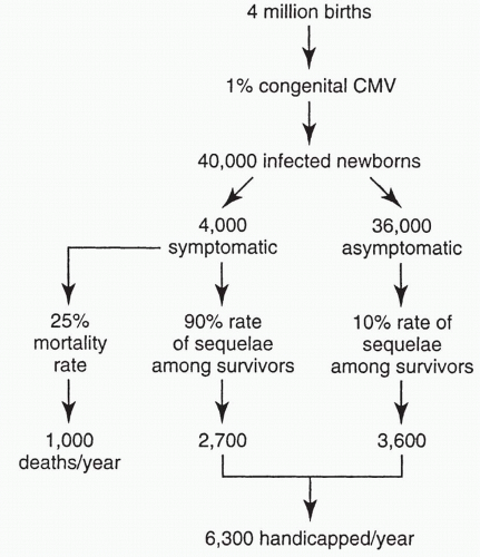
FIGURE 15.2 Public health impact of congenital cytomegalovirus infection in the United States.


Neonatal Infection.

The spectrum of disease caused by CMV in the fetus and neonate is very broad. Of the congenitally infected infants, 90% are completely asymptomatic at birth. Clinically apparent disease occurs in 10% of infants with congenital CMV and ranges from isolated organ involvement to the classic multiorgan system disease. Approximately 50% of the symptomatic infants have the typical generalized pattern of the characteristic cytomegalic inclusion disease. In severely infected neonates, the clinical features include petechiae (79%), hepatosplenomegaly (74%), jaundice (63%), thrombocytopenia, microcephaly (50%), deafness, chorioretinitis (12%), optic atrophy, and cerebral calcifications (Fig. 15.3). The cerebral calcifications of CMV are characteristically periventricular in the subependymal region. A characteristic tetrad of findings has been described in infants who have survived fulminant clinically apparent CMV infection. These are (i) mental retardation, (ii) chorioretinitis, (iii) cerebral calcification, and (iv) microcephaly or hydrocephaly. Such severe symptomatic CMV disease occurs in an estimated 1 in 10,000 to 1 in 20,000 newborns.

The long-term prognosis for symptomatic congenital CMV infection is poor. The mortality rate is 20% to 30%, and more than 90% of the survivors develop significant neurologic sequelae. These sequelae include microcephaly, psychomotor retardation, neuromuscular disorder, unilateral and bilateral hearing loss, chorioretinitis, optic nerve atrophy, and dental defects.

Ninety percent of newborns with congenital (intrauterine acquired) CMV infection are asymptomatic. However, 5% to 15% of newborns with asymptomatic CMV infection at birth will go on to develop late sequelae such as sensorineural hearing loss, subnormal intelligence, and behavioral problems.


Laboratory


Maternal Infection.

Several reliable IgG antibody tests are available for CMV. These include indirect hemagglutination, enzyme-linked immunosorbent assay (ELISA), indirect fluorescent antibody, and neutralization tests. Because approximately 40% of adults have antibody, a single positive result does not necessarily indicate recent or current infection. Demonstration of seroconversion is the best documentation of primary infection. If infection has occurred within the previous 4 to 8
months, IgM-specific antibody can be detected in the serum. Moreover, IgM may remain positive for up to 18 months and in 10% of women with recurrent CMV, IgM can be detected.


FIGURE 15.3 An infant with congenital cytomegalovirus infection. Infant had jaundice, hepatosplenomegaly, and petechiae.

Gestational age at time of serologic screening affects diagnostic sensitivity. In one study, screening in the first half of pregnancy had a diagnostic sensitivity of 64% while screening in the second half had a sensitivity of 85%. Specificity and positive predictive value were 100% in both halves of pregnancy.

As in diagnosing many infections, avidity assays are now available for CMV. Low avidity indicates recent infection whereas high avidity indicates recurrent infection.

Another way to establish the presence of CMV infection is by isolating the virus. Virus isolation does not differentiate primary and recurrent infections. On the other hand, diagnosis of asymptomatic recurrent CMV depends on viral isolation from urine or cervix, because no change in antibody level occurs in normal hosts with recurrent infection. CMV from swabs or urine may require 2 to 6 weeks before cytopathic effects of the virus are seen in tissue culture. Polymerase chain reaction (PCR) detection of CMV is available and provides a sensitive and specific diagnostic test for the presence of CMV.


Infections in Neonates.

The characteristic periventricular calcifications may be helpful in clinically differentiating congenital CMV infection from these other perinatal infections, but laboratory confirmation is necessary to document CMV infection. Although serology can be used as an aid in the diagnosis of congenital CMV infection, virus isolation is more sensitive and direct. As in adults, newer methods such as an indirect hemagglutination test, ELISA, and a fluorescent antibody test have replaced the CF test.

Most neonates with congenital CMV have antibody to CMV when tested with the newer methods. Approximately 80% of congenitally infected infants have IgM-specific antibody in the serum during the first few months of life.

Virus isolation is the best method available for documenting newborn CMV infection. Specimens can be taken from the urine, nasopharynx, conjunctiva, and spinal fluid. PCR is also available.

Characteristic cytomegalic inclusions may be seen in tissues collected at autopsy or by biopsy, for example, of the kidney.

Laboratory abnormalities found in neonates with symptomatic congenital CMV infection include elevated cord IgM (abnormal in 84%), atypical lymphocytosis (present in 80%), elevated SGOT (in 78%), thrombocytopenia (in 61%), hyperbilirubinemia (in 61%), and increased CSF protein (in 47%). Similar abnormalities can be detected in the in utero infected fetus and are included as part of the assessment for prenatal diagnosis of congenital CMV.


Prenatal Diagnosis

Prenatal diagnosis of in utero acquired (congenital) CMV infection of the fetus is available using ultrasound, amniocentesis, and cordocentesis (percutaneous umbilical cord sampling). On ultrasound, the most common findings include microcephaly (10%); hepatosplenomegaly (18%); ventriculomegaly (32%); or calcified lesions in the periventricular region of the brain, liver, or placenta (40%); intrauterine growth retardation/oligohydramnios (55%); ascites; pericardial or pleural effusion; hypoechogenic bowel; and hydrops (Fig. 15.4).

Direct fetal sampling via fetoscopy and cordocentesis has detected elevated levels of anti-CMV IgM. Subsequent studies demonstrated that amniocentesis with culturing of amniotic fluid is an excellent method for detection of in utero CMV infection. With amniotic fluid culture, these authors correctly diagnosed all cases of congenital CMV infection. The accuracy of amniotic fluid cultures for CMV is not unexpected, because the fetal kidney is one of the major sites for CMV involvement. Amniotic fluid culture for CMV should be offered in pregnant women with documented primary CMV infection or when ultrasonography suggests intrauterine growth retardation, hydrops, ascites, and CNS abnormalities. As noted above, viral titers correlate with risk of symptomatic infection, but no clinically useful cutoff value can be determined.


FIGURE 15.4 Cytomegalovirus infection: Fetal ultrasound showing intracranial calcification.

Another group assessed the relationship of CMV viral load in amniotic fluid with fetal or neonatal outcome using PCR and quantitative PCR. CMV infection was noted in 16 (23%) fetuses and neonates, five of whom were symptomatic. Using quantitative PCR, these authors demonstrated that the presence of >105 genome equivalents predicted development of symptomatic infection.

Fetal blood sampling may provide additional information about the fetal condition. Grossly abnormal laboratory findings such as severe thrombocytopenia, anemia, or signs of hepatic involvement were associated with a rapidly fatal outcome after birth. With cordocentesis for fetal blood sampling used to complement ultrasound and amniocentesis, assessment of in utero CMV infection includes specific and nonspecific tests for fetal CMV infection (Table 15.4). The combination of these
tests resulted in antenatal diagnosis of CMV in 13 of 16 infected fetuses (sensitivity, 81%) in one study. Amniocentesis diagnosed 12 of 13 antenatally identified cases of CMV infection. Of these 12 cases, four had a negative result on the first amniocentesis before 20 weeks; 4 to 8 weeks later, the results of a second amniocentesis were positive. Diagnostic sensitivity of amniocentesis increases with the time interval after primary screening. Thus, when the interval is <8 weeks, the sensitivity is 50%, but increases to 91% after 13 weeks. Thus, with a strong suspicion (i.e., documented maternal primary CMV) if the initial assessment is negative, the testing should be repeated later. Detection of CMV IgM antibody in fetal blood had a sensitivity of 69% (9 of 13 cases). More recently, another group assessed amniocentesis, fetal blood sampling and serial ultrasounds in the prenatal diagnosis of CMV. Nearly 23% (26 of 114) of fetuses were infected with prenatal diagnosis identifying 20 of these cases, resulting in a sensitivity of 77% and specificity of 100%. In this study, amniocentesis best diagnosed fetal infection while ultrasound examination and fetal blood sampling identified fetuses at risk for severe sequelae.


TABLE 15.4 ▪ DIAGNOSIS OF FETAL CMV INFECTION


Amniocentesis


Virus isolation by culture (monoclonal antibody early AG13)


PCR


Fetal blood sampling (PUBS)


Indirect


CBC and platelet count


Liver function test (g-glutamyltransferase)


Total IgM


CMV-specific IgM


Direct


Viral culture


Viral antigen


PCR


CBC, complete blood count; CMV cytomegalovirus; IgM, immunoglobulin M; PCR, polymerase chain reaction; PUBS, percutaneous fetal blood sampling.


Treatment

In the last few years, specific treatments of CMV infection have developed. Antiviral agents such as adenosine arabinoside, cytosine arabinoside and ganciclovir have been used for severe clinical infection, including one report of clearance of CMV. However, these drugs are quite toxic. Ganciclovir has recently been demonstrated to be effective in the treatment of CMV retinitis in HIV-infected patients. Another agent, foscarnet, is also approved for treatment of CMV retinitis. Use of these therapies should be undertaken in consultation with experts in perinatal infection.

Recently, an international group has reported benefits in decreasing symptomatic infection at birth by use of CMV specific IVIG in pregnancies complicated by documented CMV infection and abnormal ultrasound findings. In this nonrandomized trial, use of the CMV specific IVIG lead to a rate of symptomatic infection at birth of 7% (1 of 15) in fetuses with abnormal ultrasounds vs. a rate of 100% (7 of 7) in those not given the IVIG. Methodologic problems with the study are important however, and this approach to treatment remains to be established.

The development of a CMV vaccine has been suggested as a means of preventing congenital CMV infection with its associated morbidity and mortality. Use of such a vaccine could prevent nearly all of the neonatal deaths (n = 1,000) and approximately 90% of the severe neurologic sequelae associated with congenital CMV secondary to primary maternal infection that occur each year in the United States. However, no CMV vaccine is currently available.


Screening and Counseling

Although reliable tests are available for detecting IgG class antibody to CMV in maternal serum, mass screening is not recommended. Such a screening program would be expensive, because detection of primary infection requires serial testing of women who are seronegative initially or testing for IgM. (As noted, tests for CMV-specific IgM have not been evaluated as a screening tool.) Then, even if primary infection is documented, present information does not allow straightforward decisions. Because most maternal infections are asymptomatic, the time in gestation of the infection is nearly always unknown. Prospective studies have shown that infections early in pregnancy are more likely to cause serious fetal injury than those late in pregnancy. Once maternal primary CMV infection has been documented, a combination of ultrasound, amniocentesis, and fetal blood sampling is necessary to identify the 30% to 40% of fetuses infected with CMV. A major limitation of a screening program is that many cases of congenital infection occur in immune women. Because there is no marker to detect these at-risk fetuses, all of these cases would be missed by a screening program.

One team of experts concluded that “there is inadequate information to serve as a basis for recommendations regarding termination of pregnancy after a primary CMV infection. Similarly, there is no information regarding how long conception should be delayed after primary infection.”

The annual primary CMV infection rate (as determined by seroconversion) was no higher among pediatric house staff (2.7%) and pediatric nursing staff (3.3%) than it was among young women in the community (2.5% during pregnancy and 5.5% between pregnancies). The higher than expected rate of conversion between pregnancies suggests that exposure to young infants by family or other social exposure may be the most important factor in horizontal transmission. Toddlers are often the most likely source of CMV for both the mother and the fetus or infant. As a result, the CDC currently recommends that day care workers be counseled regarding risk and instructed in careful, frequent hand washing. Use of gloves to handle material contaminated with body fluids is also useful.

The pooled risk ratio for CMV infection was statistically significant (risk ratio, 2.7; 95% confidence interval [CI], 1.33 to 5.52). In studies published before widespread adoption of universal precautions, pediatric nurses may have been at increased risk for CMV infection because of occupational exposure. More recently, among employees of a children’s hospital, the seroconversion rate to CMV was 2.2% per year, and there was no statistically significant difference in the incidence of CMV infection by job type, number of hours per week of patient contact, or nursing unit. This incidence of CMV was similar to that expected in the general population. Standard infection control measures for handling potentially contaminated material in pediatric units would seem to provide sufficient protection from CMV infection.


Hepatitis

Acute viral hepatitis is a systemic infection predominantly affecting the liver. Distinct hepatotropic viral agents cause hepatitis types A, B, C (which was formerly known as non-A, non-B hepatitis), D, and E. Other transmissible agents causing secondary hepatitis include CMV, Epstein-Barr virus, varicella-zoster (VZ) virus, coxsackievirus B, HSV, and rubella virus. The major impact of hepatitis on the fetus and neonate relates to hepatitis B and hepatitis C. These viruses are discussed fully in Chapter 8.


Varicella-Zoster Infection

Varicella-zoster (V-Z) virus, a member of the herpes virus group, is a DNA virus and thus exhibits viral latency. Initial (primary) infection with V-Z virus results in varicella (chickenpox). This common childhood disease is usually marked by typical skin lesions, which progress from macules and papules to vesicles that occur in successive crops and evolve into pustules that form crusts and scabs. A highly contagious disorder, it is acquired by most persons in the United States prior to reproductive age and is generally self-limited. Among adults who contract the disease, constitutional and pulmonary symptoms may be more severe. Adults are 40 times more likely to develop a severe complication of varicella than a child.


Zoster (shingles) is the result of reactivation of the latent virus. It generally occurs in the older adult population or immunocompromised patients. Characteristically, zoster presents as painful vesicular lesions that occur in a pattern of distribution that follows segmental dermatomes.

This discussion focuses on the effects of V-Z virus in pregnancy, the management and prevention of V-Z infections during the perinatal period, and the possibly increased severity of the infection in pregnancy. Two major problems exist when V-Z infection occurs in pregnancy. First, the infection itself poses a risk for significant morbidity and mortality for mother and neonate. Second, V-Z virus has a teratogenic effect, and infection early in pregnancy may result in congenital anomalies.


Epidemiology

Prior to availability of the varicella vaccine, over 80,000 cases of chickenpox were reported annually in the United States, making this disease the third most frequently reported infectious disease in the United States. Because of extensive underreporting, it was estimated that 2 to 3 million cases of chickenpox occur annually in the United States. Over 90% of the population has been infected before adulthood. Consequently, chickenpox is uncommon among women of childbearing age and thus uncommon in pregnancy. Based on several large studies, the incidence of varicella in pregnancy ranges from 1 to 7 per 10,000 pregnant women. Thus, approximately 3,000 cases of varicella in pregnancy occur annually.


Clinical Presentation

V-Z virus is highly contagious. It is spread by respiratory droplets and close personal contact. The incubation period of varicella ranges from 10 to 20 days, but usually is 13 to 17 days. In children, fever and rash occur simultaneously, while in adults fever and generalized malaise precede the rash by several days. Characteristically, the rash begins on the face and scalp and spreads to the trunk (Figs. 15.5, 15.6). The extremities tend to be minimally involved. Skin lesions begin as macules then progress to a vesicular stage followed by pustules, crusts, and scabs. Itching is a common and prominent feature of the disease. For 2 to 5 days, new crops of lesions occur, and the various stages (i.e., vesicles, pustules, scabs) are present simultaneously. Patients are contagious from 1 to 2 days prior to the onset of rash until the lesions have dried and crusted over.


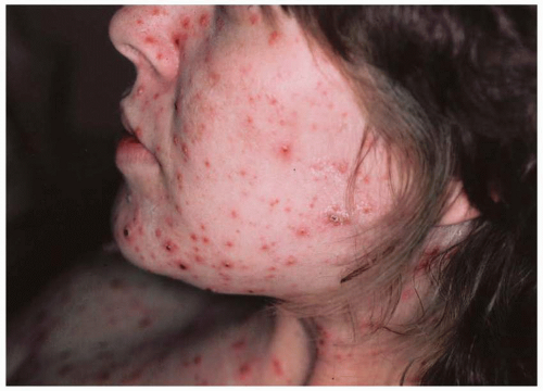
FIGURE 15.5 Rash of varicella: Crops of lesions in various stages (vesicles, pustules, scabs).


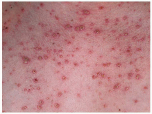
FIGURE 15.6 Vesicular-pustular rash of varicella: close-up.

Adults are 40 times more likely to develop a complication during acute varicella infection (Table 15.5). The most common complication of chickenpox is secondary bacterial infection of the skin lesions, usually caused by Staphylococcus aureus or Group A Beta-hemolytic streptococcus. Necrotizing fasciitis and varicella gangrenosis are the most serious soft tissue complications. The most serious complication of chickenpox is varicella pneumonia, which occurs in about 5% to 10% of adults with chickenpox and is associated with a significant mortality risk.

Varicella pneumonia develops several days following onset of the rash. The severity of varicella pneumonia ranges from a mild illness to severe life-threatening disease characterized by cough, tachypnea, dyspnea, hemoptysis, chest pain, and cyanosis. On radiographic examination, the chest x-ray demonstrates diffuse, nodular peribronchial infiltrates, which in severe cases can progress to adult respiratory distress syndrome (Fig. 15.7). Pneumonia may lead to hypoxemia and tachypnea with respiratory failure occurring in severe cases. Respiratory failure is more likely to develop in cigarette smokers, pregnancy, and immunosuppressed patients. It is one of the most common causes of death in varicella.


Diagnosis of Varicella

The characteristic features of the varicella vesicular rash are usually sufficient to establish the clinical diagnosis. Where the
diagnosis is uncertain, the V-Z virus can be indirectly identified by PCR or rapid antigen tests based on immunofluorescence techniques. Culture is available for direct identification but requires 3 to 5 days and is rarely used. Skin scrapings are the preferred specimen. Rapid antigen tests are the best test for detection of acute V-Z infection.


TABLE 15.5 ▪ COMPLICATIONS OF ACUTE VARICELLA INFECTION (CHICKENPOX)


Skin and Soft Tissue


Central Nervous System


Secondary bacterial infection


Benign cerebellar ataxia


(S. aureus, Group A beta hemolytic strep)


Necrotizing fasciitis


Meningoencephalitis


Varicella gangrenosis


Vasculitis


Pneumonia


Other Rare Complications


Nephritis


Hemorrhagic Manifestations


Myocarditis


Thrombocytopenia; DIC


Pericarditis


Vesicular hemorrhage


FIGURE 15.7 Varicella pneumonia.

Serologic tests are less useful for acute V-Z. IgM and IgA antibodies may be present within 1 to 2 days after appearance of the rash. However, their absence does not exclude V-Z. The Fluorescent Antibody to Membrane Antigen (FAMA) test is the gold standard for identification of antibodies (IgG) to V-Z which correlate with previous infection. However, it is difficult to perform and not widely available. Thus, ELISA is most commonly used and is sufficiently sensitive and specific for detection of immunity to V-Z.


Varicella in the Mother

Varicella is an unusual infection among adults and probably occurs with no greater frequency among pregnant women (Fig. 15.8). Older age and immunosuppression are the major risk factors associated with severe varicella and mortality. In the past, pregnant women were believed to be at greater risk of developing severe or fatal varicella than were nonpregnant adults. More recent studies from the United States and Europe have not confirmed this finding. Among over 1,500 women with chickenpox during pregnancy only one death occurred. The current consensus holds that while adults are at increased risk of developing varicella pneumonia, this risk is not greater among pregnant women. A recent review of the literature on varicella pneumonia in pregnancy, reported that among 198 cases of chickenpox in pregnant women, 57 developed varicella pneumonia. All 16 deaths occurred in the pneumonia group, for a pneumonia mortality rate of 28% (overall mortality rate for varicella was 10%) (Table 15.6). Thus, it seems clear that uncomplicated chickenpox poses no severe risk to pregnant women. While pneumonia (in the past) was associated with a significant mortality rate, pneumonia is an uncommon complication, and our current ability to manage severe respiratory distress and failure is much enhanced. Moreover, acyclovir in high doses is effective against V-Z. While the mortality rate for varicella pneumonia during pregnancy has
improved since the introduction of high dose acyclovir therapy (1985), mortality still remains a substantial concern.


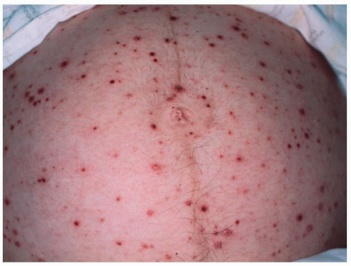
FIGURE 15.8 Rash of varicella on abdomen in pregnant woman.


TABLE 15.6 ▪ MATERNAL MORTALITY ASSOCIATED WITH VARICELLA DURING PREGNANCY


Study


No. of Cases


No. with Pneumonia


No. of Deaths


Garcia, 1963 (11)


2


0


0


Abler, 1964 (12)


18


0


0


Pearson, 1964 (9)


16


1


1


Newman, 1965 (13)


9


0


0


Harris and Rhoades,a 1965 (14)


17


17


7


Siegel and Fuerst, 1966 (6/19)


11


0


0


Pickard, 1968 (15)


1


1


1


Mendelow and Lewis, 1969 (16)


2


2


1


Geeves et al, 1971 (17)


1


1


0


Paryani and Arvin, 1986 (18)


41


4


1


Esmonde et al, 1989 (20)


3


3


1


Cox et al, 1990 (21)


5


5


1


Broussard et al, 1991 (22)


1


1


0


Smegmo and Asperilla, 1991 (6)


21


21


3


Baren et al, 1996 (23)


28


1


0


Figueroa-Damian and Arrendondo-Garcia, 1997


22


0


0


Total


198


57 (28%)


16 (28%)b


a Includes review of the literature before 1963.

b Mortality among cases with pneumonia. Reproduced with permission from Gershon AA. Chickenpox, measles, and mumps. In: Remington JS, Klein JO, eds. Infectious diseases of the fetus and newborn. Philadelphia: WB Saunders; 1990.


Management of Acute Varicella (Chickenpox) in Pregnancy

In adults (pregnant and nonpregnant) the management of uncomplicated chickenpox is primarily aimed at symptomatic treatment and prevention of secondary bacteria infection. For patients with severe varicella or in whom complications occur, acyclovir treatment should be instituted. The recommended dosage of acyclovir treatment of severe varicella is 10 to 15 mg/kg intravenous 3 times daily for 7 days.

Clinical trials in immunocompetent adults with acute varicella have demonstrated that starting oral acyclovir within 24 hours of rash onset is associated with decreased time to cutaneous healing and diminished symptoms and fever. With concerns regarding increased severity of varicella pneumonia in pregnant women, some authorities suggest that risk/benefit analysis favors giving oral acyclovir to pregnant women in the second half of pregnancy presenting with 24 hours rash onset.

Whether acyclovir is given or not, the clinical progress of acute varicella in pregnancy should be closely monitored, with complete reassessment and immediate hospitalization if respiratory symptoms or other systemic complications develop. See Box 15.1 for the criteria for hospitalization of patients with acute varicella (chickenpox).

The clinician must maintain a high index of suspicion that pneumonia may complicate varicella in pregnant women. Pulmonary symptoms begin on the second to sixth day after appearance of the rash and usually consist of a mild nonproductive cough. If the disease is more severe, there may be pleuritic chest pain, hemoptysis, dyspnea, and frank cyanosis. Physical examination reveals fever, rales, and wheezes. The chest x-ray characteristically reveals a diffuse nodular or military pattern, especially in the perihilar regions (Fig. 15.7). With varicella in pregnancy, the patient should be warned to contact the physician immediately if even mild pulmonary symptoms develop. Hospitalization with full respiratory support, if necessary, should then be made available.

Because varicella infection in pregnancy is associated with significant morbidity and mortality, prevention of varicella among susceptible exposed patients is paramount. For a pregnant woman with exposure to varicella, infection is very likely if she is not immune. Up to 90% of adults in developed countries are immune to varicella. Even most pregnant women with a negative or indeterminate history of chickenpox have detectable antibody. For those with no history of chickenpox, it is appropriate and cost-effective in the face of maternal exposure to test for maternal antibody by any of the following: fluorescent antibody to membrane antigen (FAMA), ELISA, immune adherence hemagglutination, and the enhanced neutralization test (Fig. 15.9).


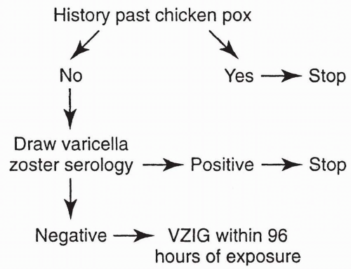
FIGURE 15.9 Protocol for management of pregnant women exposed to varicella.

Then, in susceptible women, varicella-zoster immune globulin (VZIG) may be given. When given intramuscularly, within 96 hours of exposure, it is likely that VZIG ameliorates the course of the maternal disease. There is no certainty, however, that passive immunization prevents fetal infection. The recommended dose of VZIG is 125 units per 10 kg up to a maximum of 625 units (five vials) intramuscularly. The average pregnant woman requires the maximum dose, at a cost of approximately $625.


Effects of Varicella in Early Pregnancy

Maternal V-Z infection has been reported to result in spontaneous abortion, stillbirth, and congenital anomalies. It is now appreciated that maternal varicella infection in the first trimester of pregnancy can produce a congenital varicella syndrome, which consists of cutaneous scars, limb hypoplasia, rudimentary digits, ocular abnormalities (optic atrophy, microphthalmia, cataracts), cerebral cortical atrophy, mental retardation, and growth retardation (Fig. 15.10). The risk of
developing the congenital varicella syndrome after exposure in the first or second trimester is reviewed in Table 15.7. Overall, only 1% of infants exposed to V-Z virus during the first or second trimester developed stigmata of congenital varicella, with a range of 0% to 9%. In a large prospective study in Germany and the United Kingdom of 1,373 women who had varicella prior to 36 weeks’ gestation, nine cases of congenital varicella syndrome were identified, for a risk rate of 1%. Interestingly, the highest risk (2.0%) was noted between 13 and 20 weeks’ gestation (7 of 351 pregnancies; 95% CI, 0.8-4.1%). Before 13 weeks, only 2 of 472 pregnancies (0.4%) (95% CI, 0.05-1.5%) were identified.


FIGURE 15.10 Congenital varicella syndrome.


TABLE 15.7 ▪ RISK OF CONGENITAL VARICELLA-ZOSTER SYNDROME WITH ACUTE MATERNAL VARICELLA-ZOSTER INFECTION DURING THE FIRST TRIMESTER OF PREGNANCY


Study


No. of Infants Exposed


Infants No. of with Congenital Varicella-Zoster Virus (%)


Siegel (28)


27


2 (7.4)


Paryani and Arvin (18)


11


1 (9)


Enders (29)


23


0


Balducci et al (8)


35


0


Pastuszak et al (30)


49


1 (2)


Enders et al (31)


472


2 (0.4)


Total


617


6 (1)


It has been suggested that many of the congenital malformations related to V-Z virus may not be caused by acute varicella in the fetus but rather are the result of sequelae of recurrent herpes zoster in the fetus. In particular, recurrent herpes zoster infection would explain the cutaneous and limb abnormalities that follow nerve distribution. On the other hand, acute varicella is responsible for the CNS and neurologic lesions present in the congenital varicella syndrome. The frequency of clinical findings in infants with congenital varicella syndrome is presented in Table 15.8. Cicatricial skin lesions, eye abnormalities, and hypoplastic limb are the most common findings. Nearly one fourth of the affected infants died. Although the risk of congenital varicella syndrome is small, administration of VZIG to pregnant women without evidence of previous varicella as soon as possible (but within 96 hours of exposure) is recommended, as this may protect the fetus during the viremia (see previous section). Once the mother has the rash, there would be no reason to administer VZIG, as viremia would have already occurred. VZIG is prepared from donors with high antibody titers to V-Z virus.


TABLE 15.8 ▪ CLINICAL DATA IN INFANTS WITH DEVELOPMENTAL DEFECTS BORN TO 37 WOMEN WITH VARICELLA-ZOSTER INFECTIONS DURING EARLY PREGNANCY


Defect


No. of Cases (%)


Cicatricial skin lesions


26 (70)


Eye abnormalities


23 (62)


Cataract


9 (24)


Chorioretinitis


10 (27)


Microophthalmia


10 (27)


Hypoplastic limb


17 (46)


Cortical atrophy/mental retardation


11 (30)


Early death


9 (24)


Reproduced with permission from Gershon AA. Chicken pox, measles, and mumps. In: Remington JS, Klein JO, eds. Infectious diseases of the fetus and newborn. Philadelphia: WB Saunders; 1990:395-445.


As with other perinatal infections, ultrasonography, amniocentesis, chorionic villus biopsy, and cordocentesis have been proposed as techniques for diagnosing in utero varicella infection. While virus-specific IgM antibody can be detected in cord blood by 19 to 22 weeks’ gestation and V-Z virus can be recovered from amniotic fluid and identified by an in situ hybridization in placental tissue, detection of either antibody or virus does not provide information about the severity of fetal infection nor do they correlate with damage to fetus. Ultrasound assessment appears to be the best method available for assessing the severity of fetal involvement with varicella infection. An ultrasound study reported findings in 37 cases with maternal varicella infection in early pregnancy. Abnormal findings were noted in five fetuses, including hydrops, hyperechogenic foci in the liver, and hydramnios. All five fetuses with abnormalities on sonography had varicella embryopathy at postdelivery examination or autopsy. All sonographic abnormalities were observed prior to 20 weeks. Ultrasound has also been successful in identifying abnormal limb development; 14 of 17 fetuses with hypoplastic limbs also had either brain damage or died in early infancy. More recently, PCR of amniotic fluid was performed for perinatal diagnosis of V-Z infection in 107 pregnant women with varicella prior to 24 weeks’ gestation. Nine (8.4%) of specimens were positive. The reported incidence of congenital varicella-zoster was three (2.8%); all were PCR positive and two had abnormal sonographic findings at 21 to 22 weeks. The third infant had bilateral microophthalmia in the face of a normal ultrasound at 24 weeks.

Because the latency period from maternal infection to appearance of sonographic demonstrated fetal anomalies may be from 5 to 19 weeks, serial ultrasounds may be required to detect varicella embryopathy. The common ultrasound findings seen with congenital varicella are listed in Box 15.2.


Varicella in the Newborn

Acquisition of maternal antibody usually protects the fetus. However, if an infant is born after the maternal viremia but before the mother has developed an antibody response, the
fetus is at high risk for life-threatening, neonatal varicella infection. Infants at risk are those whose mothers contract varicella within 2 days of birth or within the first 5 days after delivery. Congenital varicella infection has been reported in 10% to 20% of term infants born to mothers with varicella within 4 to 5 days of delivery, and the case fatality was 20% to 30%. Infants born 5 or more days after maternal illness onset developed either mild varicella or no infection at all.


Management of varicella in the newborn should focus on prevention. Ideally, delivery should be delayed until 5 to 7 days after onset of maternal varicella. This will allow transfer of protective IgG antibody from the mother to the fetus. However, if delay is not possible, then the neonate should receive VZIG passive immunization as soon as possible following delivery.

VZIG modifies or prevents varicella in normal children, and has been recommended for use in preventing severe neonatal varicella. Thus, infants at risk (born to mothers who develop varicella between 5 days before and 2 days after delivery) should receive VZIG as passive immunization. The dose is 125 units. Recently, the outcome of 281 newborns whose mothers had chickenpox during the perinatal period was studied. All infants had received VZIG shortly after birth. However, 169 (60%) of the children were noted to be infected: 134 (48%) with chickenpox and 35 (13%) without clinical features. While VZIG did not prevent neonatal varicella, it did prevent fatal outcome. The authors concluded that VZIG was still indicated for newborn infants whose mothers have chickenpox within 7 days prior to or after delivery. This differs from the recommended 5 days before delivery in the United States.

The CDC has published guidelines for the prevention of varicella. A live attenuated virus vaccine (Varivax) has been approved in the United States since 1995. See Chapter 25 for a detailed discussion of varicella vaccine. Because Varivax is a live attenuated virus it is recommended that pregnant women nor women attempting to conceive not receive varicella vaccine. A Pregnancy Registry for Varivax has been established and from May 1995 through December 1998 there were 371 women vaccinated within 3 months before or during pregnancy prospectively reported. There were zero cases of congenital varicella reported (95% CI, 0.0-0.01). This finding plus the 1% rate of congenital varicella resulting from mild varicella virus infection resulted in the recommendation that exposure to varicella is not an indication for pregnancy termination. Also, because the vaccine virus is not transmissible, presence of a pregnant woman in a household is not a contraindication for vaccination of household members.


Effect of Zoster on Pregnancy

Herpes zoster is caused by the same virus as is varicella. It occurs very rarely in pregnancy. As V-Z virus is not viremic during herpes zoster in immunocompetent persons there is no risk for transmission to the fetus. Moreover, because it is reactivation of latent V-Z virus and maternal antibodies are present in normal healthy women, zoster poses no threat to the fetus or neonate.


Measles (Rubeola)

Rubeola is an acute illness that most commonly occurs in childhood. It is the most communicable of the childhood exanthems. Rubeola is characterized by fever, coryza, conjunctivitis, cough, and a generalized maculopapular rash that usually appears 1 to 2 days after the pathognomonic Koplik spots in the oral cavity. The rubeola virus is a paramyxovirus that contains RNA as its nuclear protein.


Epidemiology

The virus is spread chiefly by droplets expectorated by an infected person and gains access to susceptible people via the nose, oropharynx, and conjunctival mucosa. The incubation time is between 10 and 14 days. Measles is most communicable during the prodrome and catarrhal stages of the infection. Approximately three fourths of exposed susceptible contacts acquire rubeola.

Before the availability of live measles vaccines, epidemics of measles occurred at intervals of 2 to 3 years in the United States. The use of attenuated measles vaccine since 1963 has had a major impact in decreasing the number of measles cases in the United States.

Before the introduction of the measles vaccine, there were 0.4 to 0.6 cases of measles per 10,000 pregnancies. This figure is probably even lower since the measles vaccine was introduced.


Clinical Manifestations

The prodrome of fever and malaise begins 10 to 11 days postexposure and is followed within 24 hours by coryza, sneezing, conjunctivitis, and cough. This catarrhal phase is exacerbated over the next several days, and a marked conjunctivitis and photophobia occur. The pathognomonic Koplik spots appear at the end of the prodrome. These are tiny, granular, slightly raised white lesions surrounded by a halo of erythema, which are located on the lateral buccal mucosa. The rash appears 12 to 14 days after exposure. It begins on the head and neck, particularly postauricularly, and subsequently the maculopapular rash spreads to the trunk, upper extremities, and finally the lower extremities.

The respiratory tract is the most frequent site for complications of measles. Otitis media and croup are frequent occurrences, but bacterial pneumonia is the complication most frequently associated with mortality. The most common bacterial organisms involved in rubeola pneumonia are Streptococcus pneumoniae, Haemophilus influenzae, Staphylococcus aureus, and Streptococcus pyogenes. Encephalitis, a less common but serious complication, is estimated to occur with a frequency of 1 per 1,000 cases of measles. Other complications of measles include thrombocytopenic purpura, myocarditis, and subacute sclerosing panencephalitis, which is a progressive neurologic disease associated with chronic rubeola infection of the CNS.


Maternal Effects of Measles

It is unclear whether pregnant women with measles are at greater risk for serious complications and death than nonpregnant adults, and measles in pregnant women is only rarely associated with pneumonia or other complications.


Fetal Effects of Measles

Measles in pregnancy is associated with an increased rate of prematurity, particularly when the disease occurs late in gestation, but maternal measles is not associated with an increased risk of spontaneous abortion.

Only gold members can continue reading. Log In or Register to continue

Stay updated, free articles. Join our Telegram channel

Jul 8, 2016 | Posted by in INFECTIOUS DISEASE | Comments Off on Perinatal Infections

Full access? Get Clinical Tree

Get Clinical Tree app for offline access