Fig. 2.1
Immunohistochemistry demonstrates basal cells (stained brown with marker 34betaE12) in benign glands, while the surrounding malignant glands are negative
2.3 Immunohistochemistry
The immunostains commonly used are p63 (nuclear stain) or a high molecular weight cytokeratin, e.g. 34betaE12 or CK5/6 (cytoplasmic stains). Alpha-methylacyl-CoA racemase (AMACR) is overexpressed in the cytoplasm of neoplastic epithelial cells. A combination of two or more of the above markers is commonly used. The presence of strong luminal positive staining of epithelial cells for AMACR in the complete absence of staining for basal cell markers would confirm the presence of a carcinoma at a suspicious focus.
2.4 Gleason Grading
Based on the degree of differentiation of the tumour, it is graded from 1 (well differentiated) to 5 (poorly differentiated), referred to as Gleason grades (or patterns). The Gleason score is the sum of the most prevalent and the second most prevalent grades in a given tumour. Gleason score is a strong predictor of the behaviour of prostate cancer. The Gleason grades are described as follows:
Grade 1. The tumour is perfectly circumscribed and contains closely packed acini that closely resemble normal prostate tissue. This is a very rare pattern, and circumscription is not assessable on needle core biopsies.
Grade 2. The tumour is well circumscribed but with some irregularity of the margin and contains acini closely resembling normal prostate tissue. This is also an uncommon pattern, and circumscription is not assessable on needle core biopsies.
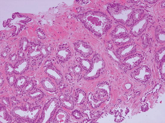
Grade 3. The tumour has irregular margins and may infiltrate between normal prostate tissues. The acini are of variable shapes and sizes and closely set but separate as individual acini with luminal spaces (Fig. 2.2).
Grade 4. The tumour has a more complex architecture often with cribriform (sieve-like) structures and with glandular fusion although luminal spaces are still identifiable (Fig. 2.3).
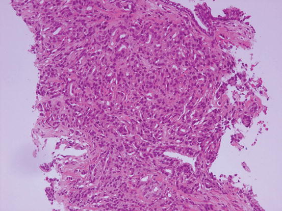
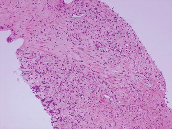
Grade 5. The tumour bears little resemblance to normal prostate tissue with presence of either a dispersed single cell infiltrate (Fig. 2.4) or sheets of tumour cells, sometimes accompanied by necrosis.

Fig. 2.2
Gleason grade 3. Discrete, variable-sized malignant glands with distinct luminal spaces

Fig. 2.3
Gleason grade 4. Fusion of glands with complex architecture but recognisable luminal spaces

Fig. 2.4
Gleason grade 5. Singly scattered cells lacking glandular formations and luminal spaces
Only grades 3, 4 and 5 are used in reporting prostate needle core biopsies with a range of Gleason scores from 6 to 10. The most prevalent grade in the tumour is referred to as the primary grade or pattern. The next most prevalent grade is the secondary grade or pattern, and if another pattern is also present, it is referred to as the tertiary grade or pattern. If only one pattern is present, it is regarded to be both the primary and secondary grade, e.g. 3 + 3 = 6. In the modified Gleason scoring system, any grade five tumour is included in the Gleason score in reporting needle biopsies.
Following hormone therapy, the tumour may undergo morphological changes (Fig. 2.5), and the Gleason score may be difficult to apply. An estimated score may be suggested.
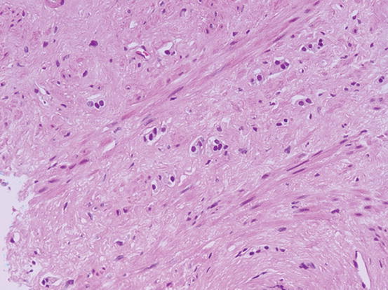

Fig. 2.5
Effect of hormone therapy. The tumour cells appear bland and are difficult to detect especially if present in small numbers
Stay updated, free articles. Join our Telegram channel

Full access? Get Clinical Tree


