PATHOLOGICAL MANIFESTATIONS OF AGING
PEARLS
❖ The death rate for certain diseases increases more steeply than the overall death rate, which arouses the suspicion that aging predisposes one to the development of the condition or a fatal outcome.
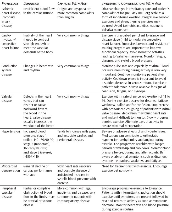
❖ There are no clinically significant effects on heart function that can solely be ascribed to aging. Therefore, the numerous pathologies seen in the cardiovascular system (eg, ischemic heart disease, cardiomyopathy, conduction system disease, valvular disease, hypertension, myocardial degeneration, and peripheral vascular disease) account more for the decrements in the function of the system.
❖ The most common types of diseases affecting the respiratory system are pneumonia, which compromises gas exchange and serves as a source for sepsis, and obstructive and resistive lung disease, which affect the amount of airflow in the lungs.
❖ The most commonly seen conditions affecting bone in older adults include osteoporosis, osteopenia, Paget disease, and joint changes (eg, degenerative arthritis or osteoarthritis, rheumatoid arthritis).
❖ Diseases of the neuromuscular system (eg, confusion/delirium, dementias, Alzheimer disease, cerebrovascular disease, Parkinson disease), like those of the cardiovascular system, are much more responsible for the decrements seen in aging than the effects of aging.
❖ Vestibular problems in older adults are numerous and include benign paroxysmal positional vertigo, acute vestibular neuritis or labyrinthitis, Ménièr e disease, and bilateral vestibular disorders.
❖ Two life-threatening conditions, hypothermia and hyperthermia, need to be recognized and treated in older adults. The regulation of body temperature will greatly impact an individual’s homeostatic well-being during activity and exercise and must be considered when prescribing exercise.
Aging is considered a normal physiological process because of its universality. As much as the aging process may influence the predisposition to disease, aging in and of itself is not considered to be pathological. This distinction seems conceptually clear; however, the fine line between aging and disease is often blurred when applied to specific cases, and some degree of decreasing biological, physiological, anatomical, and functional capabilities occurs as one ages. (See Chapter 3.) Some degree of atrophy is evident in all tissues of the body. A variety of degenerative processes are called “normal aging” until they proceed far enough to cause clinically significant disability.
Recent epidemiologic genetic study findings indicate the pathological significance of age-related changes such as vascular stiffening and loss of muscle mass,1 which are genetically driven and were, until recently, considered normal aging. Genes with effects on unrecognized pathologies could be detected through genome scans to identify loci affecting broader outcomes.
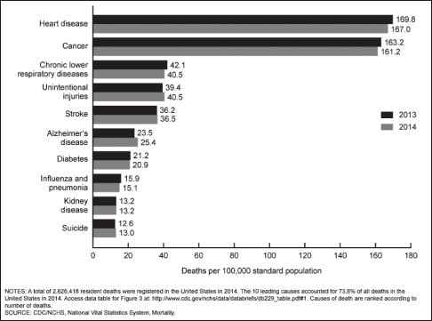
Figure 5-1. Leading causes of death for persons age 65 and older in the United States for 2013 and 2014. (Adapted from the Centers for Disease Control and Prevention/National Center for Health Statistics and Kochanek KD, Murphy SL, Xu J, Tejada-Vera B. Deaths: final data for 2014. Natl Vital Stat Rep. 2016;65[4]:1-122.)
Genetic factors may influence not only physiological functions but also their rates of change with age, which can influence whether and when disease occurs. Rates of change with age in many physiological functions directly affect the risk of age-related morbidity (eg, changes in bone density, vital capacity, cognitive function, lens opacity, and blood pressure). Another important group includes rates of decline in homeostatic functions, such as glucose tolerance, blood pressure stability, and balance. Rates of change in cellular biochemical properties implicated in age-related pathologies could also be studied because data are needed on the predictive validity of such changes for mortality, age-related diseases, or functional status.
The incidence of many diseases is influenced markedly as age advances (Figure 5-1).2 The death rates for atherosclerosis, myocardial degeneration, hypertension, and cancer all increase more steeply than the overall death rate, which arouses the suspicion that aging predisposes an individual either to the development of the condition or to a fatal outcome. With some conditions, such as respiratory infections, the incidence is not increased in older adults, but the likelihood of fatality from the insult is greater than in younger persons.3 In a child or young adult, death is most commonly caused by some form of accident; however, in older adults, the main problems are coronary heart disease, cerebrovascular accidents, respiratory diseases, diabetes, peripheral vascular diseases, and neoplasms.4
The purpose of this chapter is to review those pathologies that are manifested in the older population, but it does not cover a detailed consideration of every possible geriatric pathology; rather, it examines some of the more common conditions that afflict older adults and affect functional activities of daily living (ADLs).
AGING AS A DISEASE
There is always the tacit implication that aging, like growth and development, is a normal physiological process lying outside the domain of disease. Although aging may not be considered a disease process, the time-dependent loss of structure and function in all organ systems leads to pathological end states. There is a general decline in structure, function, and the number of many kinds of cells with age. Cellular aging is accompanied by denaturation of extracellular proteins. The collagen and elastin of the skin become irreversibly crystalline and broken. The hyaline cartilage on articular surfaces of joints becomes fibrillar and fragmented, and the beautifully ordered structure of the lens of the eye becomes brittle and chaotic as lens protein is gradually denatured.
The aging process proceeds slowly and ubiquitously over the life course, resulting in a loss of structure and function within every organ or tissue. Countless microtraumas occur and accumulate in small increments as imperceptible injuries. Over a lifetime, the skin elastin is exposed to microinsults from ultraviolet rays from the sun, repetitive mechanical stresses cause degeneration of articular cartilage, and reactions with metabolites diminish the opacity of the lens of the eye. The most important aging changes occur at the molecular level, as reviewed in Chapter 2. Small injuries occurring within the cell result in the loss of genetic memory and progressive cross-linking of collagen, the chief structural protein in the body.
Mortality experience in 2014 according to the National Vital Statistics Report (June 30, 2016) reveals the following highlights:
▷ In 2014, a total of 2,626,418 resident deaths were registered in the United States.
▷ The age-adjusted death rate was 724.6/100,000 US population.
▷ Life expectancy at birth was 78.8 years.
▷ The 15 leading causes of death in 2014 were the following:
- Heart diseases
- Malignant neoplasms
- Chronic lower respiratory diseases
- Accidents (unintentional injuries)
- Cerebrovascular diseases
- Alzheimer disease
- Diabetes mellitus
- Influenza and pneumonia
- Nephritis, nephrotic syndrome, and nephrosis (kidney disease)
- Intentional self-harm (suicide)
- Septicemia
- Chronic liver disease and cirrhosis
- Essential hypertension
- Parkinson disease
- Asphyxiation pneumonitis
Some pathologists studying the pathologies common as we age defined disease as the reaction to injury.5 If aging is a gradual accumulation of incompletely repaired injuries due to microtrauma through the life course, it may not be normal, despite its universality. Perhaps “aging” is a pathological process resulting from tissue reactions to imperceptible, avoidable injuries.
Physical and occupational therapists could play a major role in preventing the disabilities that result from these insidious microtraumas. Preventive strengthening and conditioning exercises, positioning, joint and tissue mobilization, and the numerous modalities that could be used all impact functional capabilities, especially in an older population. From the authors’ clinical perspective, preventing disabilities that can result from pathological processes greatly improves the level of function and the quality of life. There are certainly changes that occur in aging that do not need to be inevitable.
CARDIOVASCULAR MANIFESTATIONS OF AGING
There are no clinically significant effects on heart function that can be ascribed to aging alone. Cardiovascular dysfunction attributed to the aging process closely mimics the decline in cardiac function seen with inactivity.6–8 Arteriosclerosis is a generally used term to describe any form of vascular degeneration associated with a thickening and loss of resilience in the arterial wall.9 Atherosclerosis is a more specific type of degeneration that is associated with an accumulation of fat in the intimal lining of the blood vessels and an increase of connective tissue in the underlying subintima.9 Almost all animal species show some degree of atherosclerosis, and, for this reason, it has been considered an inevitable accompaniment of aging. The pathological consequences depend on the site. Weakness in the aorta can cause an aneurysm, ischemic heart disease can result from atherosclerotic changes in the coronary arteries, and cerebrovascular accidents can result from involvement of the cerebral vessels.10
Other cardiovascular pathologies increase in incidence with age in addition to ischemic heart disease, including cardiomyopathies, conduction system diseases, valvular heart disease, and peripheral vascular disease.11 The result is that the heart is less effective as a pump and has less reserve to meet increased activity needs. Functional abilities of the individual become more and more restricted as the severity of heart disease increases.
Ischemic Heart Disease
In older adults, anginal pain is not a consistent symptomatic indicator of ischemia of the cardiac tissue. Older adults more commonly report dyspnea (ie, shortness of breath). Clinically, shortness of breath is a much more reliable indicator of ischemia than anginal pain in the older adult.12 There is a general correlation between ST segment depression on electrocardiography and the onset of anginal symptoms,9 although in older adults marked ST segment depression occurs with dyspnea without the development of the characteristic anginal pain.12
Early intervention in ischemic heart disease takes several forms. The most important is to reduce the risk factors that predispose individuals to the development of coronary artery disease, such as cigarette smoking, high blood pressure, and serum cholesterol. Secondary prevention of heart attacks, once coronary artery disease is established, requires the reduction of risk factors as well as the use of aspirin to prevent platelet aggregation, which may initiate obstruction of a coronary artery, and beta blockers, which appear to limit the extent of muscle injury. Management of symptomatic coronary artery disease is similar in all age groups and consists of medical and surgical interventions. Because the pathophysiology of coronary artery disease is the mismatch between the metabolic demands of the heart muscle and the ability of the coronary arteries to supply blood, interventions are directed at decreasing the metabolic needs of the heart muscle or increasing the ability of the coronary arteries to carry blood. The metabolic demands of heart muscle can be reduced by lowering the pressure against which the heart has to push, by reducing the rate at which the heart contracts, and by reducing the overall metabolic demands of the body by correcting hyperthyroidism, anemia, low oxygen, or elevated temperature. Calcium channel blocking agents and beta blockers reduce the metabolic demands on heart muscle, and nitroglycerin reduces the pressure against which the heart has to pump by causing dilatation of the coronary vessels. Beta blockers13 and calcium channel blockers14 often improve function by reducing myocardial contractility; however, this may leave the older adult vulnerable to cardiac failure. It is still debated how far the effectiveness and toxicity of these drugs are modified by aging.15 Exercise is still the treatment of choice for cardiovascular diseases.
There are a number of reasons why exercise is helpful to the patient with angina.9 Development of an enhanced “collateral” flow has not been established.16 However, the cardiac oxygen demand is decreased by a decrease in both heart rate and blood pressure at a given workload. The lengthening of the diastolic phase facilitates coronary perfusion.9 Although progress is slow in older adults, dramatic gains of maximum oxygen intake can be achieved if the exercise periods are of sufficient lengths.9,17 Many ADLs can be brought below the anginal threshold by exercise training. Strengthening of the skeletal muscles may help to reduce blood pressure and, therefore, to reduce the likelihood of developing anginal symptoms during functional ADLs.
Cardiomyopathy/Congestive Heart Failure
Cardiomyopathies are conditions in which the heart muscle hypertrophies, and cardiac function is impaired, often resulting in congestive heart failure.10 The muscle of the heart weakens because of poor nutrition, toxins, infections, or genetic factors.5 The weakening results in dilation of the heart and can lead to congestive heart failure because the heart cannot contract strongly enough to empty a sufficient amount of blood into the peripheral vasculature to meet the body’s needs. Hypertrophy of cardiac muscle tissue can be the end result of hypertension, outflow obstruction, or genetic factors.10 The cardiovascular changes imposed by cardiomyopathies impair function through several pathological mechanisms. The hypertrophied heart is stiff and does not easily fill with blood. As a result, the heart contracts vigorously, but there is little forward circulation to show for the effort, and the body’s energy and oxygen needs are not met. In hypertrophic cardiomyopathy, the muscle abnormally contracts, actually creating an obstruction to the outflow of blood from the heart. The more strongly the heart contracts, the greater the obstruction is.
Exercise has historically been contraindicated in the patient with cardiomyopathy; however, that perspective is rapidly changing as the benefits of exercise in this condition are documented and acknowledged.18–20 Medical treatment is directed at correcting or ameliorating the pathophysiology of the underlying cause of heart failure. Rehabilitative efforts need to be directed toward maintaining and improving the maximal functional capabilities of the older adult and preventing the debilitating effects of immobility.
Two evidence-based strategies are cited below.
▷ Karlsdotter AE, Foster C, Porcari JP, Palmer-McLean K, White-Kube R, Backes RC. Hemodynamic responses during aerobic and resistance exercise. J Cardiopulm Rehabil. 2002;22:170-177.21
- Left ventricular function remains stable during moderate-intensity resistance exercise even in patients with congestive heart failure.
- This form of exercise can be used safely in a rehabilitation program.
▷ Keteyian SJ, Levine AB, Brawner EG, et al. Exercise training in patients with heart failure. Ann Intern Med. 1996;124(12):1051-1057.22
- Compensated heart failure due to left ventricular dysfunction benefits significantly from regular moderate aerobic exercise.
- The program is 3 times a week for 12 weeks.
- How it works
- Increase in cardiac output
- Reduction in vascular resistance
- Amelioration of muscle metabolism on the cellular level
- Increase in cardiac output
Conduction System Disease
Conduction system diseases affect the rate and rhythm of the heart’s contractions.10 The propagation of the electrical wave that results in the coordinated contraction of the heart muscle is initiated in the 2 pacemaker sites in the heart and carried initially along specialized pathways that spread the wave throughout the heart, also known as the conduction system. These pacemakers and pathways can be damaged by many different agents, including those that result in cardiomyopathies and myocardial infarction.23 The most common consequences of pacemaker dysfunction are extremely rapid (tachycardic) contractions, poorly coordinated (dysrhythmic) contractions, or extremely slow (bradycardic) contractions that are less effective in moving blood and result in diminished cardiac output.9 Low cardiac output can result in confusion, fatigue, poor exercise tolerance, and congestive heart failure. Rapid reductions in cardiac output can cause syncope.
Tachycardias and poorly coordinated rhythms, such as atrial fibrillation, are usually treated with medication to control the rate and convert the rhythm back to normal. Occasionally, electrical cardioversion is required.10 Bradycardia is usually managed by surgical implantation of an artificial pacemaker, which can be set to trigger a heartbeat at a predetermined rate. Age, per se, is not a contraindication to pacemaker therapy, and the surgery is minor and well tolerated.
Valvular Disease
The heart valves, which function to keep the blood flowing in one direction, tend to withstand many microtraumas throughout the life course. Defects of the heart valves are of 2 types. Stenosis or narrowing of the valve restricts blood flow,10 and insufficiency or regurgitation results in the backward flow of blood. Both conditions increase the workload on the heart and greatly reduce its efficiency.9 Two valves are most frequently involved: the mitral valve between the left atrium and ventricle and the aortic valve, which moves the blood into the systemic circulation.
Rheumatic valve disease caused by earlier episodes of rheumatic fever is the most common cause of mitral stenosis and insufficiency in older adults.23 Congestive heart failure, arrhythmias, and embolization of blood clots from the heart to the brain and other organs are the most common complications of mitral valve disease. These patients require attentive medical management, which includes the use of anticoagulants to prevent emboli, diuretics to control congestive heart failure, and digitalis or other medications to control the heart rate.10 Nutritional support is often required to ensure compliance with a low-sodium diet. Because of the potentially serious side effects from too much anticoagulant (bleeding and hemorrhagic stroke) and digoxin (arrhythmias), the narrow range of effective dosages, and the deleterious effect of inadequate dosage, exercise must be gradually implemented and progressed slowly. Protective intervention should focus on skin protection and maintenance of maximal functional capabilities, with close monitoring of the individual’s vital signs and subjective responses of perceived tolerance to increasing activity levels.
Hypertension
Hypertension is another common condition affecting the cardiovascular system. It is clear that older adults with systolic blood pressures above 160 mm Hg and diastolic pressures above 95 mm Hg are at increased risk for stroke, congestive heart failure (hypertensive cardiomyopathy), and renal failure. Isolated systolic hypertension carries a similar risk10 because much of the cardiovascular morbidity and mortality in older adults is related to hypertension.2 This is true for both isolated systolic hypertension and systolic and diastolic blood pressure elevations.24 In addition to accelerated atherogenesis (myocardial infarction and congestive heart failure), hypertension adversely affects cardiac performance, renal function, and cerebral blood flow. It also increases aortic aneurysm rupture and dissection and increases the incidence of cerebrovascular bleeding.25
Treatment to lower blood pressure significantly reduces the risks of developing these complications. Medical and dietary management are important in controlling hypertension. In older adults, most recommendations are for drug treatment for blood pressure readings in excess of 160/95 mm Hg.25 There is little proof that antihypertensive drug therapy alters the course of asymptomatic older individuals without evidence of end organ damage from isolated systolic hypertension.
Nevertheless, isolated systolic hypertension doubles the risk of cardiovascular complications so that the risk, expense, and inconvenience of drug therapy must be compared with the benefits of lower systolic blood pressure. Compliance with medication and the early identification and avoidance of drug-induced side effects, such as dizziness, hypokalemia, depression, syncope, and confusion, are major challenges to the health care team. Complications of antihypertensive therapy are more frequent in the older adult, both because the diminution of renal function increases the incidence of drug toxicity and because the older patient with less sensitive baroreceptor responses is more susceptible to the orthostatic complications of volume depletion. Assuming the upright posture gradually may help avert dizziness and syncope. Exercise has been shown to have positive effects in reducing high blood pressure.
Three evidence-based treatment studies are cited below.
▷ Young D, Appel L, Lee S, Miller E. The effects of aerobic exercise and T’ai Chi on blood pressure in older people: results of a randomized trial. J Am Geriatr Soc. 1999;47:277-284.26
- Blood pressure improved in both groups, more so in the aerobic group.
- Aerobic capacity improved in only the aerobic exercise group.
▷ Vaitkevicius PV, Ebersold C, Shah MS, et al. Effects of aerobic exercise training in community based subjects aged 80 and older: a pilot study. J Am Geriatr Soc. 2002;50:2009-2013.27
- Persons aged 80 years and older can improve aerobic capacity and reduce systolic blood pressure in an aerobic exercise program.
▷ Kelley GA, Kelley KS. Progressive resistance exercise and resting blood pressure: a meta-analysis of randomized controlled trials. Hypertension. 2000;35:838-843.28
- Progressive resistive exercise is efficacious for reducing resting systolic and diastolic blood pressure.
Myocardial Degeneration
The general decline of cardiac performance with age and inactivity affects the ability of older individuals to function at their maximum. The recognized changes in cardiac function include a decrease in right ventricular work rate and a variable change of left ventricular work rate depending on the relative magnitudes of the reduction in maximum cardiac output and the increase of systemic blood pressure.9,29 While a young person readily accepts a sustained increase of cardiac work rate, in old age an equivalent relative stress may give rise to cardiac failure, particularly if there are other circulatory problems such as a high systemic blood pressure, a minor disorder of the heart valves, or an excessive intake of fluids. Complaints of shortness of breath in older adults frequently reflect problems with getting enough oxygen to the working muscle through a failing circulatory system.
Peripheral Vascular Disease
Peripheral vascular disease is frequently the result of untreated hypertension, cigarette smoking, diabetes mellitus, and elevated serum cholesterol.23 Atherosclerosis and other forms of peripheral vascular disease can lead to partial or complete obstruction of the main arterial supply to the limbs. The consequences are intermittent claudication with walking and skin lesions, which may lead to amputation.9 When early intervention to reduce risk factors is unsuccessful, management is through the modification of diet to reduce weight and cholesterol, medications to enhance blood flow and reduce blood pressure, and behavior modifications to reduce cigarette consumption. Exercise is particularly helpful in treating peripheral vascular disease from a preventative perspective.9,30–33
Evidence-based studies for peripheral artery disease are cited in the next 2 columns.
▷ Brandsma JW, Robeer BG, van den Heuvel S, et al. The effect of exercises on walking distance of patients with intermittent claudication: a study of randomized clinical trials. Phys Ther. 1998;78:278-288.34
- An analysis of the literature was performed.
- All studies showed that walking exercises improved walking distance.
- Patients should be encouraged to exercise to the point of maximum claudication pain.
▷ Hunt D, Leighton M, Reed G. Intermittent claudication: implementation of an exercise program. Physiotherapy. 1999;85(3):149-153.35
- Nonweightbearing warm-up and cooldown and stretching exercise for lower extremity.
- Main session (past pain)
- Alternating heel raises
- Simultaneous heel raises
- Step-ups on low bench
- Toe walking
- Alternating heel raises
- Patients were encouraged to walk (past pain).
▷ Gardner AW. Exercise training for patients with peripheral artery disease. Phys Sports Med. 2001;29(8):25-35.36
Recommended Exercise Program for Patients Who Have Peripheral Artery Disease
| Exercise Component | Detail |
| Frequency | 3 sessions per week |
| Intensity | Progression from 50% of peak exercise |
| Capacity to 80% by program’s end | |
| Duration | Progression from 15 minutes of exercise per session to more than 30 minutes by program’s end |
| Mode | Walking, nonweightbearing tasks (bicycling) |
| May be used for warm-up and cooldown | |
| Type of exercise | Intermittent walking to near maximal, claudication pain |
| Program length | At least 6 months |
▷ Gelin J, Jivegard L, Taft C, et al. Treatment efficacy of intermittent claudication by surgical intervention, supervised physical training compared to no treatment in unselected randomised patients I: one year results of functional and physiological improvements. Eur J Vasc Endovasc Surg. 2001;22:107-113.37
- Invasive treatment increased walking capacity, leg blood flow, and pressure. Supervised physical exercise training offered no therapeutic advantage compared with untreated controls.
▷ Langbein WE, Collins EG, Orebaugh C, et al. Increasing exercise tolerance of persons limited by claudication pain using polestriding. J Vasc Surg. 2002;35(5):887-893.38
- This randomized controlled trial determined that 24 weeks of polestriding significantly improves measures of exercise tolerance limited by intermittent claudication pain. (Polestriding simulates cross-country skiing.)
▷ Gardner AW, Katzel LI, Sorkin JD, et al. Exercise rehabilitation improves functional outcomes and peripheral circulation in patients with intermittent claudication: a randomized controlled trial. J Am Geriatr Soc. 2001;49:755-762.39
- Exercise is beneficial well into a 12-month maintenance program (3 times a week for 30 minutes).
▷ Tsai JC, Chan P, Wang CH, et al. The effects of exercise training on walking function and perception of health status in elderly patients with peripheral arterial occlusive disease. J Intern Med. 2003;252:448-455.40
- Significant improvements in claudication (walking time, pain, and function).
- Treatment
- 12 weeks
- A 5-minute warm-up and cooldown
- Begin for 10 minutes and increase the grade of the treadmill
- Encouraged to walk to 30 minutes and a grade of 2 to 3 pain (moderate)
- 12 weeks
▷ Ambrosetti M, Salerno M, Tramarin R, Pedretti RFE. Efficacy of a short-course intensive rehabilitation program in patients with moderate-to-severe intermittent claudication. Ital Heart J. 2002;3(8):467-472.41
- Treatment and outcomes are the same as in the study by Tsai et al40 but are 4 weeks long.
PULMONARY MANIFESTATIONS OF AGING
Pneumonia
Pneumonia is the most common infectious cause of death in older adults42 and the most common infection requiring hospitalization. It is often the means of death for patients with other serious conditions, such as diabetes, cancer, stroke, congestive heart failure, dementia, and renal failure. The increased incidence of pneumonia with aging is due in part to the weakening of the local pulmonary defenses; however, the high mortality of pneumonia is largely due to its more subtle presentation in older adults. Typical symptoms such as a productive cough, fever, and pleuritic chest pain are frequently absent, but subtler symptoms, such as confusion, alteration of sleep-wake cycles, increased congestive heart failure, anorexia, and failure to thrive, are more common. A typically lower core temperature in an older, inactive adult results in a failure to recognize that the individual has a fever. Misdiagnosis and late diagnosis are common and contribute to the high mortality of pneumonia in older adults.42
Successful treatment of pneumonia requires early recognition and institution of proper antibiotic therapy. The identification of causative bacteria in the examination of a sputum sample is the single most important diagnostic test for determining initial antibiotic therapy. Unfortunately, such samples are often difficult to obtain in a dehydrated and confused older patient, and, as a result, therapy is often empirical and not as specifically directed or effective as possible. Hydration, nutritional support, chest physical therapy, and treatment of complicating illnesses are often required in addition to antibiotics.
Obstructive and Resistive Lung Disease
Conditions that cause obstruction to airflow within the lungs are called obstructive airway diseases, whereas conditions that result in resistance to airflow are called resistive airway diseases. They share the common characteristic of increased resistance to airflow within the airways.43
The treatment goal of individuals with chronic obstructive pulmonary disease (COPD) is to prevent smoking and maintain optimal functioning for as long as possible. This usually involves chest physical therapy, medication, oxygen therapy, and environmental changes designed to conserve energy and reduce exertion. The depression that accompanies chronic illness of all types can be particularly significant in patients with COPD, many of whom feel that they have brought it on themselves by smoking. Because of its complexity, COPD is an excellent example of a health problem that requires an interdisciplinary approach.
See page 83 for several evidence-based studies showing the efficacy of rehabilitation for COPD.
Cardiovascular Pathologies in Older Adults and Therapeutic Considerations
▷ Alfaro V, Torras R, Prats MT, Palacios L, Ibáñez J. Improvement in exercise tolerance and spirometric values in stable chronic obstructive pulmonary disease patients after an individualized outpatient rehabilitation program. J Sports Med Phys Fitness. 1996;36:195-203.44
- Individualized outpatient rehabilitation programs are able to improve exercise tolerance in stable patients with COPD affected by dyspnea during exercise through reconditioning of both skeletal and respiratory muscles and improved gas exchange during exercise, thus reducing the ratio of dead space to tidal volume.
▷ Casaburi R, Porszasz J, Burns MR, Carithers ER, Chang RS, Cooper CB. Physiologic benefits of exercise training in rehabilitation of patients with severe chronic obstructive pulmonary disease. Am J Resp Crit Care Med. 1997;155:1541-1551.45
- Rigorous exercise training for patients with COPD yields a more efficient exercise breathing pattern and improved exercise tolerance.
- Program = 3 days a week for 8 weeks, 80% work rate, 45 minutes.
▷ Rosenbaum R, Bach JR, Penek J. The cost/benefits of outpatient-based pulmonary rehabilitation. Arch Phys Med Rehabil. 1997;78:240-244.46
- The program consisted of education, training, group therapy, and individualized home exercise.
- Patients cycled 2 times a day for 7 days a week (active range of motion) and used an inspiratory muscle trainer 2 times a day.
- Weekly patient education on energy conservation, relaxation, smoking, etc.
- Results: patients improved on cardiopulmonary parameters and ADLs.
- The cost of 10 sessions was $650.
MUSCULOSKELETAL MANIFESTATIONS OF AGING
Fibromyalgia
Fibromyalgia is a common form of nonarticular rheumatism with diffuse musculoskeletal aching and multiple tender points at characteristic sites.47
Short-term exercise and educational programs can produce immediate and sustained benefits for patients with fibromyalgia.48
Evidence-based studies for fibromyalgia are cited in the next column.
▷ Clark SR. Prescribing exercise for fibromyalgia patients. Arthritis Care Res. 1994;7(4):221-225.49
- Avoid eccentric exercise
- Avoid high-intensity exercise
- Prescribe stretching after exercise
- Exercise at 60% to 70% of maximum heart rate
- Use active range of motion as a warm-up
▷ Jentoft ES, Kvalvik AG, Mengshoel AM. Effects of pool-based and land-based aerobic exercise on women with fibromyalgia/chronic widespread muscle pain. Arthritis Rheum. 2001;45(1):42-47.50
- Pool and land exercise beneficial
- Once a week for 20 weeks
- Improve strength and fatigue
- Improve cardiovascular parameters
- Decrease pain
▷ Rooks DS, Silverman CB, Kantrowitz FG. The effects of progressive strength training and aerobic exercise on muscle strength and cardiovascular fitness in women with fibromyalgia: a pilot study. Arthritis Rheum. 2003;47(1):22-28.51
- 60 min/session 3 times a week for 20 weeks
- 4 weeks of active range of motion in the pool
- 16 weeks of land cardiovascular exercise, progressive resistive exercise, and flexibility that progressed as the person tolerated
▷ Buckelew SP, Conway R, Parker J, et al. Biofeedback/relaxation training and exercise interventions for fibromyalgia: a prospective trial. Arthritis Care Res. 1998;11(3):196-209.52
- 120 patients were assigned to 1 of the following 4 groups:
- Biofeedback/relaxation only
- Combined biofeedback/relaxation and exercise
- Exercise only
- Education only (no exercise or biofeedback)
- Biofeedback/relaxation only
- The first 3 groups listed above improved in self-efficacy. The exercise and combination group improved in physical activity and kept the benefits the longest.
▷ Pfeiffer A, Thompson JM, Nelson A, et al. Effects of a 1.5 day multidisciplinary outpatient treatment program for fibromyalgia. Am J Phys Med Rehabil. 2003;82:186-191.53
- Rx = education, self-management, occupational therapy, and physical therapy
- Rx group = improved in function
▷ Bailey A, Starr L, Alderson M, Moreland J. A comparative evaluation of a fibromyalgia rehabilitation program. Arthritis Care Res. 1999;12(5):336-340.54
- Fibro-Fit was effective in improving physical impairments and function
- 12 weeks, 3 times a week
- Physical therapy, occupational therapy, and social work
- Orientation, self-management, goal setting, stretching, strengthening, and aerobics
- Counseling—sleep, stress, fatigue, nutrition, and coping
- 12 weeks, 3 times a week
SKELETAL MANIFESTATIONS OF AGING
Osteoporosis
Osteoporosis is a heterogeneous condition characterized by an absolute decrease in the amount of normal bone (ie, loss of bone). Osteoporosis is defined as a metabolic bone disease “characterized by low bone mass and microarchitectural deterioration of bony tissue leading to enhanced bone fragility and a consequent increase in fracture risk.”55 Osteoporosis results when the production of new bone mass is exceeded by the reabsorption of old bone; in other words, osteoporosis is the failure of bone formation to keep pace with bone resorption. This is termed coupling. The result is that bone becomes structurally weakened.
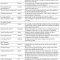
Bone mineral density (BMD) as measured by dual x-ray absorptiometry is expressed as absolute BMD (g/cm2) and may be designated by either the number of standard deviations from the mean of age-matched controls (known as z score) or the number of standard deviations from the young normal mean (T score).56 The World Health Organization (WHO) developed guidelines for the clinical diagnosis of osteoporosis, which are based on the T score, with a T score of less than –1.0 being defined as osteopenic and a T score of less than –2.5 being referred to as osteoporosis. An outline of these diagnostic criteria for osteoporosis is shown in Table 5-1.57
The WHO guidelines for T scores are as follows: (1) a T score above –1.0 is defined as normal; (2) a T score between –1.0 and –2.5 is defined as osteopenic; (3) a T score below –2.5 is defined as osteoporotic; and (4) a T score below –2.5, plus one or more fragility fractures, is defined as severely osteoporotic. This classification is particularly important for the physical or occupational therapist prescribing exercise and determining levels of risk for fractures and frailty. The authors of the book have added preventive “actions” to those prescribed by WHO in Table 5-1, in addition to expanding the criteria to provide suggested prescriptions for rehabilitative interventions.
Osteopenia
The term osteopenia describes an evenly systemic decrease in bone density below an expected level. Therefore, bone loss is not unilateral or limited to one area of the skeleton. Osteopenia is the prelude to osteoporosis. Table 5-1 indicates the BMD range that differentiates osteopenia from osteoporosis.
Paget Disease
Paget disease (also called osteitis deformans) is a disorder of bone remodeling characterized by increased bone resorption and increased formation. It usually presents after the fourth decade of life, and its prevalence increases with age to affect 1% to 2% of individuals over age 60 in the United States, with a slight male predominance.58 It is usually a focal disease involving one bone (monostotic), but it can involve several bones (polystotic). In rare cases, Paget disease can become generalized, especially in familial disease. Osteogenic sarcoma is a dreaded complication of Paget disease in older adults.58
Increased and abnormal osteoclastic bone resorption is the initiating event in Paget disease. There is accumulating evidence that a slow virus plays a role in its pathogenesis. Increased interleukin 6 production may be a contributing factor to the development of Paget disease. Genetic mechanisms are also likely to be important in familial cases, with a possible linkage to chromosome 6 or 18.58,59
The rate of bone resorption in Paget disease can be as much as 10- to 20-fold the normal rate. This is reflected in increased biochemical indices of bone resorption, including urinary excretion of collagen catabolites, like hydroxyproline and collagen cross-linked peptides, and osteoclast products, notably serum acid phosphatase. Osteoblastic new bone formation responds appropriately to the increased resorption, and this is reflected in increased osteoblast products such as alkaline phosphatase and osteocalcin. The increased cellular activity produced highly vascular and cellular bone, and collagen is deposited by overworked osteoblasts in an abnormal and disorganized pattern of “woven bone.”59
Because the tight coupling of formation and resorption is maintained in Paget disease despite the increased skeletal turnover, systemic mineral homeostasis is usually unperturbed, and serum calcium is usually normal. However, when patients with Paget disease are immobilized, as can occur with fracture or surgery, they may become hypercalciuric or even hypercalcemic. This occurs because muscle stimulation of bone formation is decreased, and bone resorption proceeds relatively unopposed. Hypercalcemia may also signal the presence of hyperparathyroidism, which is commonly reported with an increased level of calcium in the serum plasma of a patient with Paget disease.59
Table 5-1. Modified World Health Organization Osteoporosis Criteria, With Action and Rehabilitation Prescription
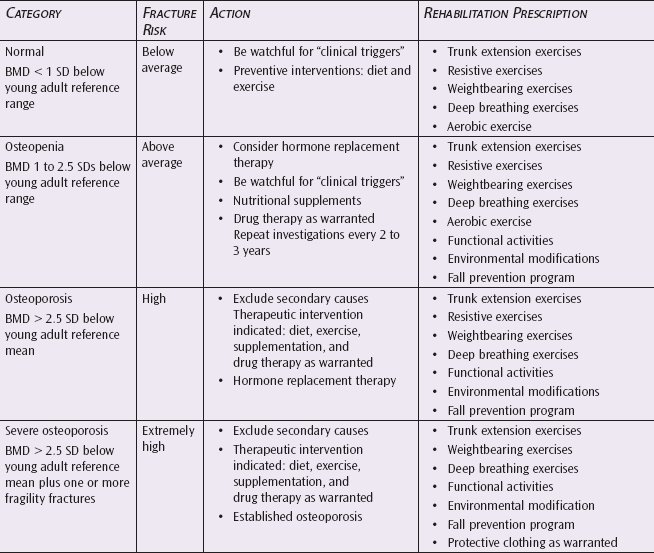
BMD, bone mineral density; SD, standard deviation. Adapted from Briggs et al.57
Joint Changes
Degenerative Arthritis/Osteoarthritis
There are several different types of arthritis, but all lead to a common final pathway known as degenerative joint disease (DJD) or osteoarthritis.60 Recurrent joint trauma leads to intra-articular damage, resulting in the release of proteolytic enzymes, which often causes bleeding. A cycle is set up with increased cartilage damage, bleeding, and the release of potentially destructive enzymes. Over time, cartilage is eroded, new bone growth is stimulated, and the joint gradually loses its ability to respond to trauma, making it even more susceptible to additional trauma and damage.61 Pain develops because of irritation of the periosteum, joint capsule, and fibrotic changes of the periarticular muscles. Unlike rheumatoid arthritis, the synovial membrane in osteoarthritis is not the primary site of involvement. However, over time, it can become fibrotic as a result of the primary degenerative process.60
The joints most often affected by DJD are the hands, knees, hips, and the lumbar and cervical spine. It is manifested clinically by stiffness and pain that increase with use. Impaired mobility makes it difficult to accomplish routine ADLs.62
The onset of symptoms in osteoarthritis can occur insidiously or suddenly. Generally, joint destruction occurs gradually and progresses slowly. Pain is described as a deep ache, can occur at rest, and often awakens the individual at night with nocturnal discomfort. Stiffness of the involved joint(s) after periods of inactivity occurs and usually is resolved in a relatively short period of movement. Loss of flexibility is associated with soft tissue contractures, intra-articular loose bodies, osteophytes, and loss of joint surface congruity.63
The development of effective anti-inflammatory medications and improvement of surgical interventions in joint disease has changed the current management of end-stage DJD. Joint replacement can be very effective in restoring function and limiting pain, and joint fusion and anti-inflammatory medications are often effective in pain control.
Rehabilitative treatment is focused on the protection of the involved joint from excessive mechanical stresses with the use of an assistive ambulatory device, foot orthotics that provide shock absorption and proper joint positioning, and patient education on nutrition, hydration, and reduction of wear and tear on the joint. Maximizing joint function through flexibility and strengthening exercises is important.
Rheumatoid Arthritis
Rheumatoid arthritis can occur at any age and is characterized by the abrupt onset of symmetrical joint swelling, erythema, and pain. Inflammation of the synovial membrane results in the release of proteolytic enzymes, which perpetuate inflammation and joint damage.61 A biochemical marker for rheumatoid arthritis is a positive rheumatoid factor, which is an antibody that reacts with immunoglobulin antibodies found in the plasma. Rheumatoid factor is also often found in the synovial fluid and synovial membranes of individuals with the disease. It is hypothesized that the interaction between rheumatoid factor and immunoglobulin triggers events that initiate an inflammatory reaction. The leukocytes, monocytes, lymphocytes, and phagocytes attracted in the immune system response lead to the release of lysosomal enzymes, which cause articular cartilage destruction and synovial hyperplasia. These changes also result in the development of destructive vascular granulation tissue called pannus. This tissue proliferates, gradually diminishing the joint space.62 The pannus contains inflammatory cells that destroy the cartilage, bone, and periarticular tissues, leading to joint instability, deformity, or ankylosis.
Symptoms are usually insidious and progress slowly as the disease progresses. Complaints of joint pain, muscle fatigue and weakness, weight loss, and general loss of stamina are common. Inflammation and musculoskeletal symptoms are localized to the specific joint although multiple joints are usually involved. Morning stiffness is more pronounced and of longer duration than in osteoarthritis. Intense pain can occur following periods of rest. The involved joints tend to be the small joints of the hands and feet, wrists, shoulders, elbows, hips, knees, and ankles. Essentially, every joint is involved in this autoimmune, systemic condition. Eventually deformities occur affecting mobility and basic ADLs. Rheumatoid arthritis is a systemic disease; therefore, other signs and symptoms are often present including fever, fatigue, malaise, poor appetite, weight loss, nutritional deficiencies, weakness, anemia, an enlarged spleen, and lymphadenopathy (disease of the lymph nodes).60
The response to therapy is usually quite good. Treatment needs to focus on reducing pain, maintaining mobility, and minimizing joint restrictions, edema, and joint damage. Given the ease with which older adults develop muscle atrophy with disuse, aggressive physical therapy is essential to maintain strength and joint mobility during phases of remission, and occupational therapy is required to focus on joint protection and basic ADLs. During exacerbation, physical and occupational therapy should be directed toward pain management through decreasing swelling in the joint (ice and electrical stimulation), maintaining joint mobility and decreasing discomfort (oscillation joint mobility techniques and active motion), and encouraging participation in functional ADLs. Foot orthotics are recommended to reduce shock and reposition the foot to lessen the stresses on the involved lower extremity joints.64
NEUROMUSCULAR MANIFESTATIONS OF AGING
Confusion/Delirium
Cognitive disorders of all types account for nearly two-thirds of nursing home admissions and a significant majority of those older persons who are incapacitated by illness.65 Severe cognitive dysfunction may not impair an individual’s longevity, especially with meticulous attention to treatable complicating illnesses. As a result, older adults with cognitive dysfunction make up the largest group of individuals with functional disabilities.
Those conditions resulting in alterations in cognitive function can be divided into several groups on the basis of reversibility and chronicity. Acute cognitive dysfunction of rapid onset without underlying damage to brain tissue carries the best prognosis for recovery. This usually results from toxic or metabolic derangements, which affect the normal functioning of the brain, or from psychiatric illness, such as depression, which also has a metabolic basis. The term delirium is used to describe these acute confusional states. Susceptibility to toxic/metabolic delirium (toxic encephalopathy) is not limited to older adults; however, the more limited metabolic reserve of the aging brain makes older adults more sensitive than younger persons to minor stresses.
Confusion, restlessness, agitation, poor attention span, reversal of sleep-wake cycles, hallucinations, and paranoia can all be manifestations of delirium. Subtle changes resulting from correctable toxic/metabolic abnormalities can persist for extended periods before they are recognized as resulting from potentially reversible causes. Appropriate interventions are frequently delayed. It is particularly important for these reversible conditions to be identified by the health care team because failure to intervene in a timely manner can result in permanent cognitive dysfunction. Equally important is the risk that inappropriate treatment will result in further functional impairment or a greatly increased risk of injury. Pathological brain dysfunction resulting from toxic/metabolic causes usually has a good prognosis for recovery when the underlying abnormality is corrected. Table 5-2 lists the more common causes of toxic confusion/delirium in older adults.
Table 5-2. Common Causes of Toxic/Metabolic Confusion/Delirium in Older Adults
DRUGS | METABOLIC ABNORMALITIES |
| Alcohol | Hypoglycemia |
| Psychotropics (tranquilizers, antipsychotics, and antidepressants) | Hyponatremia |
| Over-the-counter sleep, cold, and allergy medications | Hypocalcemia |
| Analgesics | Hypothermia |
| Antihypertensives | Hypothyroidism |
| Beta blockers (propranolol) | Hypoxia |
| Antiparkinsonian medications | Vitamin B12 deficiency |
| Anticonvulsants (phenobarbital, phenytoin, and carbamazepine) | Hepatic failure (elevated ammonia) |
| Digoxin | Renal failure (elevated blood urea nitrogen and creatine) |
| H2 blockers (cimetidine) | Elevated cortisol |
| Amphetamines | Cortisol deficiency, pulmonary failure (elevated carbon dioxide) |
Adapted from Ghosh.66
Interventions are directed at identifying and correcting the underlying metabolic abnormality. Although the prognosis for return to baseline central nervous system function is usually good with correction of the abnormality, the patient’s overall prognosis is often determined by the underlying disease that causes the metabolic abnormality rather than the degree of brain dysfunction. However, during a confusional state, the individual is more prone to accidental injury, complications such as aspiration pneumonia, and further cognitive dysfunction due to inappropriate use of sedatives that may aggravate rather than relieve the agitation. The occurrence of any of these complications may worsen the overall prognosis for recovery. Acute toxic/metabolic delirium may coexist with chronic progressive forms of brain dysfunction, such as Alzheimer disease (AD).
Dementias
Dementias are characterized by a slow onset of increasing intellectual impairment, including disorientation, memory loss, diminished ability to reason and make sound judgments, loss of social skills, and development of regressed or antisocial behavior.66 Frequently, depression is superimposed on dementia as a reaction to the perceived loss of intellectual skills, which leads to further cognitive impairment.67
AD and multi-infarct dementia are the 2 most common forms of irreversible dementia. Each has a fairly characteristic pattern of onset and findings. AD is usually slowly progressive and begins insidiously. It is not associated with focal neurologic deficits or abrupt changes in severity. Patients typically begin with short-term memory deficits that progress to severely regressed behavior, an inability to learn or remember new tasks, and the loss of ability to perform ADLs.68 Multi-infarct dementia is usually of more rapid onset, occurs in younger individuals, and progresses in a stepwise fashion with abrupt worsening and subsequent plateaus of function. Frequently, there are focal neurologic deficits such as paresis and paresthesia.69 Often, the individual is hypertensive, diabetic, or both. He or she may also show evidence of generalized atherosclerosis.70
It is important to distinguish between Alzheimer and multi-infarct dementias. The prevention of recurrent cerebral infarction may arrest the progression of multi-infarct dementia, which has as its pathophysiological basis irreversible brain damage resulting from repetitive ischemic injury caused by emboli or bleeding. Normalization of blood pressure is the most effective intervention known. Other types of reversible dementia, such as those resulting from hypothyroidism, vitamin B12 deficiency, and normal pressure hydrocephalus, can become “fixed” and unresponsive to treatment unless identified and treated at an early stage. Early identification of these correctable dementias is essential. Unfortunately, although no such therapeutic imperative currently exists for AD, recent research on AD has produced promising results, and the potential for delaying and perhaps preventing the onset of this dreaded dementia looms on the horizon.
Regardless of the etiology of dementia, once reversible causes have been ruled out, the main tasks of the clinical team are to minister to the patient’s emotional needs, assist in the act of grieving for lost function, alter the environment so that the patient’s remaining skills can be used, augment the patient’s capacity to successfully undertake ADLs, educate the family, provide emotional and physical support for the family and caretakers, and provide the patient and family with a realistic prognosis. Any superimposed illness can cause a rapid and prolonged decline in mental status, which may totally resolve as the underlying illness is treated.
Alzheimer Disease: A Special Consideration
Striking with cruel randomness across an increasingly older population, AD afflicts some 4.7 million Americans, most of them over the age of 65.71 They may range from a former president to a neighbor next door, but the ailment is always the same—it clutters the brain with tiny bits of protein, slowly robbing victims of their mental power until they are no longer able to do even the simplest chores or recognize their closest friends and kin. So far, medical science has been stymied, unable to treat the disease or slow its fatal progression. However, recent research is encouraging. Strategies to prevent or delay the onset of symptoms, as well as to prevent the decline into the advanced stage of AD, are being explored. Although these strategies do not yet exist in a proven and clinically applicable form, the science is progressing rapidly.72 There may yet be a light at the end of that long, dark tunnel called AD.
AD is the form of dementia that is most common in the older population. Currently, it has been diagnosed in approximately 4.7 million Americans, and if the present trend continues, it is projected that the number will increase to 14 million by the year 2050. Women are more likely to suffer from AD, with a women to men ratio of 2.8:1.0 for those aged 75 years and older. AD is the fourth leading cause of death following heart disease, cancer, and stroke.73–75 One in every 10 persons over the age of 65 has AD, and approximately half of those over the age of 85 are diagnosed with it.73,74
AD is a progressive, degenerative disease that affects the hippocampus, neocortex, and transcorticol pathways of the spinal cord, resulting in impaired memory and thinking, behavioral changes, and progressive return of primitive motor patterns that were encephalized during late fetal and early childhood development. The classic appearance of neurofibrillary plaques and tangles progressively impedes synaptic connections and results in neuronal death.72,76 The neuritic plaques are composed of amyloid precursor protein (APP), which is encoded for by a gene on chromosome 21. Smaller fragments of APP called amyloid beta peptides have also been identified.77 The gene carries the code for APP and appears to be one link in the chain of events that leads to deposits of beta amyloid. The neurofibrillary tangles are composed of paired helical filaments that consist of tau proteins and develop in the cytoplasm of pyramidal cells.77 Neuritic plaques and neurofibrillary tangles are located in the areas of the cerebral cortex linked to intellectual function and sensory integration (just posterior to the Wernicke area) and the hippocampus and neocortex, the 2 most primitive areas of the brain.71,78,79
Some scientists believe that as many as half of all cases of AD may have a genetic component. In addition to the abnormality on chromosome 21, chromosome 14 defects have also been identified in individuals with early-onset AD. Further support of this theory is the recent discovery of a gene on chromosome 19 that appears to be defective in many people with the more common, late-onset form of AD. It has also been found that one form of the apolipoprotein E4 gene is inherited at an increased rate among patients with late-onset AD.77,80 This apolipoprotein is involved in cholesterol metabolism. Chromosome 1 has also been identified to carry a “presenile” gene associated with early-onset AD.80
The protease theory of AD is also being explored. An enzyme called protease has been identified and isolated in individuals with AD and may play a key role in creating the biochemical chaos in the brain that causes AD. This enzyme has become the target of many drug designers. Just as protease inhibitor medications are currently used to block the activity of the autoimmune deficiency virus by targeting proteins (eg, beta-secretase), it is hypothesized that protease inhibitors may also be effective in blocking amyloid plaque formation.79 Protease has long been postulated to act as chemical scissors that help snip away excess protein from brain cells, thereby inhibiting the buildup of protein debris that accumulates into amyloid plaques.
Nongenetic, environmental factors, such as an infectious agent (eg, virus), nutritional components, or toxic environmental substances (eg, metals or industrial chemicals), are also being evaluated for their potential roles in the development of this disease. An example of potential nutritional factors in AD is provided by the Nun Study. Previous studies suggested that low concentrations of folate in the blood are related to poor cognitive function, dementia, and AD-related neurodegeneration of the brain. Nutrients, lipoproteins, and nutritional markers were measured in participants in the Nun Study who later died between the ages of 78 and 101 years (mean = 91 years). At autopsy, several neuropathic indicators of AD were determined including atrophy of the neocortex (frontal, temporal, and parietal) and the number of neocortical lesions (senile plaques and neurofibrillary tangles). There was a strong correlation between serum folate and severity of atrophy of the neocortex. Low serum folate was associated with atrophy of the cerebral cortex.81 Other nutrients, such as the low circulating levels of antioxidants, have been identified as potentially contributing to the development and progression of the disease.
Scientists have now learned that AD begins at least 20 years before symptoms appear, and prevention and early intervention could potentially improve the quality of life of people predisposed to this disease. Some of the major scientific discoveries related to AD over the past few years include the following:
❖ Genes associated with AD have been identified on 4 chromosomes.
❖ Diagnostic techniques have improved to a 90% accuracy rate (without autopsy).
❖ The Food and Drug Administration has approved several drugs for the treatment of AD, an effective first step toward effective therapies.
❖ Mounting evidence indicates that readily available treatments, such as estrogen, vitamin E, folate, ibuprofen, and exercise, may help slow or prevent AD.
❖ Associations between higher levels of education and a reduced risk of AD have been observed.77
❖ Scientists have learned that AD is not caused by a single factor but probably by a number of genetic and environmental factors.
Sister Mary, the gold standard for the Nun Study, was a remarkable woman who had high cognitive test scores before her death at 101 years of age. What is more remarkable is that she maintained this high status despite having abundant neurofibrillary tangles and senile plaques. Findings from Sister Mary and all 678 participants in the Nun Study may provide unique clues about the etiology of aging and AD, exemplify what is possible in old age, and show how clinical expression of some diseases may be averted.82
Cerebrovascular Disease
In contrast to the dementing illnesses that result in “global” brain dysfunction, cerebrovascular disease more commonly results in focal brain dysfunction.70 There are several different types of cerebrovascular disease, each with a different pathophysiological mechanism, prognosis, and treatment. The mechanisms include the rupture of small blood vessels from hypertension, abrupt blockage of vessels by emboli from the heart or atheromatous plaques in the large arteries leading to the brain, and spontaneous formation of blood clots within the blood vessels due to local increases in coaguability. The pathophysiology of cerebrovascular disease is the interruption of blood flow to brain tissue with resultant cell damage or death from ischemia.9 Decreases in the heart’s ability to pump blood can lead to ischemia, as can blockage of the blood vessels to or within the brain from atheromatous plaque, emboli, or inflammation of the lining of the blood vessels. Uncontrolled hypertension, diabetes mellitus, smoking, and elevated cholesterol contribute to cerebrovascular disease directly by affecting the entire circulatory system. (See Chapter 11 for evidence-based medicine strategies.)
Preventive interventions must be specifically directed at the underlying pathophysiology. Hypertension can be controlled by medication, diet, and exercise. The prevention of emboli usually requires the use of anticoagulants such as aspirin, dipyridamole, and warfarin. The risk of bleeding, both into the brain and into other organs, increases with the use of these agents and often limits their use in certain patients. If emboli result from cardiac arrhythmias, prevention results from a return to normal sinus rhythm through the use of electrical cardioversion or anti-arrhythmics such as quinidine, procainamide, and digoxin.23 Because of the heightened risk of intracerebral bleeding, anticoagulants are avoided in the presence of hypertension and in cerebrovascular accidents resulting from bleeding into brain tissue.
Recurrent, small cerebrovascular accidents can result in multi-infarct dementia. More commonly, however, limited areas of the brain are damaged and result in more focal disabilities, including loss of motor or sensory function over the right or left side of the body and alterations in vision, speech, and the ability to interpret sensory inputs. The extent of the deficit following a stroke depends on the location and function of the injured part of the brain, the degree of damage, and the availability of the unaffected regions of the brain that can assume the lost function. Residual effects can be so subtle as to be functionally negligible or so extensive that only the most basic brain functions, such as the control of respiration and blood pressure, are preserved.
Parkinson Disease
Parkinson disease is the most prevalent type of parkinsonism, a clinical syndrome caused by lesions in the basal ganglia, predominantly in the substantia nigra, that produce deficits in motor behavior.83 Parkinsonism is a clinical rather than an etiologic entity because it is associated with several pathological processes that damage the extrapyramidal system. Its many causes are divided into the following 4 categories83:
- Primary or idiopathic Parkinson disease
- Secondary parkinsonism (associated with infectious agents, drugs, toxins, vascular disease, trauma, and brain neoplasm)
- Parkinson-like syndromes
- Heredodegenerative diseases
Parkinson disease is a progressive, degenerative disease of unknown cause resulting in the loss of melanin-containing brain cells in the substantia nigra and locus coeruleus and a decrease of dopamine in the caudate nucleus and putamen. The term Parkinson disease is reserved for those cases of unknown etiology.84 Parkinson disease makes up approximately 80% of cases of parkinsonism.83 The syndrome results in a reduction in muscle power, rigidity, and slowness of movement (akinesia), which are crucial to the characteristic tetrad known as TRAP—resting tremor, cogwheel rigidity, bradykinesia/akinesia, and postural reflex impairment.
Peripheral Neuropathies
With aging, the number and size of peripheral nerve fibers diminish with a concomitant decrease in conduction velocity.85 There is often a clinically insignificant decrease in touch and vibration sense. However, the peripheral nerves are easily affected by nutritional deficiencies, toxins, and endocrine disorders.23 The resulting neuropathies can cause marked loss of position sense, resulting in instability, falls, chronic pain, and dysesthesia (painful and persistent sensation induced by even a gentle touch of the skin).
The common nutritional deficiencies that lead to neuropathy are folic acid (caused by poor diet or folic acid antagonists, such as diphenylhydantoin and sulfonamides), vitamin B12 (caused by pernicious anemia due to malabsorption of B12), and alcohol-related deficiencies of thiamine, pyridoxine, and other B vitamins.43,86 The neurologic manifestations of folate deficiency overlap with those of vitamin B12 deficiency and include cognitive impairment, dementia, depression, and, less commonly, peripheral neuropathy and subacute combined degeneration of the spinal cord. In both deficiency states, there is often dissociation between the neuropsychiatric and the hematologic complications. There is a similar overlap and dissociation between neurologic and hematologic manifestations of inborn errors of folate and vitamin B12 metabolism. Low folate and raised homocysteine levels are risk factors for dementia, including AD, and depression.86 Folates and vitamin B12 have fundamental roles in central nervous system function at all ages, especially in purine, thymidine, nucleotide, and DNA synthesis; genomic and nongenomic methylation; and, therefore, tissue growth, differentiation, and repair. There is interest in the potential role of both vitamins in the prevention of disorders of central nervous system development, mood, dementia including AD, and aging.
Chronic inflammatory neuropathies represent a heterogeneous group of disorders that affect patients’ functional status and quality of life. Toxic neuropathies can result from heavy metal exposure (such as lead and arsenic), medications (such as nitrofurantoin, disulfiram, or diphenylhydantoin), or uremia. Replacement of the deficiency and removal of the toxin are the cornerstones of therapy. Prognosis is good for resolution.87 Although medication, toxic, and vitamin-related neuropathies are rare causes of neuropathy, they are important to recognize because they are treatable and preventable. It is often difficult to conclusively demonstrate that a particular agent is the cause of neuropathy, but specific electrodiagnostic and clinical patterns produced by these agents are important for making these determinations.88
Diabetic neuropathies (DNs) are one of the most prevalent chronic complications of diabetes and a major cause of disability, high mortality, and poor quality of life. Given the complex anatomy of the peripheral nervous system and types of fiber dysfunction, DNs have a wide spectrum of clinical manifestations. The treatment of DNs continues to be challenging, likely because of the complex pathogenesis that involves an array of systemic and cellular imbalances in glucose and lipid metabolism.89 DN can take several forms. There is distal sensory polyneuropathy, which affects the hands and feet with diminished sensation and burning pain; proximal motor neuropathy resulting in proximal muscle wasting and weakness; and diffuse autonomic neuropathy resulting in orthostatic hypotension, neurogenic bladder, obstipation (intractable constipation), and bowel immotility.88–90 In addition to these diffuse forms of neuropathy, single nerves can be affected. The resulting mononeuropathies can cause loss of ocular muscle function and painful nerve root and branch dysfunction wherever an involved nerve travels.89 Treatment is symptomatic and may involve analgesics, specific physical therapy, and possible splinting. Relief from painful dysesthesias may be obtained in some cases with the use of diphenylhydantoin, amitriptyline, or carbamazepine. Tight control of the blood sugar appears neither to prevent nor to lessen DN.90 Rarely, another endocrine disease, hypothyroidism, can present with neuropathy. It responds to thyroid hormone replacement. Other causes of neuropathy in older adults include paraneoplastic syndromes (lung, ovary, and multiple myeloma) and amyloid.91
NEUROSENSORY MANIFESTATIONS OF AGING
Vestibular Problems
Aging affects every sensory system in the body, including the vestibular system. Although its impact is often difficult to quantify, the deleterious impact of aging on the vestibular system is serious both medically and economically. The deterioration of the vestibular sensory end organs has been known since the 1970s; however, the measurable impact from these anatomical changes remains elusive. Tests of vestibular function either fall short in their ability to quantify such anatomical deterioration, or they are insensitive to the associated physiological decline and/or central compensatory mechanisms that accompany the vestibular aging process. When compared with healthy younger individuals, a paucity of subtle differences in test results has been reported in the healthy older population, and those differences are often observed only in response to nontraditional and/or more robust stimuli. In addition, the reported differences are often clinically insignificant insomuch that the recorded physiological responses from older adults often fall within the wide normative response ranges identified for normal healthy adults. An understanding of the effects of age on the vestibular system is imperative if clinicians are to better manage older patients with balance disorders, dizziness, and vestibular disease.92
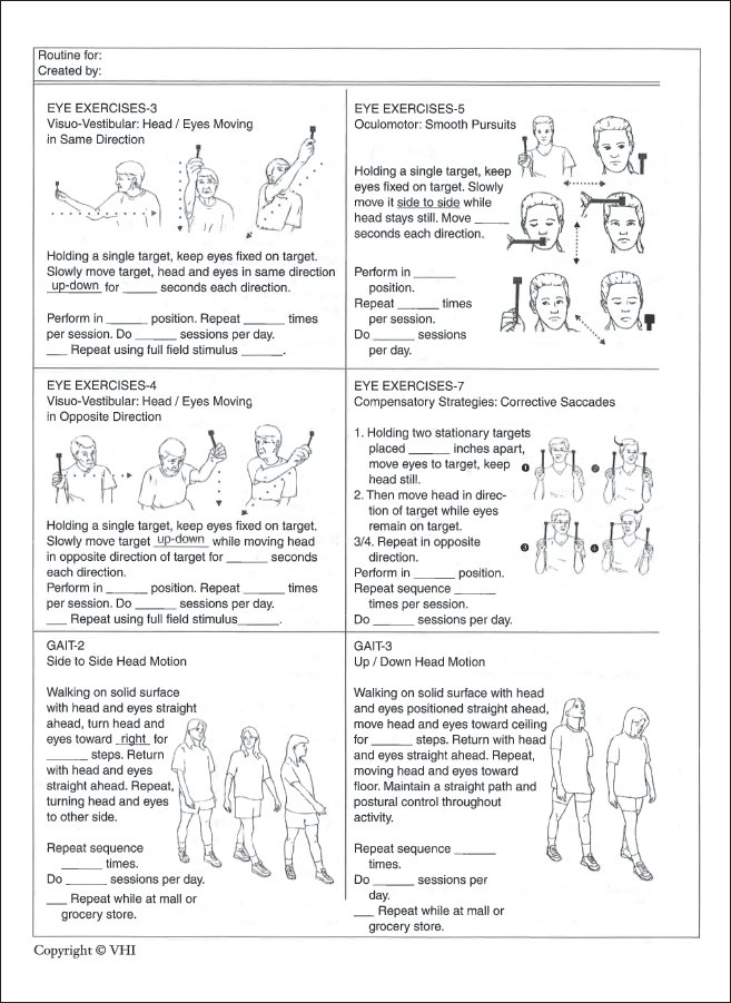
Vestibular complaints have been reported in over 50% of older people.92 The central mechanisms that are involved in the control of balance do not appear to change excessively with age but are more likely to be affected by degenerative neurologic diseases such as AD or Parkinson disease. However, there are age-related changes in the peripheral vestibular system. Hair cell receptors decrease in number, and there is a loss of the vestibular receptor ganglion cells. The myelinated nerve cells of the vestibular system decrease by as much as 40%. There is a reported increase in the incidence of benign paroxysmal positioning vertigo (BPPV) with complaints of dizziness with head movements. This may be due to an increase in the deposits in the posterior semicircular canal. Partial loss of vestibular function in older adults can lead to complaints of dizziness, with less ability of the nervous system to accommodate to positional changes.92,93
Coupled with the vestibular losses, there is concomitant loss in vision and somatosensation, which severely affects sensory input necessary for the maintenance of balance. In addition, there are longer response latencies and delayed reaction times. Vision changes include loss of acuity, decreased peripheral fields, and loss of depth perception. The loss of input from this combination is slow, with compensation developing through the years. Therefore, compensatory strategy in response to postural instability is not the same as in the person with acute vestibular insufficiency. There is an overall loss of functional reserve so that the threshold for clinical loss is lowered. This is demonstrated by the increased number of falls in older people with a history of falls compared with age-matched groups with no history of falls when they are tested with increased challenges to balance.93 There is an apparent decrease in the ability to integrate the conflicting sensory information to determine appropriate postural responses. Because there are also changes in motor output, the loss of balance, in addition to the lack of sensory organization, may be due to a poor response to vestibulospinal stimulation.93,94
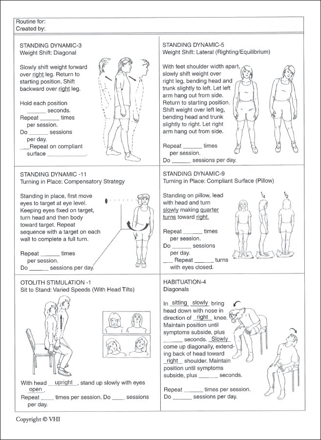
Various pathological conditions will affect the peripheral vestibular system to produce vertigo or disequilibrium. BPPV is the most common cause of vertigo with changes in head position in an older population. Generally, BPPV is associated with the deposition of otoconial material in the cupula of the posterior semicircular canal. The otoliths adhere to the cupula in some cases and retard its return to a resting position after head rotation or obstruct the flow of endolymph, producing symptoms from the affected posterior semicircular canal by impeding or ceasing stimulation to the vestibular nerve. It can be unilateral or bilateral. Prolonged inactivity can also lead to symptoms of BPPV. Habituation exercises (placing the person in positions that provoke vertigo) and balance exercises have been found to be very effective in the treatment of this disorder. These exercises are discussed in Chapter 12.
Acute vestibular neuritis, also known as labyrinthitis, is the second most common cause of vertigo in older adults.92–94 It is associated with a viral infection causing inflammatory changes of branches of the vestibular nerve. In older adults, onset is usually preceded by upper respiratory or gastrointestinal tract infections. The chief complaint is the acute onset of prolonged severe rotational vertigo that is exacerbated by movement of the head. Symptoms include spontaneous horizontal rotatory nystagmus beating toward the good ear, postural imbalance, and nausea.94 Antiviral medications are used in this condition, and habituation exercises help to quickly resolve this condition once the infection clears.
Ménièr e disease is a disorder of the inner ear that can cause hearing problems and vestibular symptoms in older adults.94 The patient complains of a sensation of fullness of the ear, a reduced ability to hear, and tinnitus. These symptoms are accompanied by rotational vertigo, postural imbalance, nystagmus, and nausea and vomiting that can last for extended periods of time. A phenomenon identified in Ménièr e disease is endolymphatic hydrops, a condition in which malabsorption of endolymph results in an increase in endolymphatic fluid pressure in the endolyphatic duct and sac. Medical intervention includes a salt-restricted diet and the use of diuretics to maintain the fluid balance in the ear. Vestibular suppressant medication is sometimes used during the acute phases, and the patient is advised to avoid caffeine, alcohol, and tobacco. In severe cases, surgical intervention is to insert a shunt to drain excess fluid from the ear; however, the effectiveness of this procedure is questionable.94 Ménièr e disease presents a challenge to rehabilitation efforts. Until the fluid imbalance is controlled, patients with this disease do not respond well to typical vestibular rehabilitation programs. Emphasis should be on safety and balance exercises.95 Challenging the balance to recruit the other systems, such as the cervico-ocular reflex and proprioceptive and visual mechanisms for controlling posture during stance and ambulation, may be effective.
Bilateral vestibular disorders may occur secondary to other diseases in older adults or could be drug induced. Conditions that may lead to vestibular problems include meningitis, labyrinthine infections, osteosclerosis, Paget disease, polyneuropathy, bilateral tumors (acoustic neuromas in neurofibromatosis), endolymphatic hydrops, bilateral vestibular neuritis, cerebral hemosiderosis, ototoxic drugs, inner ear autoimmune disease, or congenital malformations of the inner ear.94,95 Autoimmune conditions such as rheumatoid arthritis, psoriasis, ulcerative colitis, and Cogan syndrome (iritis accompanied by vertigo and sensorineural hearing loss) can lead to progressive, bilateral sensorineural hearing loss often accompanied by bilateral loss in vestibular function. Additionally, alcohol may cause acute vertigo because the dehydration created by alcoholic substances can change the specific gravity of the endolymph. Other agents that may cause vertigo include organic compounds of heavy metals and aminoglycosides.94–96 Controlled physical exercises can improve the condition in patients with bilateral vestibulopathy by recruiting nonvestibular sensory capacities such as the cervico-ocular reflex and proprioceptive and visual control of stance and gait.
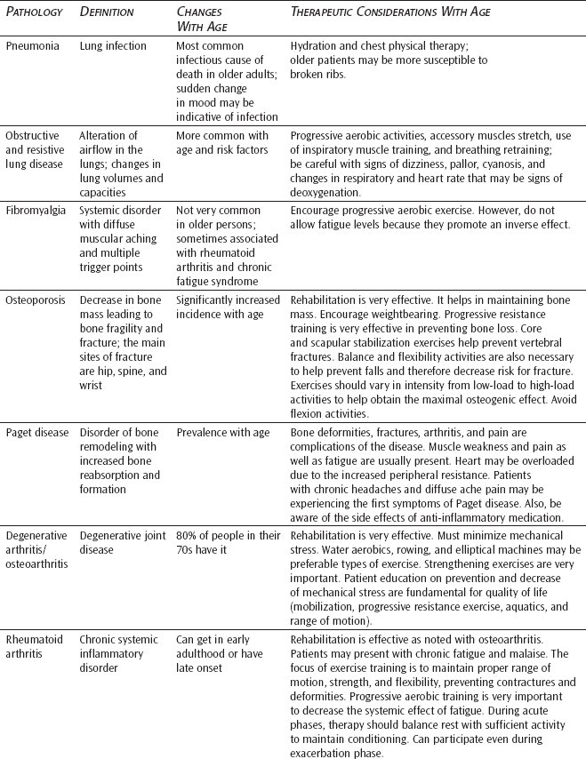
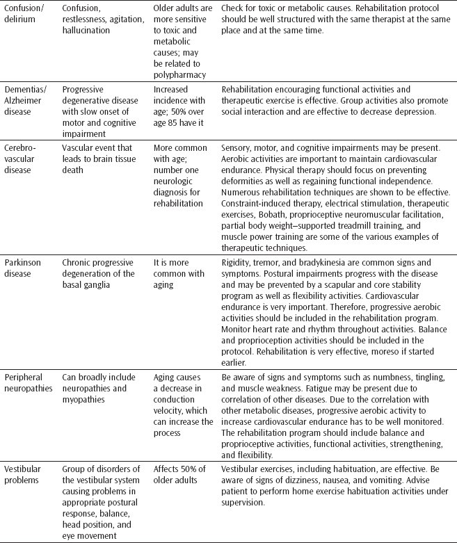
Pathologies in Older Adults and Therapeutic Considerations
Disturbances of Touch and Vision
Skin Pathologies
The skin is the largest organ of the body and functions to protect the interior of the body from the effects of pathogens, toxins, environmental extremes, trauma, and ultraviolet irradiation. With age, and often as the result of the accumulated effects of repeated injury, the skin changes. It grows and heals more slowly, becomes more sensitive to most toxins, and is less able to resist injury.97 Skin aging is characterized by different features including wrinkling, atrophy of the dermis, and loss of elasticity associated with damage to the extracellular matrix protein elastin.98 It becomes less effective as a barrier to infections. The specialized appendages, such as sweat and sebum glands, pressure and touch sensors, and hair follicles, atrophy. This results in dryness, a decrease in ability to alter body temperature through sweating, and loss of hair. The small blood vessels in the skin diminish with age, which contributes to less effectiveness as a barrier to infection, diminished reserve for repair, and altered ability to assist in thermoregulation.98
There are several skin diseases that are common in older adults and have significant effects on function. These include malignant tumors, herpes zoster, and decubitus ulcers. A great deal of data have accumulated to demonstrate an association between cutaneous aging and the development of skin cancers, infections, and ulcers.97 Many of the factors that appear to predispose individuals to the development of pathological manifestations in older adults are similarly operative in the development of skin problems.99 These include cumulative exposure to carcinogens, diminished DNA repair capacity, and decreased immunosurveillance. In addition, the reduced epidermal density in the skin that is seen with senescence is likely to play a role in the development of skin lesions, infections, and ulcerations.
Macular Degeneration
Age-related macular degeneration causes loss or blurriness of central vision creating a blind spot. Peripheral vision remains intact. Age-related macular degeneration results in an inability to see fine details, such as colors or a person’s facial features.100 Age-related macular degeneration is a progressive, degenerative disorder of the retina, and it is the leading cause of new cases of blindness in people age 65 and older.101
Immediately beneath the sensory retina lies a single layer of cells called the retinal pigment epithelium. These cells provide nourishment to the portion of the retina where they are in contact. In age-related macular degeneration, the maintenance of this contact is threatened. A small hemorrhage may break through and accumulate between the retinal pigment epithelium and the sensory retina. This leads to disruption of the photoreceptor cells’ nutrition and their death, with attendant loss of central vision.102 This type of age-related maculopathy is referred to as the wet type because of the leaking vessels and edema or blood that detaches the retina. The dry type consists of disintegration of the retinal pigment epithelium because of nutritional loss. The light-sensitive cells of the macula break down over time, and a yellow deposit called drusen accumulates under the retina.103
Cataracts
A cataract is opacity of the lens that reduces visual acuity (to 20/30 or less).104 An early sign is the complaint of glare from bright lights at night or even during the day, the result of rays of light being scattered by the opacities. Over time, the lens opacities progress and eventually interfere with vision so that reading becomes difficult even with glasses. The hallmark of all cataracts is painless, progressive loss of vision. Although the cause has not been elucidated, there appears to be a strong nutritional link related to low levels of antioxidants.105 Cataract surgery, the removal of the lens from the eye, is the treatment of choice.106
Glaucoma
Glaucoma is a disorder characterized by increased intraocular pressure that can lead to irreversible damage to the optic nerve, with the accompanying impairment of blindness. It is a pathological process in the eye that anatomically or functionally blocks the outflow channels. The most common type of glaucoma in older adults is open-angle glaucoma, accounting for 90% of all cases.107 Onset is insidious and usually asymptomatic, causing slow loss of visual field affecting both eyes and occurring more commonly in black individuals.108 Secondary glaucoma is associated with diabetes (diabetic retinopathy), uveitis, and ocular tumors that affect the optic nerve.108
The leading causes of visual impairment are age related, but appropriate care can preserve useful vision for most older adults.109 Vision loss due to macular degeneration cannot be delayed in all patients; however, it can sometimes be postponed through laser therapy. Low vision rehabilitation can maximize the usefulness of remaining eyesight. Cataract surgery is highly successful. Early detection and treatment of glaucoma can prevent vision loss. Laser treatment is remarkably effective in treating diabetic retinopathy.109 Otherwise, the low vision state is best addressed with vision-enhancing devices, adaptive equipment, and patient education available through occupational therapy. Referral to a low vision rehabilitation program is needed for a comprehensive evaluation and intervention. Individual adaptation and supportive services often result in a significant improvement in function and quality of life for those older adults with low vision.
Hearing Pathologies
The hearing losses that occur with presbycusis affect the higher, pure-tone frequencies.110 Basically, the individual loses the hard sounds of language. This leads to a decrease in the ability to understand speech in which parts of words or whole words are lost because of higher tones as well as the interference of background noise. The use of hearing aids and surgical implants has provided relief for some, but the process of presbycusis is such that these steps can only blunt the effects of the problem. Because most cases of presbycusis are of mixed etiology, as has already been noted, intervention will not completely correct the loss. Thus, the clinical focus should be on improving and maintaining as much of the hearing capability as possible and helping the older person and the family adapt to the limitations necessitated by substituting other forms of communication and environmental stimuli to compensate for the loss that remains.111
The clinician needs to be mindful of the effects of hearing loss on all aspects of the older patient’s life. Failure to consider the effects of hearing loss when evaluating such problems as depression, confusion, possible attention span deficits, and a variety of other clinical problems may lead to less-than-adequate clinical intervention.
Tinnitus is the diagnosis given to a variety of “ear noise” disorders. A small percentage of older adults suffer from this condition to varying degrees,110 and it is an annoying problem. Patients often report constant or intermittent noises, such as buzzing, ringing, or hissing, that result in a distortion of accurate reception of environmental sounds and voices. If patients complain of tinnitus, considerations for a quiet treatment environment should be made to decrease the bombardment of external noise sources superimposed on the internal sources.
Otalgia is ear pain that results from an otologic process or may be referred along neural pathways, including the trigeminal, glossopharyngeal, vagus, and cervical nerves. Inflammation of the pinna, external auditory canal, tympanic membrane, or middle ear can result in otalgia. With Eustachian tube obstruction, negative pressure in the middle ear may produce painful retraction of the tympanic membrane. In older adults, it is common for pain in the temporomandibular joint to be referred to the ear.
Massive accumulation of cerumen (wax) is frequently seen in older adults. The individual is usually dehydrated, complains of hearing loss and a feeling of fullness in the ear, and often reports dizziness.
Effusion in the middle ear, usually related to obstruction of the Eustachian tube, is called serous otitis media. In older adults, this condition generally occurs unilaterally, and the patient perceives a sensation of aural fullness and hearing loss.
Dizziness, encompassing the sensations of vertigo, dysequilibrium, and unsteadiness, is a common complaint in older adults with ear disorders. Evaluation of balance problems should include an assessment of hearing pathologies.
GASTROINTESTINAL MANIFESTATIONS OF AGING
Dysphagia
Dysphagia is difficulty in swallowing. It commonly results from neuromuscular disorders, such as a cerebrovascular accident, Parkinson disease, diabetes, or other neuropathies. Malnutrition results from decreased intake; aspiration of oral contents is a common accompaniment that frequently leads to pneumonia. Siebens and colleagues112 have identified a fairly high incidence of swallowing problems involving the mouth, pharynx, and upper esophageal sphincter in the older population.
True esophageal dysphagia, in which the transport of the ingested material down the esophagus is impaired, is common in older adults.113 Carcinoma of the esophagus, which occurs with increasing frequency in older adults, usually presents with dysphagia. The most common symptom is the sensation of food “hanging up” in the esophagus. It has a poor prognosis for cure and usually requires extensive palliative treatment. Hiatus hernia, another cause of dysphagia, is also increasingly common in older adults. Few, however, are symptomatic, and medical management with antacids and H2 blockers is effective.114 It is important to understand that achalasia can initially present in older adults and that other motility disorders, such as diffuse esophageal spasm and scleroderma, do occur in these individuals.113 Another cause of esophageal dysphagia that is unique to the older population is dysphagia aortica, in which the transport of material down the esophagus is impaired by a markedly tortuous and enlarged aorta, heart, or both.115
The role of the physical and occupational therapist in treating dysphagia is to coordinate the team efforts of speech pathology, dietary, and nursing to provide a comprehensive positioning and feeding program. The physical therapist is involved in evaluating and treating head and trunk control; neck range of motion; neck weakness; sitting balance; abnormal postural reflex activity interfering with head control, sitting balance, or both; gross facial muscle test; ability to handle secretions; voluntary deep breathing ability, breath control, and voluntary cough; and gross motor upper extremity ability. The occupational therapist is involved in some of the same interventions and additionally provides adaptive equipment as warranted. Specific emphasis needs to be placed on wheelchair and bed positioning and respiratory status.
Medical Pathologies in Older Adults and Therapeutic Considerations
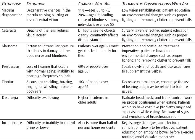
BOWEL AND BLADDER PROBLEMS
Urinary Incontinence
Urinary incontinence can affect both men and women. It afflicts more than half of nursing home residents and is often the reason for admission.116 The causes of incontinence can be divided into 2 broad categories: established and transient.117 Established incontinence is usually the result of neurologic damage or intrinsic bladder or urethral pathology. By contrast, incontinence caused by transient causes, such as a medication or diet, is generally reversible if the underlying problem can be addressed adequately.
Incontinence is not a normal sequela of aging, and characteristics of what is commonly called overactive bladder include decreases in bladder capacity, urethral compliance, maximal urethral closure pressure, and urinary flow rate.117 In both sexes, postvoid residual volume and the prevalence of involuntary detrusor contractions probably increase, while urethral resistance increases in men.118
The various types of urinary incontinence identified are stress, urge, mixed, overflow, and functional incontinence. Stress incontinence refers to the loss of bladder control due to the physical stress of increased pressure in the abdomen from such activities as coughing, sneezing, laughing, jogging, or straining on a lift or during a bowel movement. Urge incontinence is defined as the sudden urge to urinate without the ability to hold the urine long enough to reach a bathroom. Mixed incontinence is a combination of stress and urge incontinence. Overflow incontinence is the accidental loss of urine from a chronically full bladder. This may occur as the result of a cystocele (a vaginal hernia or bulge due to weakened vaginal muscles), an enlarged prostate, or a tumor, all of which block the flow of urine through the urethra. Other causes of overflow incontinence might include damage to the bladder nerves from diabetes, loss of adequate estrogen or progesterone,119 or a herniated lumbar disc. Functional incontinence is the inability to get to the bathroom because of physical limitations or the inability to manage clothing once the individual has made it to the bathroom. In the older adult, a combination of these conditions may exist.
The causes of transient incontinence may be denoted by the mnemonic DIAPPERS as follows120:
❖ Delirium
❖ Infection (especially urinary tract infection)
❖ Atrophic vaginitis
❖ Pharmaceuticals
❖ Psychological factors (eg, depression and poor motivation)
❖ Excess fluid output (eg, diuretics and diabetes)
❖ Restricted mobility (eg, Parkinson disease and arthritis)
❖ Stool (constipation or impaction)
Any condition that impairs cognition, mobility, or the ability to hold urine can contribute to functional incontinence. Although such causes potentially may be reversible, in reality, many patients’ functional status may not improve, and, therefore, incontinence becomes established. Many of the causes of established incontinence involve urinary tract dysfunction. These include overactivity of the bladder with involuntary contraction, failure of the bladder to contract at the appropriate time or as strongly as it should, low resistance to urinary flow when it should be high (stress incontinence), and high resistance to urinary flow when it should be low (urinary obstruction).121 Detrusor instability is characterized by a sudden and urgent need to empty the bladder. The volume emptied is variable but may be large. Often in older adults, the detrusor muscle contracts, but the bladder does not empty completely, leaving residual urine and an increased risk for urinary tract infection.117
There are numerous interventions for urinary incontinence that rehabilitation therapists can offer the older person with this condition.122,123 Behavioral treatments are considered appropriate for patients with stress, urge, and mixed incontinence. Physical therapy may include biofeedback, therapeutic exercise, neuromuscular reeducation, therapeutic activity, and gait training. Instruction in pelvic floor exercises, commonly known as Kegels, is helpful in regaining strength of the pelvic floor musculature.122 Occupational therapy may be involved in training for functional activities that facilitate toileting as well as the modification of clothing (ie, replacing buttons or zippers with Velcro) to enhance the ease of disrobing and eliminate incontinence resulting from functional limitations.
Evidence-based treatment strategies are cited in the next column.
▷ Burgio KL, Locher JL, Goode PS, et al. Behavioral versus drug treatment for urge urinary incontinence in older women. JAMA. 1998;280(23):1995-2000.124
- Randomized and controlled trial = safe and effective.
- Behavioral training
- Visit 1—anorectal feedback
- Visit 2—urge strategies (when urged, pause, relax, and contract pelvic muscles; then proceed to toilet)
- Visit 3—muscle biofeedback if not 50% better with above
- Visit 4—fine-tune
- Home program—15 Kegels, 3 times a day, for 10 seconds, lying/sitting/standing, interrupting or slowing urine flow
- Visit 1—anorectal feedback
▷ Bo K, Talseth T, Holme I. Single blind, randomised controlled trial of pelvic floor exercises, electrical stimulation, vaginal cones, and no treatment in management of genuine stress incontinence in women. BMJ. 1999;318:487-493.125
- 4 groups
- Pelvic floor exercise
8 to 12 contractions, 3 times a day
Once a week with physical therapy in a group (supine, sitting, etc)
- Electrical stimulation
Intermittent stimulation 30 minutes a day
- Vaginal cones 20 minutes a day
- Control—untreated
- Pelvic floor exercise
- All groups had a once-a-week meeting with the physical therapist.
- Improvements in leakage and strength were greater in the exercise-only group; no side effects (discomfort and bleeding).
▷ Spruijt J, Vierhout M, Verstraeten R, Janssens J, Burger C. Vaginal electrical stimulation of the pelvic floor: a randomized feasibility study in urinary incontinent elderly women. Acta Obstet Gynecol Scand. 2003;82:1043-1048.126
- This study on electrical stimulation in older adults showed no difference in exercise or exercise and electrical stimulation. Electrical stimulation has high physical and emotional cost.
Stay updated, free articles. Join our Telegram channel

Full access? Get Clinical Tree


