Papillary Urothelial Neoplasms and Their Precursors


Papillary Urothelial Neoplasms and Their Precursors


The World Health Organization (WHO)/International Society of Urological Pathologists (ISUP) classification of noninvasive papillary urothelial lesions is outlined in Table 3.1 (1). The diagnosis of lesions within this classification takes into account architectural and cytologic features within the urothelium (Table 3.2).


PAPILLARY UROTHELIAL HYPERPLASIA

Papillary urothelial hyperplasia is a likely precursor lesion to low-grade papillary urothelial neoplasms (2, 3). Typically, they are discovered on routine follow-up cystoscopy for papillary urothelial neoplasms and less frequently in the workup for microhematuria or urinary obstructive symptoms. In the latest fourth edition of the WHO classification, the term “urothelial proliferation of uncertain malignant potential” is being recommended for such lesions, acknowledging the fact that some may represent “shoulder” lesion of previously resected papillary tumors and up to 20% harbor the same genetic abnormalities as low-grade papillary carcinomas. In most cases at cystoscopy, a focal elevated or roughened lesion is identified that is variably described as “bleb-like,” “papillary,” “raised,” “sessile,” or “frondular.”

Histologically, papillary hyperplasia consists of undulating urothelium arranged into mucosal narrow papillary folds of varying heights (Figs. 3.1, 3.2, 3.3, 3.4) (efigs 3.1-3.7). Both the urothelium within papillary hyperplasia and the adjacent flat mucosa are often thicker than normal. In addition to the diagnostic mucosal folds, in some cases, there are also tent-shaped, somewhat broader folds that lack the edema and inflammation typical of polypoid cystitis. The cytologic findings in typical papillary hyperplasia are similar to that in normal urothelium. Some cases show increased vascularity in the stroma at the base of the papillary folds. An occasional case may also demonstrate early slight branching beginning at the top of one of the folds of the papillary hyperplasia. Papillary hyperplasia is distinguished from
papillary urothelial neoplasms by lack of true fibrovascular cores within the hyperplastic mucosa, lack of complex arborization, and hence the absence of the appearance of “detached” papillary fronds (Figs. 3.5, 3.6). It is distinguished from low-grade papillary urothelial carcinoma by lack of cytologic abnormality.


TABLE 3.1 The World Health Organization/International Society of Urological Pathology Consensus Classification of Papillary Urothelial (Transitional Cell) Lesions


Hyperplasia


Papillary hyperplasia (urothelial proliferation of uncertain malignant potential)


Papillary neoplasms


Papilloma


Papillary neoplasm of low malignant potential


Papillary carcinoma, low grade


Papillary carcinoma, high grade


Large studies with sufficient follow-up to evaluate the clinical significance of de novo (without concurrent or prior history of urothelial neoplasia) papillary hyperplasia are lacking. Nevertheless, even though de novo papillary hyperplasia will not inevitably progress to urothelial neoplasia, it is reasonable to suggest that patients should be followed up more closely than individuals in the general population. The finding should be reported as it correlates with a cystoscopic abnormality. If papillary hyperplasia is diagnosed in someone with a history of urothelial neoplasia, it most likely indicates early recurrence of papillary neoplasia and warrants continued close follow-up. Molecular data also demonstrate that at least a proportion of papillary hyperplasia represents precancerous lesions of the bladder that subsequently progress to papillary bladder cancer (4).

Less commonly, the overlying urothelium in a lesion with the architectural pattern of papillary hyperplasia reveals varying degrees of cytologic atypia ranging from dysplasia to CIS (5) (Fig. 3.7) (efigs 3.8, 3.9). One hypothesis is that, in some cases, CIS or dysplasia may evolve into a papillary lesion, with further progression to high-grade papillary cancer. This process is analogous to papillary hyperplasia without cytologic atypia progressing to low-grade papillary urothelial neoplasms. In cases of CIS or dysplasia with the architecture of papillary hyperplasia, reasonable diagnoses are “CIS with early papillary formation” or “dysplasia with early papillary formation” (6). Patients will be treated for CIS or dysplasia, respectively, yet the diagnoses correlate with the cystoscopic appearance of a slightly papillary lesion.


TABLE 3.2 Histologic Features of Papillary Urothelial Lesions


Papilloma


Papillary Neoplasm of Low Malignant Potential


Low-Grade Papillary Carcinoma


High-Grade Papillary Carcinoma


Architecture of papillae


Delicate


Delicate; occasionally fused


Fused; branching; delicate


Fused; branching; delicate


Organization of cells


Identical to normal


Polarity identical to normal; any thickness; cohesive


Predominantly ordered, yet minimal crowding and minimal loss of polarity; any thickness; cohesive


Predominantly disordered with frequent loss of polarity; any thickness; often discohesive


Cytology


Identical to normal


May be uniformly enlarged


Enlarged with variation in size


Enlarged with variation in size


Nuclear size


Nuclear shape


Identical to normal


Elongated; round-oval; uniform


Round-oval; slight variation in shape and contour


Moderate-marked pleomorphism


Nuclear chromatin


Fine


Fine


Mild variation within and between cells


Moderate-marked variation within and between cells with hyperchromasia


Nucleoli


Absent


Absent to inconspicuous


Usually inconspicuousa


Multiple prominent nucleoli may be present


Mitoses


Absent


Rare; basal


Occasional, at any level


Usually frequent, at any level; may be atypical


Umbrella cells


Uniformly present


Present


Usually present


May be present


a If present, small and regular and not accompanied by other features of high-grade carcinoma.


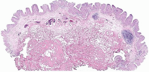
FIGURE 3.1 Papillary urothelial hyperplasia.


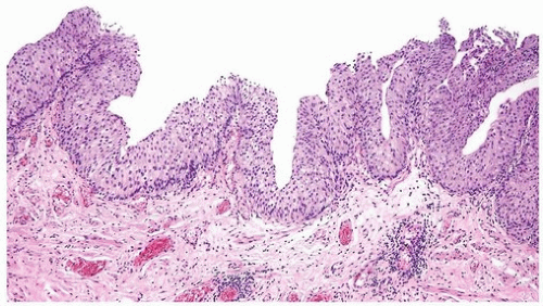
FIGURE 3.2 Papillary urothelial hyperplasia.


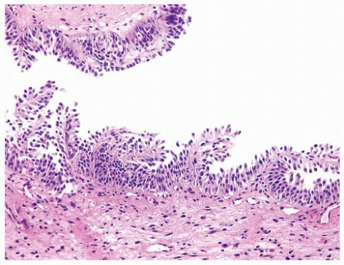
FIGURE 3.3 Papillary urothelial hyperplasia.


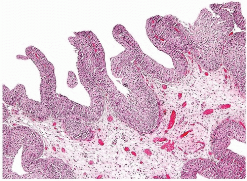
FIGURE 3.4 Papillary urothelial hyperplasia with tall folds. Some would regard as early papilloma.


PAPILLARY UROTHELIAL NEOPLASMS


Background

The classification and grading of papillary urothelial neoplasms has been a long-standing source of controversy (7). There are numerous grading systems, most of which have poor interobserver reproducibility, with most cases falling into an intermediate category (8, 9, 10, 11, 12, 13, 14, 15). The most commonly used grading systems for bladder tumors have been those proposed by the World Health Organization (WHO). In 1973, the WHO system proposed that tumors be categorized into benign urothelial papillomas and three
grades of carcinoma (grades 1, 2, and 3) (10). A major limitation of the WHO (1973) grading system was the lack of precise definition of the various grades and lack of specific histologic criteria. The following statement is the sole description of the difference between WHO grades 1, 2, and 3 as written in the original WHO (1973) system: “Grade 1 tumors have the least degree of anaplasia compatible with the diagnosis of malignancy. Grade 3 applies to tumors with the most severe degrees of cellular anaplasia, and Grade 2 lies in between.” In December 1998, members of WHO and ISUP published the WHO/ISUP consensus classification of urothelial (transitional cell) neoplasms of the urinary bladder (1). This new classification system arose out of the need to develop a more detailed classification system for bladder neoplasia and because many of the tumors classified
as “transitional cell carcinoma, grade 1” had no potential for malignant behavior once completely excised. In 2004, WHO revised its classification of urothelial tumors, adopting the WHO/ISUP system, resulting in a unified grading system for urothelial tumors (16). Because these systems are identical, in this text, the grading system is referred to as the WHO/ISUP system. The WHO 2016 blue book continues to endorse this classification system.


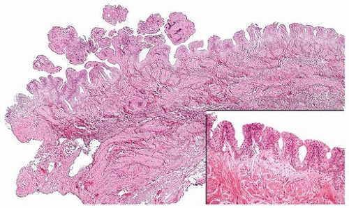
FIGURE 3.5 Papillary urothelial hyperplasia (right and inset) adjacent to urothelial papilloma (left).


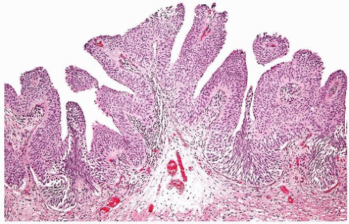
FIGURE 3.6 Papillary urothelial hyperplasia starting to develop branched fronds and “detached-appearing” papillary fronds diagnostic of early urothelial papilloma.


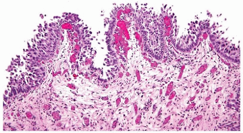
FIGURE 3.7 CIS with early papillary formation.


WHO/ISUP System

The WHO/ISUP system is a modified version of the scheme proposed by Malmström and colleagues (13). One of the major contributions of the WHO/ISUP system is a detailed histologic description of the various grades, using specific cytologic and architectural criteria. These criteria are based on the architectural features relating to the histology of the papillae and the overall organization of the cells. Cytologic features encompassed in the WHO/ISUP system include nuclear size, nuclear shape, chromatin content, nucleoli, mitoses, and umbrella cells. Papillary tumors may show heterogeneity of grade. Tumors are graded based on the highest grade exhibited, although it remains to be defined what percentage is minimally needed to place tumors in a higher category when the highest grade is focal (see section “High-Grade Papillary Carcinoma”). The terminology used in the WHO/ISUP system for carcinomas is consistent with that used in urine cytology. Criteria and terminology for papillary urothelial neoplasms with an inverted growth pattern are described in Chapter 4.

Representative data on the prognosis of the 2004 grading system come from Pan et al. who studied 1,515 patients who underwent TURB for non-muscle-invasive bladder cancer. The recurrence rates for papillary urothelial neoplasms of low malignant potentials (PUNLMPs), low-grade, and high-grade carcinomas were 18%, 35%, and 34%, respectively (17). Tumor stage progression occurred in 2%, 6%, and 29% of patients with PUNLMP, low-grade, and high-grade papillary carcinoma, respectively. None of the patients with PUNLMP presented with an associated invasive component or died of the disease. Of patients with an initial diagnosis of low- and high-grade papillary carcinoma, 2% and 22%, respectively, eventually died of these cancers. PUNLMPs had a significantly lower recurrence rate than the low- and high-grade cancers, and the differences between all three tumor grades were significant in terms of stage progression and death due to cancer. In a subsequent study from this group, they developed nomograms to predict progression-free and cancer-specific survival factoring grade, stage, and intravesical therapy (18). Similar findings showing differences between PUNLMP, low-, and high-grade noninvasive papillary urothelial carcinoma in recurrence, progression, and death have been published by other authors (19, 20, 21, 22, 23).


Relation of WHO (1973) to WHO/ISUP

A major misconception in the application of the WHO/ISUP classification is that there is a one-to-one translation between it and the WHO (1973) classification system (Fig. 3.8). Only at the extremes of grades in the WHO (1973) classification does this one-to-one correlation hold true. Lesions called papilloma in the WHO (1973) classification system are also called papilloma in the WHO/ISUP system. At the other end of the grading extreme, lesions called WHO (1973) grade 3 are by definition highgrade carcinoma in the WHO/ISUP system. However, for WHO (1973) grades 1 and 2, there is no direct translation to WHO/ISUP classification system. Rather, lesions must be analyzed using the criteria of the WHO/ISUP classification system without regard to how they were diagnosed in WHO (1973) system. Lesions formerly called WHO (1973) grade 1, which upon review show no cytologic atypia and merely thickened urothelium with, at most, nuclear enlargement and minimal disorder in orientation, would be called papillary urothelial neoplasms of low malignant potential (PUNLMP). However, other WHO (1973) grade 1 lesions with definite yet slight cytologic atypia would be diagnosed in the WHO/ISUP system as low-grade urothelial carcinomas.

Within the WHO (1973) system, grade 2 is a very broad category. It includes lesions that are relatively bland, which in some places would be diagnosed as WHO (1973) grades 1-2; these lesions in the WHO/ISUP system would be called low-grade papillary urothelial carcinoma. In other cases, WHO (1973) grade 2 lesions border on higher-grade lesions and, in many institutions, were called WHO (1973) grades 2-3; these lesions in the WHO (2004)/ISUP classification system would be called high-grade
carcinoma. In the old WHO (1973) system, a large percentage of patients with noninvasive papillary urothelial carcinomas were called grade 2, an ambiguous group that the urologists did not know how to treat. In the WHO/ISUP grading system, these grade 2 tumors can be segregated roughly equally into low- and high-grade cancers with correspondingly low and high risks of progression (24, 25, 26). When WHO (1973) grade 2 tumors are reclassified as high-grade tumors, they show molecular aberrations usually associated with cancers of higher grade and poor outcome, further supporting that the WHO (1973) grade 2 tumors form a heterogeneous group that can be subdivided into low- and high-grade cancer (27).


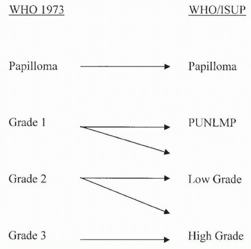
FIGURE 3.8 Relationship of WHO (1973) to WHO (2003)/ISUP classification of papillary urothelial tumors.

There have been several studies comparing WHO (1973) and the WHO/ISUP system. Most have demonstrated a superiority of WHO/ISUP in predicting recurrence and progression or interobserver reproducibility (24, 28, 29, 30, 31, 32, 33). Whether routine ancillary studies such as immunohistochemical or molecular markers will aid us in better stratifying these tumors into distinct risk groups remains to be proven.

Others have reported no difference in interobserver reproducibility of WHO/ISUP compared to WHO (1973), although in some of these works, the WHO/ISUP grade was merely translated from the WHO (1973) grade, which as described earlier is inaccurate (34, 35, 36). In another study comparing the two grading systems, there was no difference in survival, yet one would not expect a difference in survival as the vast majority of papillary urothelial neoplasms do not result in the death of the patient; the more appropriate end points to evaluate are either progression (in grade or stage) or recurrence (37).


Papilloma

Using the WHO (1973) system, some experts applied very restrictive criteria for the diagnosis of urothelial papilloma, in part based on the number of cell layers, and regarded all other papillary neoplasms as carcinomas. Others applied a broader definition of “urothelial papilloma” so as not to label all patients with papillary lesions with minimal cytologic and architectural atypia as having carcinoma; many if not all of these latter cases would now fall into the PUNLMP category.

The WHO/ISUP system has very restrictive histologic features for the diagnosis of papilloma, requiring papillary fronds to be lined by normal-appearing urothelium (Figs. 3.9, 3.10, 3.11, 3.12, 3.13, 3.14) (efigs 3.10-3.45). Most papillomas present as single lesions that are relatively small. However, multifocal tumors and larger lesions may be only occasionally seen.

Most papillomas have a simple nonbranching or minimally branching arrangement, slender fibrovascular stalks, and a predominantly exophytic pattern. In some papillomas, more complex anastomosing papillae, marked stromal edema within the papillae, and endophytic areas can be identified (Figs. 3.15, 3.16).

Within the limits of normal urothelial thickness, there is some variability. Uncommon cases may be composed of only one to two cell layers
of urothelium. There is no need to count the maximum number of cell layers, as long as it is not obviously thicker than the normal urothelial lining. In most cases, the urothelium is tightly cohesive, although in some cases, there may be the appearance of discohesion. Umbrella cells vary in their morphology from being inconspicuous, to being cuboidal with slightly enlarged nuclei and paler cytoplasm, to having a hobnail appearance with abundant eosinophilic cytoplasm. Occasional cases of urothelial papilloma have an umbrella cell layer with prominent vacuolization. Single, enlarged umbrella cells may also drape themselves over several underlying urothelial cells, where the umbrella cell nuclei may
demonstrate degenerative atypia. Urothelial atypia, other than that which can be seen in umbrella cells, excludes the diagnosis of papilloma. Mitotic figures are absent.


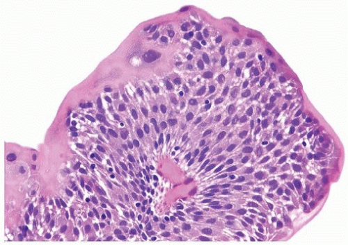
FIGURE 3.9 Urothelial papilloma with atypical umbrella cells.


FIGURE 3.10 Urothelial papilloma. There is a consistent umbrella cell layer in which the cells appear slightly enlarged: this feature does not argue against a diagnosis of papilloma.


FIGURE 3.11 Urothelial papilloma.

Papillomas are rare and more commonly, but not exclusively, occur in younger patients. Papillomas may occur in association with a prior or concurrent history of other urothelial tumors. However, if a papilloma is the first manifestation of urothelial neoplasia, only about 7% to 8% recur, typically within 5 years of the initial diagnosis (26, 34, 38, 39, 40, 41). Lesions typically recur as papilloma and less commonly as papillary urothelial neoplasm of low malignant potential (PUNLMP) or noninvasive lowgrade papillary urothelial carcinoma. Of the 4 largest series on papillomas
totaling 153 patients, only 1 patient experienced stage progression; invasive urothelial carcinoma was found extending out of the bladder 4 years after the diagnosis of papilloma (38, 40, 41, 42). A caveat to this exceptional case is that the patient was immunosuppressed following renal transplantation.


FIGURE 3.12 Urothelial papilloma with cuboidal umbrella cells with clear cytoplasm.


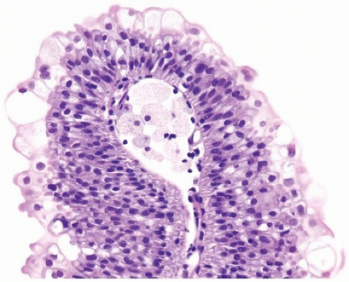
FIGURE 3.13 Urothelial papilloma with vacuolated umbrella cells. Note foam cells in a papillary core.


Papillary Urothelial Neoplasm of Low Malignant Potential

The category of PUNLMP was included to describe lesions that do not have cytologic features of malignancy, yet have thickened urothelium or have more cells per unit area (greater cellularity of urothelium)
compared to papilloma (Figs. 3.17, 3.18, 3.19, 3.20, 3.21, 3.22) (efigs 3.46-3.76). Having a category of PUNLMP avoids labeling a patient as having cancer, which has psychosocial and financial (e.g., insurance) implications, although since it is not labeled as an unequivocal benign lesion (e.g., papilloma), its diagnosis implies that the patient should be adequately followed.

Only gold members can continue reading. Log In or Register to continue

Stay updated, free articles. Join our Telegram channel

Dec 18, 2016 | Posted by in ONCOLOGY | Comments Off on Papillary Urothelial Neoplasms and Their Precursors

Full access? Get Clinical Tree

Get Clinical Tree app for offline access