Osteosarcoma


Osteosarcoma


Richard Gorlick

Katherine Janeway

Neyssa Marina


INTRODUCTION

Osteosarcoma is often compared and contrasted with Ewing sarcoma as pediatric tumors that arise in bone—but beyond that, they do not have much in common. Osteosarcoma has clear associations with a variety of genetic predisposition syndromes and a variety of well-defined epidemiologic features. This is coupled to in-depth studies of the complex genetic alterations present in the tumors. Despite all of this information, the essential features that drive the formation of progression of osteosarcoma in most individuals are poorly understood. At present, the biologic studies performed on tumor samples obtained from a patient with osteosarcoma have little to no impact on the patient’s treatment planning or regimen. A long history of observations and interventional studies in osteosarcoma have helped define subtleties in how this cancer can present clinically and how this contributes to optimizing therapy. In this chapter, we will focus on the clinical management of osteosarcoma, highlighting what constitutes the standard of therapy but also providing enough detail so that the subtleties of clinical variations among patients are appreciated.


CLINICAL PRESENTATION


Presentation

The most common initial symptom of osteosarcoma is pain.1 This pain can be intermittent but, in general, it becomes continuous and more severe with time. Most patients will relate the onset of symptoms temporally to trauma, but injury is not an etiologic factor in osteosarcoma development per se nor is it clearly tied to any particular event in the tumor’s progression, with the rare exception of a patient who presents with a pathologic fracture. In some patients, a mass may be palpable in the region of the pain, but this is variable. With time, the swelling, if present, will increase and eventually will affect adjacent joints, resulting in loss of function. Given the nonspecific symptoms at the time an osteosarcoma presents, a primary-care provider requires a high index of suspicion to detect the disease early and avoid potentially harmful diagnostic delays and misguided treatment attempts. In assessing the patient, a primary-care provider should consider factors like the age of the patient and the location of the symptoms as these have consistent patterns in osteosarcoma, as will be discussed further in the section on Epidemiology. Unfortunately, these patterns overlap with much more common conditions that afflict this age group. In the typical physically active adolescent, pain caused by osteosarcoma is often initially attributed to recent trauma, commonly related to a sports injury. The differential diagnosis of osteosarcoma also includes osteomyelitis, benign bone tumors such as osteochondroma, fibroma, osteoid-osteoma, enchondroma, giant-cell tumor of bone, bone cysts, and others, as well as other primary malignancies of bone and bone metastases.2,3

Other aspects of how the patient may present can be potentially confusing. For example, pain at an osseous site other than the primary tumor may represent metastatic involvement. That said, the most common site of metastases is the lungs. Respiratory symptoms occur very late and only with the presence of extensive lung involvement. Synchronous (at the time of presentation) and metachronous (following administration of treatment) metastases most commonly involve the lungs. In the two largest reported series of primary metastatic osteosarcomas and metastatic recurrences, pulmonary involvement was observed in 81% and 88%, respectively; the lungs were the only metastatic site in 61% and 70%, respectively.4,5,6 Distant bones are the second most frequent site of metastatic deposits, combined with lung metastases in at least half of all metastatic cases.5 Skip metastases, isolated tumor foci within the same bone as the primary tumor, represent another form of osseous dissemination.7 While they occur only in a minority of patients, they must always be sought for by appropriate imaging of the total tumor-bearing bone. Metastases to organs other than lungs or bones are rare in the absence of widespread disease.5 Weight loss is rare in patients with osteosarcoma and systemic symptoms, such as fever, diaphoresis, and malaise are uncommon in the absence of far advanced disease. Therefore, the vast majority of osteosarcoma patients do not feel systemically ill when their cancer is first diagnosed.

The nonspecific symptoms suggest that in many patients the diagnosis of osteosarcoma may be delayed. The lag-time between onset of symptoms and diagnosis ranges from 2 to 4 months for patients with localized extremity osteosarcoma in several North American and European studies, but it is considerably longer in other parts of the world.8,9 Despite this frequent delay, there is no correlation between the delay in diagnosis and the likelihood of primary metastases. This would suggest that patients who develop metastases have a biologically more aggressive tumor. Patients with axial primaries are usually diagnosed later than those with tumors located in the limbs.1,8,9

Although osteosarcoma is not often considered the most likely diagnosis in patients presenting with leg pain, arriving at the diagnosis is greatly assisted by the fact that a lesion will invariably be visualized on a plain radiograph. Although imaging is not indicated in every patient with extremity pain, a plain radiograph should be obtained if there is a suspicion for osteosarcoma.


Laboratory Evaluation

There are no known laboratory tests specific for diagnosing osteosarcoma. Serum levels of alkaline phosphatase and less frequently lactic dehydrogenase10,11 are elevated in a considerable number of patients. Elevated alkaline phosphatase levels do not necessarily correlate with disease extent but have been observed to correlate with an increased likelihood for recurrence.10,11,12,13 Several other laboratory tests must be performed before beginning chemotherapy. These aim to assess general health, and baseline function for organs affected by chemotherapy treatment. Tests to assess general health include: a complete blood count and differential, tests for serum electrolytes including calcium, magnesium, and phosphate, liver function studies, blood group typing, a coagulation profile, as well as tests for hepatitis and human immunodeficiency virus infection.

Treatment-related complications associated with osteosarcoma treatment include anthracycline-induced cardiomyopathy,14
hearing loss,15 kidney dysfunction,16 secondary malignancies,17 and sterility especially in patients receiving alkylating agents. These complications serve as an additional guide for baseline tests that should be performed on these patients. In general, these include: an audiogram, an echocardiogram, and renal function evaluation including creatinine and (estimated) creatinine clearance. Additionally, based on the risk of sterility related to chemotherapy, fertility preservation should be considered. Pubertal males are usually referred for sperm donation. Preserving fertility in prepubertal males is currently not feasible, but as technology continues to evolve, referral to a specialist should be considered especially since some institutions may offer experimental testicular tissue cryopreservation. Preserving female fertility is also important and though more complicated, it is necessary to refer female patients to reproductive endocrinology and infertility specialists to review and consider their options.


Radiologic Evaluation

Diagnostic imaging plays a major role in the management of patients with osteosarcoma. In most cases, conventional radiographs suggest the presence of a malignant bone tumor. Further imaging, usually with magnetic resonance imaging (MRI) or computed tomography (CT) of the involved area, is performed to evaluate the extent of local disease. Bone scintigraphy and chest CT scan are also performed to determine the presence and extent of metastatic disease. Additional imaging by 18F-fluoro-2-deoxy-D-glucose (FDG) positron emission tomography (PET), often combined with CT (PET/CT), thallium scintigraphy, or dynamic contrast enhanced MRI (DCE-MRI) may also be performed to further assess the local tumor and, in some cases, to evaluate the response to treatment.

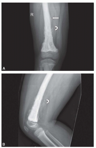
The classic high-grade intramedullary osteosarcoma presents as a large, mixed sclerotic and lucent metaphyseal lesion with fluffy, cloud-like calcific opacities characteristic of osteoid matrix production on conventional radiography (Fig. 34.1). There is usually a soft tissue mass with aggressive-appearing periosteal reaction, often with a Codman triangle, which results from the soft tissue mass breaking through the overlying ossified periosteal new bone. The periosteal new bone may be spiculated or laminated. Tumors localized in the diaphyses may have onion-skin periosteal reaction similar to Ewing sarcoma. Occasionally, a classic high-grade intramedullary osteosarcoma is completely sclerotic or lucent. The telangiectatic subtype, typically presents as a cystic, lucent lesion with subtle matrix mineralization and a geographic margin with a wide zone of transition.18 These tumors may be confused with benign aneurysmal bone cysts. CT can be helpful in determining the correct diagnosis by showing subtle matrix mineralization and either CT or MRI may show thick, solid nodular, enhancing tissue surrounding the cystic spaces and the soft tissue mass that are present in a telangiectatic osteosarcoma.18 Juxtacortical osteosarcomas involve the bone surface and include intracortical, parosteal, periosteal, and high-grade surface lesions. Parosteal osteosarcomas are the most common of these and typically involve the metaphysis of the long bones. These tumors appear radiographically as lobulated masses that attach to the cortex and are ossified centrally. Occasionally, parosteal osteosarcomas must be differentiated from myositis ossificans, which is ossified in the periphery and usually not attached to the cortex. CT is particularly helpful for making this distinction.

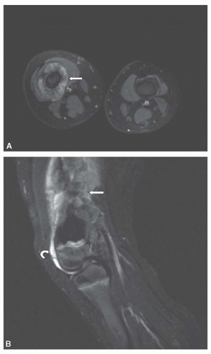
Once a malignant bone tumor is suspected, advanced imaging by CT or MRI should be performed to determine the extent of the tumor within the bone and assess the relationship of the tumor to the nearby neurovascular bundle, muscles, and joints. MRI provides superb contrast resolution and multiplanar capabilities, resulting in images that define the intraosseous and soft tissue components of the tumor and their relationship to adjacent structures (Fig. 34.2). CT is also able to define the intraosseous and soft tissue components of the tumor and is superior to MRI for the detection of small areas of mineralized matrix. A multi-institutional study19 found that CT and MRI were equally accurate for local staging without a statistically significant difference between the two imaging modalities for determining local tumor extent. However, MRI is considered the imaging study of choice based on its contrast resolution and the absence of ionizing radiation.20 Whether CT or MRI is performed for the assessment of the primary tumor, it is recommended that imaging be carried out prior to biopsy. The imaging findings can help in the selection of the optimal site for biopsy and this course of action will avoid distortion of the imaging findings by postbiopsy changes.21 Imaging of the primary tumor should include coverage of the entire bone to look for the presence of skip lesions. On CT, the extent of intramedullary involvement is determined by the presence of mineralization or soft tissue attenuation material replacing the low attenuation of normal fatty marrow. Intravenous contrast is critical to opacify and help establish the location of the blood vessels. On MRI, tumors are low signal on T1-weighted images and heterogeneous high signal on T2-weighted and STIR (short tau inversion recovery) images. T1-weighted images are more accurate than STIR images in assessing the extent of intraosseous tumor. STIR sequences are very sensitive to fluid and as a result, high-signal peritumoral edema or other benign tissue may simulate tumor (Fig. 34.2).


Figure 34.1 Plain radiograph of a patient with an osteosarcoma of the distal femur demonstrating a mixed lytic and sclerotic lesion with osteoid matrix (curved arrow) and Codman triangle (straight arrow). A PA film is shown in panel A and a lateral film in panel B.

Additional imaging is performed to detect metastatic disease, the majority of which is in the lungs. CT is superior to conventional radiography and is the imaging study of choice to detect
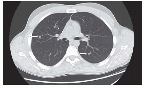
lung metastases.20 In cooperative patients, the CT scan should be performed using spiral technique during a single breathhold with 5 mm or less slice collimation. Unless there is chest wall, hilar, or mediastinal involvement, the CT should be performed without intravenous contrast. In addition, when possible, the CT should be done prior to biopsy to avoid postsedation atelectasis or other postoperative processes that could simulate or obscure lung metastases. Lung metastases are typically round or ovoid, sharply marginated, and located in the lung periphery (Fig. 34.3). The metastases may or may not be calcified and differentiating benign from metastatic nodules can be difficult, particularly in areas with endemic fungal disease, such as histoplasmosis.22 The size, number, and margins of the nodules can assist in making this distinction. Sharply marginated lesions larger than 5 mm in diameter, especially when multiple are considered to be possible metastases, while solitary nodules less than 5 mm with unsharp margins are thought more likely to be benign.23 Though there is no minimum number of nodules that excludes metastases, one study found that all patients with seven or more nodules at surgery had metastases.24 Following the size of pulmonary nodules, postchemotherapy can be helpful because benign lesions are likely to remain unchanged in size, while lesions that increase or decrease in size are more likely to be metastases.24 Criteria utilizing nodule size and number have been established to guide the decision (Table 34.1). In those uncertain cases where the diagnosis is crucial, however, nodule resection and biopsy is the only way to make the distinction. It is also important to note that there are times when granulomas and metastatic disease coexist in the same lesion and patient.25


Figure 34.2 MRI of the same patient. An axial view of a T2 fat saturated image (Panel A) and a sagittal view of a STIR image (Panel B) demonstrate the expansile lesion (straight arrow) in the distal femur with soft tissue extension (curved arrow).


Figure 34.3 Chest CT of a patient with osteosarcoma with multiple lung metastases (straight arrows).

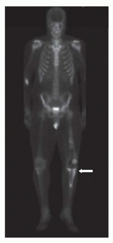
Bone scintigraphy, typically with technetium-99m-methylene-diphosphonate (MDP) is performed for the assessment of bone metastases and skip lesions (Fig. 34.4). Conventional radiographs are helpful to assess foci of increased uptake that are suspicious for bone metastases and occasionally MRI is needed to further evaluate these areas. Single-photon emission tomography (SPECT) may be performed in conjunction with planar bone scintigraphy to characterize uptake at the primary tumor site and when there is suspicion for lung metastases.26

Numerous other imaging techniques and modalities are being used or developed for use in the assessment of osteosarcoma. Thallium-201 has shown value in evaluating tumor response to chemotherapy, but has not gained widespread application. Similarly, DCE-MRI, which provides information on tissue vascularization and perfusion, capillary permeability, and the interstitial spaces, has shown promise for the assessment of tumor response and disease prognosis.27 Diffusion-weighted MRI sequences, which also can be performed in conjunction with standard MRI, are also being investigated for the assessment of tumor response.28 In addition, whole-body MRI has shown potential as a replacement for bone scintigraphy for the evaluation of bone metastases.29 Presently, however, FDG-PET and PET/CT seem to have the most potential for widespread application. FDG-PET is equal or worse than MDP-bone scintigraphy for the detection of bone metastases and worse than CT for the detection of lung metastases.30 FDG-PET, however, has shown promise in the assessment of tumor response and the detection of recurrent disease.31,32

As imaging techniques evolve and new techniques are developed, the basic imaging assessment for children with osteosarcoma will continue to rely on conventional radiography, MRI, CT, and bone scintigraphy. There is general agreement on the need for
MRI of the primary tumor at presentation and prior to local control and the use of bone scintigraphy and chest CT for the assessment of metastatic disease at presentation.3,33 There are differing opinions regarding the timing and types of imaging that should be performed later in the patients’ treatment course and during surveillance posttherapy.3,20,33 These differences will likely continue until there is objective evidence on the value of a standard protocol of imaging studies to improve the outcome of children with osteosarcoma.


TABLE 34.1 Criteria for Pulmonary Metastatic Disease from the EURAMOS I Trial


CT Chest Finding


Interpretation


One or more pulmonary/pleural Lesion(s) ≥1 cm OR three or more Lesions ≥0.5 cm maximum diameter


“Certain” pulmonary metastases


Fewer or smaller lesions


“Possible” metastatic disease


CT, computed tomography.


Figure 34.4 Bone scan of a patient with osteosarcoma. The patient’s primary osteosarcoma (straight arrow) is easily visualized. This patient did not have bone metastases.


Classification and Staging

Tumor extent (local and systemic) as well as the grade of the tumor must be taken into account when classifying and staging bone tumors. For many years, clinicians, particularly orthopedic surgeons, have relied on a staging system developed initially by Enneking and subsequently revised by the Musculoskeletal Tumor Society (MSTS), which recognizes these requirements. In the MSTS system, tumors are classified as either intra- (T1) or extracompartimental (T2) and as either low-grade (A) or high-grade (B) malignancies. Metastatic tumors are classified as T3.34 Since extension through the cortex to the periosteum (present in virtually all osteosarcomas) defines extracompartimental involvement, the MSTS staging system classifies the majority of localized pediatric and adolescent osteosarcomas as stage IIB (Table 34.2). The T-stages of the current 6th edition of the UICC TNM classification of malignant tumors,35 an advancement over the MSTS staging system, distinguishes between smaller and larger primary tumors, with a cutoff at 8 cm in the greatest dimension, and also allows for the description of skip metastases as T3 (see Table 34.3). The 6th edition of the UICC TNM classification also allows the distinction between pulmonary (M1a) and extrapulmonary (M1b) distant metastases. The resulting staging system is reminiscent of the original MSTS classification in that both grades of malignancy and local tumor extent are used for definitions.35


TABLE 34.2 Musculoskeletal Tumor Society (MSTS) Classification and Staging


Stage


T


N


M


Grade


Stage IA


T1


N0


M0


Low grade


Stage IB


T2


N0


M0


Low grade


Stage IIA


T1


N0


M0


High grade


Stage IIB


T2


N0


M0


High grade


Stage III


T3


N0


M0


Any grade


Stage IVA


Any T


N0


M1a


Any grade


Stage IVB


Any T


N1


Any M


Any grade


Any T


Any N


Any M


Any grade


T, tumor; N, node; M, metastases.


Regardless of the staging system, the vast majority of children and adolescents diagnosed with osteosarcoma will fall into two groups. First, those with high-grade osteosarcoma with tumor radiographically evident at the primary site and second, those with tumor radiographically visible at metastatic sites. Although the presence of radiographically visible metastatic disease has a major influence on prognosis, the chemotherapy typically used is similar. Indeed, virtually all patients with high-grade osteosarcoma and adequate organ function are treated with similar chemotherapy regimens which will be discussed further subsequently.


Biopsy

Although the index of suspicion of osteosarcoma can be exceedingly high based on the radiograph alone, a pathologic confirmation of that diagnosis is required. In other cases, the radiographic appearance may not be typical of osteosarcoma and a benign lesion may be suspected. As the routine management of osteosarcoma has evolved so that the resection of the entire tumor is typically performed after a period of neoadjuvant or induction chemotherapy, the initial surgical procedure will typically be an incisional biopsy, just acquiring a sample for diagnostic purposes. Incisional biopsies can be performed in different manners and include needle (closed) and open biopsies. Needle biopsies can be either fine needle or core. Indeed, the topic of whether the biopsy should be performed as a closed or open biopsy is immensely controversial. The clear advantage of a needle biopsy is that it can be performed in an interventional radiology suite under radiographic guidance or in an office setting with limited sedation, with prompt patient recovery, and at a reduced cost. The use of local anesthetic in children and adolescents for the purposes of a biopsy generally is not adequate. The clear advantage of an open biopsy is that much more tissue can readily be obtained. The debate as to what technique is preferred encompasses issues as to who performs the procedure in each setting, issues of operating room and hospital bed availability, patient discomfort and convenience, and defining how much tissue is needed to make the diagnosis.


TABLE 34.3 Sixth Edition of UICC TNM Classification of Malignant Tumors


T, N, M


Definition


Tx


Primary tumor cannot be assessed


T0


No evidence of primary tumor


T1


Tumor ≤ 8 cm


T2


Tumor > 8 cm


T3


Discontinuous tumors in the primary bone


Nx


Regional lymph nodes cannot be assessed


N0


No regional lymph node metastases


N1


Regional lymph node metastases


Mx


Distant metastases cannot be assessed


M0


No distant metastases


M1


Distant metastases


M1a


Lung


M1b


Other distant sites


T, tumor; N, node; M, metastases.


At one end of the spectrum, fine-needle biopsies are often the easiest procedure to perform but obtain the least tissue. Although some reports suggest that the accuracy of a fine-needle aspiration at diagnosing osteosarcoma may be as high as 90% in the hands of an experienced cytopathologist, it must be recognized that the radiograph alone in some osteosarcoma cases may have a similar specificity. Coupling classic radiographic findings to the presence of a malignant spindle cell on cytopathology may yield in aggregate sufficient diagnostic information.

Core biopsies allow visualization of the architecture of the tumor and tissue providing much more diagnostic material particularly when multiple cores are obtained. A risk of a core biopsy is that it enters the tumor deeply and can lead to bleeding and tumor contamination. Taking multiple cores provides additional tissue, but each additional core has a risk of compromising the area’s structural integrity. Needle biopsies, either fine needle or core, can also lead to a diagnostic delay. Permanent sections which may take several days to process can ultimately result in a specimen in which the diagnosis is indeterminate, which can occur in 25% to 30% of cases, even at experienced centers.

Open biopsies yield the most tissue and when performed by an experienced orthopedic surgeon, rarely cause complications. The debate on how much tissue is needed relates to the fact that the diagnosis of osteosarcoma is based on identifying a region of the tumor with the defining histologic appearance. Molecular analysis is typically used only to exclude other diagnoses as will be described further subsequently in the section on Pathology. Unlike other pediatric malignancies such as leukemia and neuroblastoma, biologic features of the tumors do not presently define prognosis or treatment.

Excisional biopsies are rarely performed in osteosarcoma as most of these tumors are large at presentation and not easily amenable to removal and reconstruction without significant planning. These types of extensive operations are usually not undertaken without establishing the biologic nature of the entity being removed as this heavily influences the desired surgical margins. It should be recognized that although induction chemotherapy is typical, it does not produce a survival advantage.36 In cases in which multiple biopsies obtaining large amounts of tissue do not yield a diagnosis, an excisional biopsy or pretreatment resection could be appropriate. In these cases, the surgical approaches and margins would need to appropriate for resection of an osteosarcoma. Curettage approaches used for benign bone lesions do not yield adequate surgical margins and potentially disseminate tumor locally. This can occur if a biopsy is not performed prior to excision in a lesion that is believed to be benign based on radiographic appearance and is discovered to be an osteosarcoma.

Regardless of the type of biopsy, its placement relative to the location of the tumor and the anatomic structures of the patient are also of critical importance. The location of the biopsy site is determined by a thorough prebiopsy assessment of the extent of local disease and its relationship to critical structures such as the neurovascular bundle. This must be determined on a case-by-case basis. It is strongly recommended that the biopsy be performed by the surgeon or at least by a team that is familiar with the individual who will be performing the definitive resection so that the biopsy tract can be ellipsed within the planned surgical incision. At the time of biopsy, the individual performing it must be familiar with orthopedic oncologic principles of flap development, coverage and even amputation, when definitive limb salvage is the proposed plan for a given bone tumor. Thus, the critical message is that a biopsy needs to be performed to make the diagnosis of osteosarcoma and this biopsy needs to be performed by an individual who is experienced in the diagnosis and surgical management of this entity.

EPIDEMIOLOGY


Incidence and Prevalence

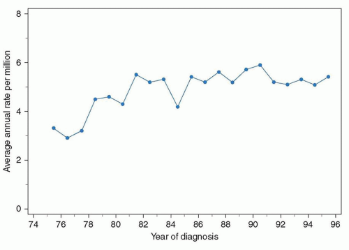
Osteosarcoma is the most common primary bone tumor in children.37 The average annual incidence of osteosarcoma in children younger than 20 years in the United States is 4.8/million (Fig. 34.5). Approximately 3% of all malignancies in this age group are osteosarcomas. Although it varies year to year, in 2000 there were 440 cases of osteosarcoma in children aged 0 to 19 years in the United States. There were an additional 135 cases in young adults aged 20 to 29. In the United States, osteosarcoma is slightly more common in African Americans, Hispanics, and Asian-Pacific Islanders. Several studies have reported that rates of osteosarcoma in black children younger than 14 years are about twice those in white children of the same age. In a report on osteosarcoma from the Surveillance, Epidemiology and End Results Program of the National Cancer Institute, United States blacks younger than 20 years continue to have a slightly higher rate of osteosarcoma.37 For the age group 15 to 29 years, the ethnic group with the highest rate of osteosarcoma is Hispanic. The relative risk of developing osteosarcoma for Hispanic children younger than 14 years of age is 1.3. Asian-Pacific Islanders have the highest reported rate of osteosarcoma in the age groups younger than 15 years and 30 to 44 years. However, the reported incidence in Hispanics and Asian-Pacific Islanders is likely to be inaccurate given the small absolute number of affected individuals and uncertainty in the denominator (i.e., the number of Hispanics and Asian-Pacific Islanders within each age group living in the United States).37 Internationally, incidence rates approximate the United States rate, with the exception of a higher incidence in some African countries. In Sudan and Uganda, the osteosarcoma incidence in children younger than 14 years is 5.3 and 6.4/million, respectively, compared with an incidence of between 2 and 3/million in this age group in the United States, Europe, and Asia.38

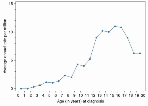
Several features characterize the epidemiology of osteosarcoma and have been taken as providing some clues as to its pathogenesis.39 Osteosarcoma occurs rarely in children younger than 5 years. After age 5, the incidence increases steadily, reaching a peak at age 15 years (Fig. 34.6). After the adolescent peak, the rate of osteosarcoma steadily declines, leveling off at a rate of 1 to 2/million. A second peak in incidence, approximately half the
magnitude of the adolescent peak, occurs in the sixth to seventh decade. Osteosarcoma in older patients is associated with Paget’s disease and prior radiation therapy, although approximately half of older patients with osteosarcoma have neither condition. The adolescent peak occurs at age 13 years in girls and between ages 15 and 17 years in boys. The age of peak incidence corresponds to the age of greatest growth velocity in each gender. This association contributes to the evidence supporting a role for growth in the pathogenesis of osteosarcoma. Osteosarcoma is slightly more common in males, particularly in the 15- to 19-year-old age group.39


Figure 34.5 Annual incidence of osteosarcoma in the United States in individuals less than 20 years of age between 1975 and 1995.

From Gorlick R, Meyers P, Osteosarcoma in Rudolph AM, Rudolph C, First L, Lister G, Gershon AA (eds.): Rudolph’s Pediatrics, 22nd Edition, New York, NY, Mcgraw-Hill, 2011.


Association with Growth

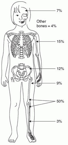
As already mentioned, the peak age for patients with osteosarcoma coincides with a period of rapid bone growth in young people, suggesting a correlation between rapid bone growth and the pathogenesis of osteosarcoma. Other evidence supporting this relationship includes its skeletal distribution. Although osteosarcoma can occur in any bone of the body, the majority occur in regions which undergo the most extensive longitudinal growth. Particularly bones around the knee and the proximal humerus are common sites in which osteosarcoma occurs (Fig. 34.7). Osteosarcoma typically occurs in the metaphyses of bones, which is the site where new bone arises from the growth plates. Greater height has been associated with a higher risk of osteosarcoma in some studies but has been refuted in other analyses and, therefore, remains somewhat controversial.40 A large study from the Children’s Cancer Group suggested that relationship with height did not exist, but many recent studies continue to suggest that taller individuals and those with earlier pubertal growth spurts are at higher risk for osteosarcoma. It has been hypothesized that this may be related to polymorphisms in growth-related genes, with an association with a vitamin-D receptor polymorphism found in a reported study.41 Other compelling evidence supporting the relationship of growth and osteosarcoma is the higher incidence of osteosarcoma in large dog breeds when compared with small breeds. In the context of canines, breed height variability can in large part be accounted for by variants in the insulin-like growth factor-1 allele.42 That said, the occurrence of osteosarcoma in the axial skeleton including the flat bones that undergo appositional bone growth rather than growth at the growth plate suggests that bone turnover or other etiologic factors may be important as well.43


Figure 34.6 Age-specific incidence rate of osteosarcoma in the United States between 1975 and 1995.

From Gorlick R, Meyers P, Osteosarcoma in Rudolph AM, Rudolph C, First L, Lister G, Gershon AA (eds.): Rudolph’s Pediatrics, 22nd Edition, New York, NY, Mcgraw-Hill, 2011.


Figure 34.7 Site of primary tumor in patients with osteosarcoma.

From Gorlick R, Persioglou M, Whelan J, Bone Tumours in Estlin E, Gilbertson R, Wynn R (eds.): Pediatric Hematology and Oncology, Hoboken, NJ, Wiley-Blackwell, 2010.


Other Etiologic Factors

Numerous other well-defined etiologic factors exist. One of the best-established etiologic factors is ionizing radiation. Although radiation exposure is a known risk factor, the interval between irradiation and osteosarcoma is typically long so this is not relevant to most pediatric patients. The excess risk of osteosarcoma associated with radiation exposure has largely been calculated on the basis of both those treated with therapeutic radiation as well as atomic bomb survivors.44 The excess risk has been found to be relatively linear and is approximately 1.8 for each gray of radiation exposure.45 The lack of a clear threshold for this malignancy as well as others has been part of the concern with regard to avoiding diagnostic radiation exposure in pediatric patients. This concern has manifested itself in efforts such as the Image Gently, Step Lightly campaign which seek to reduce unnecessary radiation exposure particularly in diagnostic CT scans.46


TABLE 34.4 Genetic Conditions Associated with Osteosarcoma Development


Hereditary Cancer Syndrome


Chromosome Location


Gene


Function


Percentage of Malignancies That Are OS


Retinoblastoma


13q14.2


RB1


Cell cycle regulation


50%


Li-Fraumeni


17p13.1


P53


DNA damage response


10%


Paget’s disease


18q21-q22


LOH18CR1


IL-1/TNF signaling


Not applicable


Rothmund-Thomson syndrome


5q31


SQSTM1


RANK signaling


5q35


MAPK8


18q24.3


RTS (ReQL4)


DNA helicase


30%


Werner syndrome


8p12-p11.2


WRN (RecQL2)


DNA helicase


<10%


Exonuclease activity


Bloom syndrome


15q26.1


15q26.1


DNA helicase


<10%


IL, interleukin; TNF, tumor necrosis factor; RANK, receptor activator of nuclear factor kappa-β.


The incidence of osteosarcoma is dramatically increased among survivors of retinoblastoma, particularly those with the hereditary form who harbor germ line mutations of the retinoblastoma gene. Patients with the hereditary form of retinoblastoma have a 19.8-fold excess risk of osteosarcoma, which makes osteosarcoma the most common secondary malignancy in this patient population. The rate of osteosarcoma in patients with unilateral sporadic retinoblastoma, generally lacking germ line mutations, is markedly lower and no higher than the general population.47 Germ line mutations in the p53 gene (the basis of the Li-Fraumeni syndrome) can lead to a high risk of developing malignancies including osteosarcoma. Patients with Rothmund-Thomson syndrome, an autosomal recessive disorder which includes a sun-sensitive skin rash, hair changes and congenital skeletal bone defects, particularly those with a RecQL4 mutation, are also at high risk of developing osteosarcoma.48 Patients with Werner and Bloom syndrome, both autosomal recessive disorders in DNA repair pathway genes, with the former associated with premature aging and the latter associated with short stature, are at a high risk of developing osteosarcoma among many other cancer predispositions.43 Other predisposing factors include a history of disorders of bone metabolism (i.e., Paget’s disease of the bone and fibrous dysplasia) (Table 34.4).

A viral etiology for osteosarcoma had been suggested on the basis of several lines of evidence. It had been suggested that contamination of poliovirus by SV40 (Simian virus 40) was an etiologic factor in osteosarcoma. It has subsequently been proven that SV40 is not a major epidemiologic factor in the development of osteosarcoma.49

Although a high proportion of individuals presenting with osteosarcoma are adolescents with primary tumors in the bones around their knees or in the proximal humerus, in the vast majority no clear etiologic factor can be identified.50 Given the potential association with germ line alterations, obtaining a careful history is essential with genetic testing reserved to those in whom a family history supports further study. Germ line alterations are rare in individuals with sporadic osteosarcoma. In initial encounters with the family members of a patient newly diagnosed osteosarcoma issues with guilt are common. It is important to reassure them that they have not contributed to the osteosarcomas development either through environmental exposures or a genetic contribution.


PATHOLOGY


Conventional Osteosarcoma

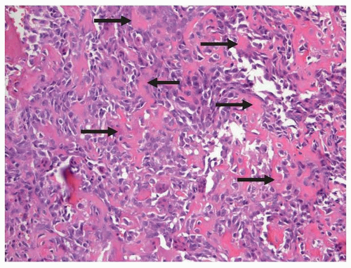
In the current WHO classification, conventional osteosarcoma is subdivided into osteoblastic, chondroblastic, and fibroblastic subtypes, on the basis of the predominant type of matrix.52 In chondroblastic osteosarcoma, the defining matrix is cartilaginous. The histologic distinction between chondroblastic osteosarcoma and chondrosarcoma may be difficult or impossible particularly in tumors with limited osteoid production and limited amounts of biopsy material. Distinction of these two entities is critical as their treatment and expected outcome are different. Fibroblastic osteosarcoma is composed of high-grade, malignant spindle cells with only scant osteoid. Giant-cell-rich and malignant fibrous histiocytoma-like osteosarcomas are also included under the fibroblastic subtype. The extreme paucity of matrix in fibroblastic osteosarcoma may result in a purely lytic appearance on imaging studies. In a manner analogous to the prior description of the chondroblastic subtype, this entity may be confused with fibrosarcoma which similarly is treated differently from osteosarcoma. Once the diagnosis of osteosarcoma is made, the value of subtyping conventional osteosarcoma is unclear, given that all of these entities are treated in the same manner and have the same prognosis. The treatment for osteosarcoma will be discussed further subsequently. Immunohistochemical stains are of little value in the diagnosis of osteosarcoma. Osteosarcoma is uniformly positive for vimentin. Osteocalcin and osteonectin, while usually positive, are of little value. Osteosarcoma may also show staining for CD99, smooth muscle actin, desmin, S100, cytokeratin, and epithelial membrane antigen. Molecular features similarly are not used to assist in making the diagnosis.52


TABLE 34.5 World Health Organization Classification of Osteosarcoma


Central (medullary) lesions


Conventional


Osteoblastic


Chondroblastic


Fibroblastic


Telangiectatic


Small cell


Low-grade central


Surface (peripheral) lesions


Parosteal


Periosteal


High-grade surface


Secondary


Figure 34.8 Conventional osteosarcoma demonstrating malignant cells associated with abundant deposition of osteoid (straight arrows). Nuclear pleomorphism is evident. (Hematoxylin and eosin.)


Other Osteosarcoma Variants

Telangiectatic osteosarcoma on imaging studies typically appear as a purely lytic, destructive tumor without peripheral sclerosis. Cysts with fluid-fluid levels are seen on radiographs suggesting an aneurysmal bone cyst. A high index of suspicion needs to be maintained for this entity as the surgical procedures typically performed for aneurysmal bone cysts would be inappropriate for osteosarcoma and may limit future surgical reconstructive options. Histologically, telangiectatic osteosarcoma is characterized by large, blood-filled spaces separated by variably thick septa correlating with its radiographic appearance. The septa are composed of anaplastic tumor cells admixed with benign-appearing multinucleated giant cells, bland mononuclear cells, and osteoid. When anaplastic cells and osteoid are inconspicuous, telangiectatic osteosarcomas may be confused with aneurysmal bone cysts.53 Telangiectatic osteosarcoma is a high-grade osteosarcoma treated identically to conventional osteosarcomas and despite historical controversy is generally believed to have the same prognosis.

Small cell osteosarcoma is composed of small, round cells with scant cytoplasm. All have osteoid, although this is often scant, and a few have foci of cartilaginous matrix. These tumors may show strong, diffuse membrane staining for CD99, and like other osteosarcomas may be positive for smooth muscle actin and cytokeratin. In biopsies with inconspicuous osteoid, small cell osteosarcoma may be mistaken for other small, round-cell tumors including Ewing sarcoma, mesenchymal chondrosarcoma, lymphoma, and metastatic neuroblastoma. Small cell osteosarcoma lacks the t(11:22) chromosomal translocations associated with Ewing sarcoma.54 Many oncologists advocate treating small cell osteosarcoma in a manner analogous to Ewing sarcoma. The limited number of cases and the varying manner in which it is treated create difficulties in assessing prognosis but historically, prognosis has been viewed as worse than conventional osteosarcoma.


Low-grade central osteosarcoma is rare particularly in children and adolescents. These osteosarcomas are called central because they arise in the intramedullary space and as such have a very different radiographic appearance as compared to surface lesions which will be described subsequently. Aggressive features may be subtle or absent on diagnostic imaging studies. Low-grade central osteosarcoma is composed of relatively bland spindle cells within fibrous stroma. Cellularity is low or moderate, and cytologic atypia is usually subtle. Histologically, these tumors may be confused with fibrous dysplasia.55 Low-grade osteosarcomas will progress and will recur locally if not adequately resected, but as would be expected from their low-grade descriptor, they do not metastasize. As such, the treatment of these tumors is surgical, with chemotherapy not part of standard management. As these tumors do not disseminate, the prognosis of patients with this diagnosis is better than those with conventional osteosarcomas. It is critically important that these tumors are resected completely as recurrent tumors can show a higher histologic grade or dedifferentiation.


Surface Lesions

Osteosarcoma can arise from the surface of the bone resulting in a variant radiographic appearance as compared to the more typical intramedullary osteosarcomas. The surface lesions, parosteal, periosteal and high-grade surface lesions are all exophytic tumors arising from the periosteal surface of the bone and eroding the underlying cortical bone with minimal or no involvement of the medullary bone. Patients with high-grade surface osteosarcomas should be treated in the same manner as patients with conventional osteosarcomas and patients with parosteal osteosarcoma should be treated in the same manner as those with low-grade central osteosarcoma. High-grade surface osteosarcoma is histologically and clinically indistinguishable from conventional osteosarcoma.

Parosteal osteosarcoma is rare before the third decade of life. This is a low-grade tumor analogous to the low-grade central osteosarcoma. Treatment is complete surgical resection and prognosis is excellent. The pathologist in diagnosing parosteal osteosarcoma needs to confirm that no foci of high-grade sarcoma is present as even if it is a minor portion of the tumor, it will behave in accordance with its most histologically aggressive component.56 Periosteal osteosarcoma is a rare form of osteosarcoma that has a predilection for the diaphyseal or metadiaphyseal regions of long bones. Histologically, this tumor has the features of an intermediate-grade chondroblastic osteosarcoma.56 As an intermediate-grade tumor, it has some risk for dissemination but this is markedly reduced as compared to conventional osteosarcoma. The successful treatment of these tumors always requires complete surgical resection. Extremely controversial is the use of chemotherapy. The patient with a periosteal osteosarcoma whose tumor is completely resected has a much higher likelihood of remaining disease-free without chemotherapy as well as it is unknown how effective chemotherapy is at reducing the risk of tumor appearing at metastatic sites. Institutional practices with regard to the chemotherapy management of periosteal osteosarcoma vary considerably.


BIOLOGY


Genetic Complexity

With the evolution of molecular techniques, osteosarcoma has been shown to be a complex tumor by genetic analyses, including conventional cytogenetics, spectral karyotyping, expression profiling and most recently, next-generation sequencing.57 Although many of the alterations present in the tumors have been clearly elucidated, the molecular complexity precludes placing the tumor’s pathogenesis into a simple conceptual framework.43 Osteosarcoma is defined as a clinical entity by the production of osteoid by the neoplastic cells. However, the production of bony matrix is a cellular behavior or a phenotype, not a genetic marker. Despite this phenotypic definition, the clinical behavior of high-grade osteosarcoma is relatively homogeneous. Unlike the majority of tumors arising in adults, osteosarcoma does not have an obvious multistep progression. Although most adult malignancies are epithelial in origin, some sarcomas common in adults also have a stepwise progression from benign enchondromas through high-grade chondrosarcomas as one example.58 Low-grade osteosarcomas cannot be identified as a precursor lesion in the majority of children, adolescents, or even adults that develop high-grade osteosarcoma. The equivalent of a premalignant dysplastic lesion or a carcinoma in situ is not known to exist for osteosarcoma, as is similarly the case for most pediatric malignancies. This fact coupled with the tumor’s molecular complexity and its phenotypic definition makes it extremely difficult to define the molecular features essential to its pathogenesis.59

Only gold members can continue reading. Log In or Register to continue

Stay updated, free articles. Join our Telegram channel

Aug 25, 2016 | Posted by in ONCOLOGY | Comments Off on Osteosarcoma

Full access? Get Clinical Tree

Get Clinical Tree app for offline access