Monoclonal Gammopathies of Undetermined Significance and Smoldering Multiple Myeloma


Monoclonal Gammopathies of Undetermined Significance and Smoldering Multiple Myeloma


S. Vincent Rajkumar

Robert A. Kyle

John A. Lust


INTRODUCTION


Nomenclature

Monoclonal gammopathy of undetermined significance (MGUS) is an asymptomatic, pre-malignant clonal plasma cell proliferative disorder.1, 2, 3, 4 It was initially referred to as essential hyperglobulinemia by Jan Waldenström, as well as several other terms such as benign, idiopathic, asymptomatic, nonmyelomatous, discrete, cryptogenic, and rudimentary monoclonal gammopathy; dysimmunoglobulinemia; lanthanic monoclonal gammopathy; idiopathic paraproteinemia; and asymptomatic paraimmunoglobulinemia.5, 6 However, because there is an indefinite risk of progression to multiple myeloma (MM) or related disorder such as Waldenström macroglobulinemia (WM) or amyloidosis (AL), the term MGUS is now the accepted nomenclature.1, 2, 7, 8 Smoldering multiple myeloma (SMM) is a clinically defined pre-malignant stage between MGUS and MM.9, 10 MGUS and SMM must be differentiated from MM, and from a number of related plasma cell disorders using the criteria listed on Table 97.1.8, 11


Detection of Monoclonal Proteins

Immunoglobulins consist of two heavy polypeptide chains of the same class and subclass and two light polypeptide chains of the same type. The various types of immunoglobulins are designated by capital letters that correspond to the isotype of their heavy chains, which are designated by Greek letters: gamma (γ) constitutes immunoglobulin G (IgG), alpha (α) is found in IgA, mu (µ) is present in IgM, delta (δ) occurs in IgD, and IgE is characterized by epsilon (ε). IgG1, IgG2, IgG3, and IgG4 are the subclasses of IgG; the subclasses of IgA are IgA1 and IgA2. Kappa (κ) and lambda (λ) are the two types of light chains. An intact immunoglobulin consists of two heavy chains of the same class and two light chains of the same type. A monoclonal increase in immunoglobulins results from a clonal process such as MGUS or MM, and a polyclonal increase in immunoglobulins is caused by a reactive or inflammatory process. The monoclonal immunoglobulin secreted by clonal plasma cells in MGUS, SMM, MM, and related monoclonal gammopathies is referred to as a monoclonal protein or M protein.


Electrophoresis and Immunofixation

Monoclonal proteins are detected using agarose gel or capillary electrophoresis of the serum and urine.12 An M-protein is usually visible as a localized band on protein electrophoresis, and as a tall narrow spike or peak in the β or γ region or, rarely, in the α2-globulin area of the densitometer tracing (Fig. 97.1A). A polyclonal increase in immunoglobulins produces a broad band or broad-based peak that migrates in the γ region. A suspected M protein on electrophoresis must be confirmed on immunofixation, which also determines the immunoglobulin heavy-chain class and its light chain type.13 In addition, immunofixation is also done when MM, WM, AL (light chain) amyloidosis, or a related disorder is suspected, because small M proteins may not be detected with electrophoresis alone. Immunofixation is performed using commercial kits or systems such as Sebia, or Pentafix (Fig. 97.1B).


Quantitative Immunoglobulins

In patients with detectable M proteins, another assay that aids in monitoring is quantitation of immunoglobulins performed with a rate nephelometer. It can accurately measure 7S IgM, polymers of IgA, and aggregates of IgG. However, levels of IgM obtained by nephelometry may be 1,000 to 2,000 mg/dl higher than those expected on the basis of the serum protein electrophoretic tracing. The quantitative IgG and IgA levels may be increased similarly.14


Serum Free Light Chain Assay

The serum free light chain (FLC) assay (FreeliteTM, The Binding Site Limited, Birmingham, UK) is an automated nephelometric assay that measures free kappa (κ) and lambda (λ) light chains that are not bound to intact immunoglobulin.15, 16 The normal serum free-κ level is 3.3 to 19.4 mg/L and the normal free-λ level is 5.7 to 26.3 mg/L.17 The normal ratio for FLC-κ/λ is 0.26 to 1.65. The normal reference range in the FLC assay reflects a higher serum level of free λ light chains than would be expected given the usual κ/λ ratio of 2 for intact immunoglobulins. This occurs because the renal excretion of free κ (which exists usually in a monomeric state) is faster than free λ (which is usually in a dimeric state).15, 16 Patients with a κ/λ FLC ratio <0.26 are considered to have a monoclonal λ free light chain and those with ratios >1.65 are defined as having a monoclonal κ free light chain. If the FLC ratio is >1.65, κ is referred to as the “involved” FLC and λ the “uninvolved” FLC, and vice versa if the ratio is less than 0.26.

The serum FLC assay can be used in place of urine protein electrophoresis and immunofixation in the initial screening algorithm for M proteins. In a study of 428 patients, Katzmann et al. found that urine studies can be eliminated by using the serum FLC assay in combination with the SPEP and immunofixation.18 However, if a monoclonal plasma cell disorder is identified on screening, a 24-hour urine collection followed by electrophoresis and immunofixation should always be done to aid in the assessment of disease progression and response to therapy over time. In addition to its role as a substitute for urine studies in the screening of plasma cell disorders, the FLC assay is used to predict prognosis in MGUS, SMM, AL, and solitary plasmacytoma.19, 20, 21

In addition, it is also used to monitor oligo-secretory MM, nonsecretory MM, light chain only form of MM, and AL amyloidosis.16, 22, 23, 24 In order to use the FLC assay to monitor disease progression, the baseline FLC ratio must be abnormal and the involved FLC level ≥100 mg/L.24, 25


MONOCLONAL GAMMOPATHY OF UNDETERMINED SIGNIFICANCE


Epidemiology


Prevalence

MGUS is the most common plasma cell proliferative disorder (Fig. 97.2). The prevalence of MGUS has been estimated in a large population-based study that included 21,463 of the 28,038 enumerated residents (77%) of Olmsted County, Minnesota, who were 50 years or older.3 MGUS was identified in 694 (3.2%) of these subjects. Age-adjusted rates were greater in men than in women, 4.0% versus 2.7% (P < 0.001; Fig. 97.3). The prevalence of MGUS was 5.3% among persons 70 years or older and 7.5% among those 85 years or older. Several other studies have reported similar prevalence estimates (Table 97.2).28 In addition, approximately 1% of the general population over the age of 50 have light chain MGUS.4

The incidence of M proteins is higher in blacks than in whites. In the study by Cohen et al., the prevalence of an M protein was 8.4% in 916 blacks and 3.6% in whites.29 Landgren et al., in a study of 4 million African American and white males admitted to Veterans Affairs Hospitals, found that the prevalence of MGUS was 0.98% in African Americans and 0.4% in whites.30 The age-adjusted prevalence ratio of MGUS in African Americans compared with whites was 3.0 (95% confidence interval, 2.7 to 3.3). The increase of MGUS in blacks may be related to genetic or environmental factors. A population-based study found that the increased risk of MGUS seen in African Americans was also seen in blacks in Ghana, suggesting that the racial disparity may be due more to genetic factors.31

Furthermore, a study of women in the southern part of the United States found that the racial disparity between blacks and whites persisted even after adjusting for socioeconomic status, again suggesting that the differences were more likely genetic rather than environmental.32


FIGURE 97.1. Monoclonal (M) protein on serum protein electrophoresis. Note tall, narrow-based peak of fast γ mobility (A). Immunofixation of serum with antisera to immunoglobulin G (IgG), IgA, IgM, κ, and λ shows a localized band with IgG and κ, indicating an IgG κ monoclonal protein (B).

One study found that only 2.7% of elderly Japanese patients had a monoclonal gammopathy.33 A subsequent population-based study in Japan found that the risk of MGUS was lower compared with the white population of Olmsted County.34


Incidence

The annual incidence of MGUS in males is estimated to be 120/100,000 at age 50, and rises to 530/100,000 at age 90 years.35 The rates for women are 60/100,000 at age 50, and 370/100,000 at age 90. The fact that the increased prevalence of MGUS with rising age is not just related to accumulation of new cases but due to an actual increase in incidence suggests that an age-related cumulative damage model is at play in the pathogenesis of MGUS.


Risk Factors

The incidence and prevalence of MGUS rises with age.3, 35 MGUS is also more common in males. Blacks have a higher risk of MGUS than whites as discussed above.29, 30, 31, 32, 36 In addition to age, race, and gender, there are other risk factors that have been identified, both genetic and environmental. First-degree relatives of patients with MGUS and MM have a two- to threefold higher risk of MGUS compared to those with no known affected relatives.37, 38, 39 Obesity and immunosuppression are also known risk factors for MGUS.32, 40, 41


FIGURE 97.2. Distribution of 1,733 cases of monoclonal gammopathy seen at the Mayo Clinic in 2011. MGUS, monoclonal gammopathy of undetermined significance.


Clinical Features

MGUS is an asymptomatic condition. It is typically detected as an incidental finding when electrophoresis and immunofixation of the serum and/or urine or the serum FLC assay are performed during the work-up of suspected MM or WM. Thus, MGUS is usually detected during the work-up of unexplained weakness or fatigue, increased erythrocyte sedimentation rate, anemia, unexplained back pain, osteoporosis, osteolytic lesions or fractures, hypercalcemia, proteinuria, renal insufficiency, or recurrent infections. MGUS is also detected during work-up of patients with symptoms suggestive of AL amyloidosis such as unexplained sensorimotor peripheral neuropathy, carpal tunnel syndrome, refractory congestive heart failure, nephrotic syndrome, orthostatic hypotension, malabsorption, weight loss, change in the tongue or voice, paresthesias, numbness, increased bruising, bleeding, and steatorrhea.

Most cases of MGUS remain undiagnosed due to the asymptomatic nature of the condition. At age 60, the proportion of prevalent cases that are clinically recognized is only 13%.35 This rate rises to 33% at age 80. When MGUS is first diagnosed, it is estimated that the condition has already been present in an undiagnosed form for a median duration of over 10 years.35

For example, it is estimated that 56% of women age 70 diagnosed with MGUS have had the condition for over 10 years, including 28% for over 20 years. Corresponding values for men are 55% and 31%, respectively.


TABLE 97.3 CLASSIFICATION OF MONOCLONAL GAMMOPATHY OF UNDETERMINED SIGNIFICANCE


Type


Risk of Progression


Non-IgM MGUSa


1% per year risk of progression to multiple myeloma, AL amyloidosis, or related disorder


IgM MGUSb


1.5% per year risk of progression to Waldenström macroglobulinemia; rare patients can progress to IgM multiple myeloma


Light chain MGUSc


Risk of progression to light chain myeloma and AL amyloidosis. Rate of progression not defined.


IgM, immunoglobulin M; MGUS, monoclonal gammopathy of undetermined significance


a Almost all patients are IgG or IgA type. Occasional patients may have IgD or IgE monoclonal proteins.

b Note that conventionally IgM MGUS is considered a subtype of MGUS. Thus, when the term MGUS is used, in general, it includes IgM MGUS.

c Because light chain MGUS was only defined in 2010, studies pertaining to MGUS prior to that time do not include patients with this entity; unless otherwise specified studies since then may also not include patients with light chain MGUS.


From Rajkumar SV. Preventive strategies in monoclonal gammopathy of undetermined significance and smoldering multiple myeloma. Am J Hematol 2012;87:453-454.


There are three distinct categories of MGUS each with a different mode of progression: IgM MGUS, non-IgM MGUS, and light chain MGUS, each with a different mode and risk of progression (Table 97.3).4, 7, 71


Prognosis


Mayo Clinic Referral Population

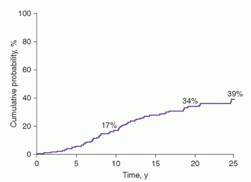
The prognosis of MGUS was first established in a study of 241 patients seen at the Mayo Clinic from 1956 through 1970.1 The actuarial rate of progression to MM or related disorder at 10 years was 17%; at 20 years, 34%; and at 25 years, 39% (Fig. 97.4).72 Of the 64 patients with progression, 44 (69%) had MM.


FIGURE 97.4. Actuarial analysis of incidence of multiple myeloma, macroglobulinemia, amyloidosis, or lymphoproliferative disease after recognition of monoclonal protein in 241 patients with monoclonal gammopathy of undetermined significance. (From Kyle RA, , Therneau TM, Rajkumar SV, Larson DR, Plevak MF, Melton LJ 3rd. Long-term follow-up of 241 patients with monoclonal gammopathy of undetermined significance: the original Mayo Clinic series 25 years later. Mayo Clin Proc 2004;79:859-866.)


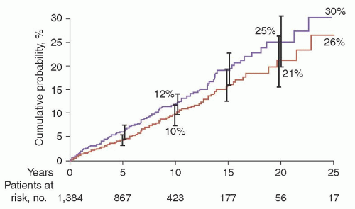
Southeastern Minnesota Study

The risk of progression has also been estimated in a larger population-based study of 1,384 persons with MGUS who resided in the 11 counties of southeastern Minnesota, the risk of progression of MGUS to MM or related disorder was found to be 1% per year.2 The median age at diagnosis of MGUS was 72 years. The M-protein level at diagnosis ranged from unmeasurable to 3.0 g/dl. On the basis of the heavy-chain type of immunoglobulins, 70% of the M proteins were IgG, 12% IgA, and 15% IgM. A biclonal gammopathy was found in 45 patients (3%). The light chain type was κ in 61% and λ in 39%. A reduction of uninvolved (normal or background) immunoglobulins was found in 38% of 840 patients in whom quantitation of immunoglobulins was determined. The 1,384 patients in this study were followed up for a total of 11,009 person-years (median, 15.4 years; range, 0 to 35 years). During follow-up, MM, primary AL, lymphoma with an IgM serum M protein, WM, plasmacytoma, or chronic lymphocytic leukemia developed in 115 patients (8%). The cumulative probability of progression to one of these disorders was 10% at 10 years, 21% at 20 years, and 26% at 25 years (
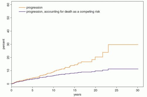
Fig. 97.5). Patients were at risk for progression even after 25 years or more of stable MGUS. Although the risk of progression is 1% per year, it must be emphasized that this does not take into account other competing causes of death in elderly patients. After adjusting for competing causes of death, the true lifetime probability of progression of MGUS for the average patient is only approximately 10% (Fig. 97.6).

The number of patients with progression to a plasma cell disorder (115 patients) was 7.3 times the number expected on the basis of the incidence rates for those conditions in the general population (Table 97.4). The risk of MM developing was increased 25-fold; WM, 46-fold; and AL amyloidosis, 8.4-fold. The risk of development of lymphoma was only modestly increased at 2.4, but this risk was underestimated because only lymphomas associated with an IgM protein counted in the observed number, whereas the incidence rates for lymphomas associated with IgG, IgA, and IgM proteins were used to calculate the expected number. The risk of development of chronic lymphocytic leukemia was only slightly increased.


FIGURE 97.5. Probability of disease progression among 1,384 residents of southeastern Minnesota in whom monoclonal gammopathy of undetermined significance (MGUS) was diagnosed from 1960 through 1994. The top curve shows the probability of progression to a plasma cell cancer (115 patients) or of an increase in the monoclonal protein concentration to more than 3 g/dl or in the proportion of plasma cells in the bone marrow to more than 10% (32 patients). The bottom curve shows only the probability of progression of MGUS to multiple myeloma, IgM lymphoma, primary amyloidosis, macroglobulinemia, chronic lymphocytic leukemia, or plasmacytoma (115 patients). The error bars indicate 95% confidence intervals. (From Kyle RA, Therneau TM, Rajkumar SV, et al. A long-term study of prognosis of monoclonal gammopathy of undetermined significance. N Engl J Med 2002;346:564-569. Used with permission.)


FIGURE 97.6. The risk of progression to myeloma or related disorder in 1,148 patients with monoclonal gammopathy of undetermined significance. The upper curve illustrates risk of progression of all patients without taking into account competing causes of death. The lower curve illustrates risk of progression after accounting for other competing causes of death. (From Rajkumar SV, Kyle RA, Therneau TM, et al. Serum free light chain ratio is an independent risk factor for progression in monoclonal gammopathy of undetermined significance (MGUS) Blood 2005;106;812—817. © the American Society of Hematology.)

The 75 patients in whom MM developed accounted for 65% of the 115 patients who had progression to a plasma cell disorder. The mode of development of MM among the patients with MGUS was variable. The M-protein level increased within 2 years of the recognition of MGUS in 11 patients, whereas the serum M-protein level was stable for more than 2 years and then increased within 2 years in 19 patients; in 9 others, the M-protein level increased gradually after having been stable for at least 2 years. In 9 patients, the M-protein level increased gradually during
follow-up until the diagnosis of symptomatic MM was made.

In 10 patients, the serum M-protein level remained essentially stable; the diagnosis of MM was unequivocal in these 10 patients because of an increase in bone marrow plasma cells, development of lytic lesions, or occurrence of anemia, renal insufficiency, or an increased level of urine M protein. Seventeen patients had an insufficient number of serum M-protein measurements to determine the pattern of increase. In patients with WM, the M-protein level showed a gradual increase in 3, stable levels were followed by a sudden increase in 2, and data were insufficient in 2.


TABLE 97.4 RISK OF PROGRESSION AMONG 1,384 RESIDENTS OF SOUTHEASTERN MINNESOTA IN WHOM MONOCLONAL GAMMOPATHY OF UNDETERMINED SIGNIFICANCE WAS DIAGNOSED, 1960-1994


No. of Patients


Relative Risk


Type of progression


Observed


Expecteda


(95% CI)


Multiple myeloma


75


3.0


25.0 (20-32)


Lymphomab


19


7.8


2.4 (2-4)


Primary amyloidosis


10


1.2


8.4 (4-16)


Macroglobulinemia


7


0.2


46.0 (19-95)


Chronic lymphocytic leukemiac


3


3.5


0.9 (0.2-3)


Plasmacytoma


1


0.1


8.5 (0.2-47)


Total


115


15.8


7.3 (6-9)


CI, confidence interval.


a Expected numbers of cases were derived from the age- and sex-matched white population of the Surveillance, Epidemiology, and End Results program in Iowa except for primary amyloidosis.

b All 19 patients had serum IgM monoclonal protein. If the 30 patients with IgM, IgA, or IgG monoclonal protein and lymphoma were included, the relative risk would be 3.9 (95% CI, 2.6-5.5).

c All 3 patients had serum IgM monoclonal protein. If all 6 patients with IgM, IgA, or IgG monoclonal protein and chronic lymphocytic leukemia were included, the relative risk would be 1.7 (95% CI, 0.6-3.7).


From Kyle RA, Therneau TM, Rajkumar SV, et al. A long-term study of prognosis of monoclonal gammopathy of undetermined significance. N Engl J Med 2002;346:564-569. (Used with permission.)


Spontaneous disappearance of M protein after the diagnosis of MGUS was rare.2 The M protein disappeared without an apparent cause in 27 patients (2%), and only 6 of these 27 patients (0.4% of all patients) had a discrete spike on the densitometer tracing of the initial electrophoresis (median, 1.2 g/dl); the rest had small M proteins detected on immunofixation only.


Follow-up in Other Series

The risk of progression of MGUS has been estimated in several other studies, and the results mirror those seen in the Southeastern Minnesota study. For example, Baldini et al. noted that 6.8% of 335 patients with MGUS had progression during a median follow-up of 70 months.73 In the Danish Cancer Registry, 64 new cases of malignancy (5 expected; relative risk, 12.9) were found among 1,229 patients with MGUS.74


Prognostic Factors

No findings at diagnosis of MGUS can reliably distinguish patients whose condition will remain stable indefinitely from those in whom MM or related malignancy develops. However, there are several known prognostic factors that assist in estimation of the risk of progression for appropriate counseling and management.


Size of M Protein

The size of the M protein at recognition of MGUS is one of the most important predictors for the risk of progression. In the study of 1,384 patients from Southeastern Minnesota, the risk of progression to MM or a related disorder 10 years after diagnosis of MGUS was 6% for patients with an initial M-protein level of 0.5 g/dl or less, 7% for 1 g/dl, 11% for 1.5 g/dl, 20% for 2 g/dl, 24% for 2.5 g/dl, and 34% for 3.0 g/dl.2

Corresponding rates for progression at 20 years were 14%, 16%, 25%, 41%, 49%, and 64%, respectively. The risk of progression in a patient with an M-protein level of 1.5 g/dl was almost twofold greater than that in a patient with an M-protein level of 0.5 g/dl, and the risk of progression in a patient with an M-protein level of 2.5 g/dl was 4.6 times that of a patient with a 0.5-g/dl spike.


Type of M Protein

Patients with an IgM or IgA M protein have a higher risk of progression compared with those with an IgG M protein.2 IgM MGUS is a unique subtype of MGUS in which patients are at risk of progression to WM rather than MM.75 Due to a confusion in terminology, some patients with WM are referred to as having non-Hodgkin lymphoma or lymphoplasmacytic lymphoma (a term commonly used by pathologists to describe the bone marrow findings of patients with WM). Rarely do patients with IgM MGUS evolve into IgM MM.71 Among 213 patients in the Southeastern Minnesota MGUS study, 23 developed “non-Hodgkin lymphoma” or WM, 3 developed chronic lymphocytic leukemia, and 3 developed AL amyloidosis.75 The risk of progression was 1.5% per year. The risk of progression of light chain MGUS relative to IgA, IgG, or IgM MGUS is not known.


Bone Marrow Plasma Cells

Cesana et al. have found that patients with MGUS who have 5% to 9% bone marrow plasma cells have a higher risk of progression compared with those with <5% bone marrow plasma cells.76 Of 1,104 patients with MGUS in this study, at a median follow-up of 65 months, 64 MGUS cases (5.8%) evolved to MM or related plasma cell disorder. Patients with greater than 5% marrow plasmacytosis had a significantly higher risk of progression compared to those with 5% or fewer plasma cells, 1.35 versus 0.64 per 100 person years, respectively, P = 0.004.


Abnormal Serum-Free Light Chain Ratio

An abnormal FLC ratio is an independent risk factor for progression of MGUS. In a study of 1,148 patients with MGUS, 379 (33%) had an abnormal FLC ratio.19 The risk of progression in patients with an abnormal FLC ratio was significantly higher than that in patients with a normal ratio (hazard ratio 3.5; P < 0.001) and was independent of the size and type of serum M protein (Fig. 97.7). The production of excess monoclonal FLC in MGUS may be a biomarker for plasma cells bearing a higher degree of cytogenetic abnormalities thereby serving as a biomarker for a higher risk of progression.


Risk Stratification

A risk-stratification model can be used to predict risk of progression in MGUS, and is useful for management.19 The model is based on the size and type of the M protein and the FLC ratio (Table 97.5). Patients with all three risk factors consisting of an abnormal serum FLC ratio, IgA or IgM MGUS, and an increased serum M-protein value (≥1.5 g/dl) have a risk of progression at 20 years of 58%, whereas the risk is 37% with any two risk factors present, 21% with one risk factor present, and 5% when none of the risk factors are present. When competing causes of death were
taken into account, the risk of progression in the low risk group is only 2% at 20 years.


FIGURE 97.7. Risk of progression of monoclonal gammopathy of undetermined significance (MGUS) to myeloma or related disorder. The top curve illustrates the risk of progression with time in patients with three risk factors: abnormal serum κ:λ free light chain (FLC) ratio (<0.26 or >1.65), a high serum monoclonal (M)-protein level (≥1.5 g/dl), and non-IgG MGUS. The second curve is the risk of progression in patients with any two of these risk factors. The third curve illustrates the risk of progression with one of these risk factors. The bottom curve is the risk of progression for patients with none of the risk factors. (From Rajkumar SV, Kyle RA, Therneau TM, et al. Serum free light chain ratio is an independent risk factor for progression in monoclonal gammopathy of undetermined significance (MGUS) Blood 2005;106;812—817. © the American Society of Hematology.)

Only gold members can continue reading. Log In or Register to continue

Stay updated, free articles. Join our Telegram channel

Oct 21, 2016 | Posted by in HEMATOLOGY | Comments Off on Monoclonal Gammopathies of Undetermined Significance and Smoldering Multiple Myeloma

Full access? Get Clinical Tree

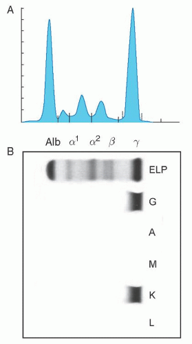
Get Clinical Tree app for offline access