(1)
Research Oncology, Guy’s Hospital, London, United Kingdom
Abstract
Examination and comparison of MBC and FBC at a molecular level reveals striking and potentially exploitable differences. Over 90% of MBC are ER+ve compared with approximately 70% of FBC. Androgen receptor mutations may be responsible for occasional MBC cases but for the majority no clear link has been shown. MIB1 is not of prognostic significance in MBC, nor is bcl2 expression. The molecular subtypes of MBC are predominantly luminal A, sometimes luminal B, rarely basal and very rarely HER2. Two MBC genomic subgroups have been described: male-complex and male-simple and the latter appears to be a male specific type. The Oncotype DX test may be useful in determining likely prognosis and suitability for chemotherapy in node negative MBC. Overexpression of cell cycle proteins such as cyclin-D and c-myc is associated with reduced lymphatic involvement and longer disease-free survival. Assembly of tissue banks of MBC will enable a greater understanding of the molecular profile and open the door to new specific therapies.
To see what is in front of one’s nose needs a constant struggle. George Orwell
Introduction
Instead of having to rely on morphological features demonstrated after staining with haematoxylin and eosin (H & E), molecular biology has yielded a panel of reagents for immunohistochemistry (IHC) to enhance the characterisation of tumours. This has not only improved diagnostic precision but also led to more sophisticated prognostic markers.
Estrogen and Progesterone Receptors
The pioneering work of Jensen which led to the identification of estrogen receptor (estrophilin) in 1960 enabled a rational approach to the endocrine treatment of breast cancer [1]. This was followed by O’Malley’s characterisation of the progesterone receptor in 1970 [2]. After being used widely to select therapies for FBC, Leclercq analysed 11 MBC samples, 7 primary and 4 metastatic for cytoplasmic estrogen receptors [3]. They measured the binding affinity of cytosol fractions for 3H-17βestradiol and the dissociation constants of binding were within the range reported for FBC. When present, receptor concentrations varied from 59 to 532 femtomoles/mg tissue protein. Competition studies indicated that the receptors were specific for estrogens and anti-estrogens suggesting that estrogen receptor (ER) was identical in MBC and FBC.
Rosen et al. assayed ER in specimens from 3 MBC cases and all were ER +ve with concentrations of 10, 16 and 105 fmoles/mg of cytosol protein [4]. Larger studies followed with Everson et al. reporting ER positivity in 29/34 (85%) of MBC cases [5]. Andres et al. investigated ER, PR, HER-2/neu and EGF-receptor status in 98 MBC specimens and found that 82 (84%) were ER+ve and 78 (80%) PR+ve [6]. The ER and PR protein levels were higher in males than females. In Cutuli’s large French MBC series ER was measured in 419 tumours and was positive in 385 (92%) and analysis for PR was positive in 356/399 (89%) [7]. Within the series the receptor phenotype was: ER+ve PR+ve (86%), ER+ve PR−ve (6%), ER−ve PR+ve (3%) and ER−ve PR−ve (5%).
Androgen Receptor
Part of our problem understanding the molecular biology of MBC is the absence of cell lines. There is in contrast a plethora of FBC established cell lines which have acted as substrates for extensive research and in particular, study of endocrine modulation of behaviour in vitro. The androgen receptor (AR) is activated by binding to testosterone or dihydrotestosterone and the complex translocates to the nucleus. Its role in MBC has been the subject of great interest and frequent disappointments.
In 1992, Wooster et al. reported two brothers with MBC and both had clinical and endocrine evidence of androgen resistance (Reifenstein syndrome) [8]. After sequencing they found a mutated AR gene within the region encoding the DNA binding domain on the X chromosome. Subsequently Lobacarro et al. screened 13 MBC tumours for the presence of germline mutations in exons 2 and 3 encoding the DNA-binding domain of the androgen receptor [9]. In one of these thirteen patients, single strand conformation polymorphism and direct sequencing detected a guanine-adenine point mutation at nucleotide 2185 that changed Arg608 into Lys in the second zinc finger of the androgen receptor. This mutation was found in a 38 year old male with partial androgen insensitivity syndrome but normal androgen-binding. The authors postulated that the androgen receptor mutation might invalidate the protective effect of androgens on male breast tissue.
Syrjäkoski et al. screened the entire coding region of the AR gene for mutations and also studied the role of repeat lengths of AR CAG and GGC in cancers from 32 Finnish MBC cases [10]. They did not find any germ-line mutations and CAG and GGC repeat lengths were similar in cases and controls so they concluded that the AR gene mutations were not a major influence on MBC risk.
Those studies measuring AR in MBC are outlined in Table 6.1 which indicates the heterogeneity of the findings [11–18]. The likelihood is that these differences are methodological but at present it is difficult to draw conclusions as to the relevance of AR expression in MBC.
Table 6.1
Frequency of AR positivity in MBC
Author | N | AR+ve |
|---|---|---|
Sasano 1996 [11] | 15 | 13 (87%) |
Rayson 1998 [12] | 77 | 73 (95%) |
Munoz de Toro 1998 [13] | 13 | 5 (39%) |
Pich 1999 [14] | 47 | 16 (34%) |
Kidwai 2003 [15] | 26 | 21 (81%) |
Kwiatowska 2003 [17] | 39 | 15 (38%) |
Murphy 2006 [18] | 16 | 14 (87%) |
Sas-Korczynska 2015 [19] | 32 | 20 (62.5%) |
Rayson et al. measured androgen receptors in 77 tumours from a cohort of 111 MBC patients treated at the Mayo Clinic between 1950 and 1992 at the Mayo Clinic and 95% of these were AR positive [12]. Because of this high positivity they were unable to assess whether AR had any influence on prognosis. In contrast, Kwiatkowska et al. reported that AR positivity was adversely associated with 5-year survival in a series of 43 MBC cases (AR+ve 33% versus 74% AR−ve [17]. Wenhui et al. provided corroborative data having measured AR, ER, PR, HER2 and Ki-67 (MKI67)) in specimens from 102 Chinese MBC cases [20]. High levels of AR expression were associated with axillary nodal spread and a significantly reduced 5-year overall By contrast. There was improved overall survival those AR-negative patients given adjuvant tamoxifen therapy.
In contrast, when Sas-Korczyska et al. performed androgen receptor assays in 32 specimens they reported that AR expression was present 20 (63%) and more frequently expressed in 17/20 (85%) of ER+ve tumours [19]. Tumours that were AR−ve were associated with a worse 5 year survival (30% versus 52%),
Johannson et al. analysed 56 fresh frozen MBC specimens using high-resolution tiling BAC arrays and compared the pattern of expression with a genomic data set of 359 FBC [21]. There was a broad spectrum of aberrations indicating the heterogeneity of MBC with genomic gains being more frequent in MBC compared with FBC but with fewer genomic losses of material. They suggested two MBC genomic subgroups called male-complex and male-simple. The male-complex type was similar to the luminal-complex FBC subgroup, whereas the male-simple appeared to be a male specific type. There are many similarities between FBC and MBC with respect to genomic imbalances, but also distinct differences as revealed by high-resolution genomic profiling. MBC can be divided into two comprehensive genomic subgroups, which may be of prognostic value. The male-simple subgroup appears notably different from any genomic subgroup so far defined in FBC.
Callari et al. surveyed the transcriptomic landscape of MBC and compared the gene expression profiles of 37 ER+ MBC biopsies with 53 ER+ FBC specimens of similar histology [22]. There were almost a thousand genes expressed differently in MBC and FBC suggesting that gender plays a major role in key functions including energy metabolism, translation regulation, and matrix remodelling together with immune system recruitment. Furthermore the analysis of genes associated with steroid receptors indicated the likelihood of a major role for AR in MBC with breast cancer being a very different phenomenon in male and females with the potential for exploitation of those differences for therapeutic purposes.
Ki67/MIB-1
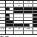
Ki67 is a monoclonal antibody which detects a nuclear antigen expressed in proliferating cells but can only be used on fresh frozen specimens. In contrast the monoclonal antibody MIB-1 which identifies recombinant components of Ki67 antigen can be used to measure proliferation in archival formalin-fixed and paraffin-embedded tissue. Pich et al. analysed 27 MBC specimens using MIB1 and allocated a score based upon the proportion of malignant cells staining with the antibody [23]. The mean MIB-1 score was 23.76% and staining was present only in the cell nuclei. There was no association between MIB-1 score and grade, stage, or ER / PR status. Those cases with a MIB-1 score ≤23.5% had a median survival of 73 months compared with 37 months for those with scores >23.5% (P = 0.01).
Wilsher et al. determined MIB-1 expression in 41 MBC and reported that 40% were positive [24]. Rayson et al. carried out IHC on 77 MBC specimens and taking a cut-off of ≥20% of cells staining, 48 (62%) were negative and 29 (38%) positive. The 5 year progression free survival was significantly worse in the MIB-1+ve cases, 48% versus 80% (p = 0.012). In contrast a series of 41 MBC cases from Kuwait were reported to have 100% Ki67 staining [25]. Wang-Rodriguez et al. examined tumour blocks from 65 MBC cases and used immunohistochemistry to determine ER, PR, p53, Her2-neu, and MiB-1 status [26]. As controls they used gynaecomastia specimens from 17 age-matched cases. Their threshold for MIB-1 positivity was >10.6% and on this basis 19 (29%) of the MBC were positive. All the gynaecomastia controls were MIB-1 negative. There was no relationship between MiB-1 expression and survival, and they concluded that MIB-1 expression was of limited value in MBC.
Kanthan et al. examined specimens from 75 cases of MBC for IHC expression of many variables including Ki67 and cyclin-D1 clinico-pathological variables such as tumor size, stage, nodal status and disease free survival (DFS) [27]. The MBC cases were predominantly MIB-1 negative. There was no relationship between MIB-1 status and tumour stage or disease-free survival leading the investigators to conclude that MIB1 does not play a major role in the behaviour of MBC. Kornegoor et al. carried out IHC on specimens from a large Dutch series of MBC, including MIB-1 among the panel of antibodies [28]. MIB-1 positivity was found in 24/131 (18%) of the tumours. Among the grade III cancers there was significant over-expression of MIB-1. There was no significant association between MIB-1 status and outcome.
Further confirmation of the lack of prognostic significance of MIB-1 status came from the work of Schildhaus et al. [29]. Of the 92 MBC tumour microarrays that they analysed 69 (75%) were negative. Although the Ki67 cases had a shorter median overall survival, 48 versus 102 months, this did not achieve statistical significance. Using a 20% cell staining threshold for positivity, Gargiulo et al. reported that 22/34 (65%) of MBC cases were MIB-1 positive [30]. Again, this was not significantly associated with survival. Despite sometimes varying percentages of MIB-1 expression, all the recent publications suggest that MIB-1 is not an important variable for determining prognosis in MBC.
Bcl2
Bcl-2 (B-cell lymphoma 2) is the product of the Bcl2 gene and is an anti-apoptotic protein. Weber-Chappuis et al. compared expression of tumour markers in 66 MBC and 190 histologically matched FBC [30]. There was a high percentage of bcl-2+ve tumours among the MBC. In the Mayo Clinic series of 111 MBC cases, there was expression of bcl2 in 104 (94%) [12]. Among the 41 MBC cases reported by Temmim there was bcl2 positivity in 32 (78%) [25]. After Abdel-Fatah et al. had shown that the combination of bcl2 and mitotic index identified significantly different prognostic groups in FBC [31], Lacle revisited this relationship in a series of 151 MBC [32]. Of the MBC cases, 142 (94%) expressed Bcl2 and this was unrelated to tumour size, grade or mitotic index. The combination of Bcl2/mitotic index was not a prognostic indicator for MBC.
Molecular Subtypes
Sorlie et al. examined patterns of expression of 534 intrinsic genes using hierarchical clustering in 115 female breast cancers [33]. Four groups emerged: luminal A (43%), luminal B (20%), HER2 (10%) and basal (46%). Subsequent analyses of MBC revealed a very different spectrum of molecular subtypes. In a large multi-centre investigation, Shaaban et al. examined the receptor profiles of tumours from 251 MBC and 263 FBC which had been matched by tumour grade, patient age, and nodal status [34]. The most common phenotype was Luminal A in both MBC and FBC. No luminal B or HER2 phenotypes were seen in MBC and basal phenotype was rare in both. Hierarchical clustering showed that whereas in FBC estrogen receptor alpha (ERα) clustered with progesterone receptor (PR); in MBC, the clustering was of ERα, estrogen receptor beta (ERβ) and androgen receptor (AR).
Further conformation came from the study by Kornegor et al. who analysed 134 cases of MBC by immunohistochemistry (ER, PR, HER2, EGFR, CK5/6, CK14 and Ki67) [28]. Of the cases, 75% were luminal A, 21% luminal B and the remainder were either basal type (4) or unclassifiable triple negative (1). Nilsson et al. reviewed tumours from 197 MBC patients and performed immunohistochemistry (IHC) on tissue microarrays and histological grading using conventional slides [35]. Most were ER positive (93%) and PR positive (77%) but only 11% were HER2 positive. Nottingham histological grade (NHG) III was seen in 41% and HER2 positivity in 11%.
Using IHC results to classify the tumours into molecular subtypes based on 5 biomarkers (ER, PR, HER2, CK5/6 and EGFR) revealed luminal A and luminal B in 81% vs. 11%. There were two cases of basal-like cancer but no cases of HER2-like tumour. There was no difference in breast cancer mortality between the luminal subgroups suggesting the prognostic impact of molecular subtyping in MBC differs from that in FBC.
The combined results of these series are summarised in Table 6.2 [28, 29, 35–39]. For comparison with FBC the data reported by Inwald et al. on over 4000 FBC cases from the Regensburg Cancer Registry are shown. There are major differences with luminal A being the predominant subtype in MBC with minimal numbers of basal cell types and no HER2 enriched subtype being seen in males.
Table 6.2
MIB1 status and outcome in MBC
Author
Stay updated, free articles. Join our Telegram channel
Full access? Get Clinical Tree
 Get Clinical Tree app for offline access

|
|---|


