Medical and Surgical Treatment of Peripheral Arterial Disease


Medical and Surgical Treatment of Peripheral Arterial Disease


Philip Joseph

Saurabh Kalra

Sonia Anand


Peripheral arterial disease (PAD) affects approximately 27 million individuals in North America and Europe, and its prevalence is increasing as life expectancy increases.1 Atherosclerosis is the most common cause of PAD, with symptoms primarily attributable to arterial stenosis and reduced blood flow. Nonatherosclerotic PAD is far less common, resulting from vasculitis, arterial vasospasm, or thromboembolism. Moreover, the development of PAD represents one manifestation of an atherosclerotic process, which in the majority of individuals affects the entire vascular tree. Consequently, patients with PAD are at an increased risk for coronary artery disease and cerebrovascular disease.

The diagnosis and management of PAD has evolved considerably over the past few decades. Advances have stemmed from improved diagnostic modalities as well as from new medical and surgical treatments, with a greater focus on overall cardiovascular risk reduction. Despite these improvements, the successful treatment of individuals with PAD remains a challenge and encompasses a multifaceted strategy involving lifestyle modification, pharmacologic therapy, and peripheral arterial interventions. This chapter provides an overview of the pathogenesis, epidemiology, and management of PAD related to the lower extremities.


PATHOGENESIS OF PAD AND LIMB ISCHEMIA


Atherosclerotic Plaque Development (see Chapter 89)

Briefly, atherosclerosis results from adverse interactions between vessel hemodynamics, blood elements, and the vascular intima.2 Covering the intimal surface of the vessels, the vascular endothelium regulates several important aspects of vascular homeostasis including vascular tone, leukocyte adhesion and migration, platelet function, and coagulation. Vascular homeostasis is achieved through several autocrine and paracrine factors mediated by endothelial cells. Nitric oxide is released by the endothelium, exerting vasodilatory, antiproliferative, and antithrombotic effects.3 Endothelial dysfunction develops in the presence of cardiovascular risk factors (such as hypertension, diabetes, smoking, and hyperlipidemic disorders) and results in the decreased availability of nitric oxide or an imbalance in vascular homeostasis favoring deleterious circulating factors. The development of endothelial dysfunction promotes atherosclerotic plaque development, hypercoagulability, and thrombosis. The nascent step in atherogenesis involves the endothelial mediated migration of low-density lipoprotein (LDL) from the blood plasma into the vessel intima.4,5 Subsequent oxidative stress and intimal inflammation lead to the increased expression of leukocyte adhesion molecules along the vascular surface. Once monocyte and T-lymphocyte adherence occurs, chemokines facilitate leukocyte migration into the vessel intima.6,7,8,9,10,11

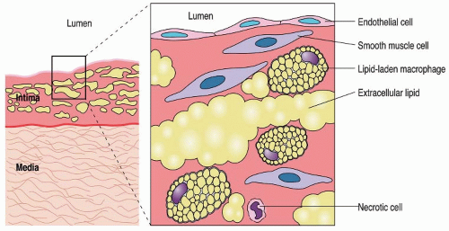
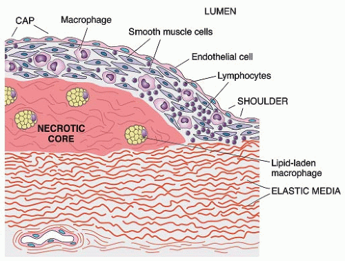
As monocytes differentiate into macrophages within the vessel intima, they internalize oxidized LDL via scavenger receptors and develop into foam cells. Macrophage colony stimulating factor has been shown to be present in human atherosclerotic lesions and hypothesized to promote macrophage replication within the intima.7 Foam cells coalesce to form early atherosclerotic plaques, commonly described as “fatty streaks” (FIGURE 94.1). Activated macrophages and T-lymphocytes cause further plaque progression through the release of inflammatory cytokines and the promotion of smooth muscle cell (SMC) migration and proliferation within the vessel intima. As foam cells, SMCs, and extracellular lipids accumulate, the atherosclerotic plaque continues to organize and grow within the intimal layer. Simultaneously, macrophage and SMC apoptosis within the plaque result in the development of a relatively acellular, collagen-based fibrous cap, which provides stability to the lipid core (FIGURE 94.2). With the continuation of these inflammatory, replicating, and apoptotic processes, the atherosclerotic plaque becomes more complex, creating an abundant extracellular matrix (comprised of interstitial collagen, proteoglycans, and elastin fibers) within the vessel wall, developing calcification, and undergoing neovascularization.7


Vascular Ischemia and Thrombosis

As the atherosclerotic plaque enlarges, an outward, “positive” remodeling phase initially predominates preventing luminal obstruction. However, luminal stenosis eventually develops as the enlarging plaque overcomes the vessel’s capacity to positively remodel. Moreover, proteolytic enzymes exuded from macrophages degrade and weaken the fibrous cap, creating a “vulnerable plaque” susceptible to rupture.12 With plaque rupture, the thrombogenic inner vessel becomes exposed to circulating platelets and coagulation factors, resulting in thrombosis. If thrombosis results in occlusion of the vessel, acute ischemia develops.7 However, repetitive, subclinical thrombotic events also occur; with subsequent healing, and fibrous tissue and collagen fiber formation, resulting in progressive plaque expansion and luminal stenosis.12 Thus, atherosclerosis progression may span many years without the development of symptoms. However, progressive obstruction of the arterial lumen eventually impedes blood flow through the affected vessel.


FIGURE 94.1 Fatty streak of atherosclerosis. The fatty streak, composed largely of foamy macrophages, is presumed to be an early stage in the formation of atherosclerotic lesions. Note the intimal thickening in the left panel and the infiltrating cells in the enlargement on the right. (From Rubin R, Strayer DS. Rubin’s pathology: clinicopathologic foundations of medicine, 5th ed. Philadelphia, PA: Lippincott Williams & Wilkins, 2008.)


EPIDEMIOLOGY OF PAD AND VASCULAR RISK FACTORS


Prevalence and Clinical Course of PAD

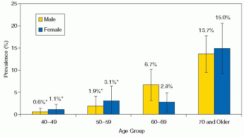
Both symptomatic and asymptomatic patients with PAD are at increased risk for adverse cardiovascular events. The prevalence of asymptomatic PAD varies with the age of the population studied, from 3% in 15,106 asymptomatic individuals between the ages of 45 and 60 years13 and 4.3% among a population >40 years of age14 to 29% of higher risk individuals over age 70 with cardiovascular risk factors.15 The prevalence of symptomatic PAD is 3% to 6% in the general population, also increasing with age (FIGURE 94.3).16


FIGURE 94.2 Fibrofatty plaque of atherosclerosis. In this fully developed fibrous plaque, the core contains lipid-filled macrophages and necrotic SMC debris. The “fibrous” cap is composed largely of SMCs, which produce collagen, small amounts of elastin, and glycosaminoglycans. Also shown are infiltrating macrophages and lymphocytes. (From Rubin R, Strayer DS. Rubin’s pathology: clinicopathologic foundations of medicine, 5th ed. Philadelphia, PA: Lippincott Williams & Wilkins, 2008.)

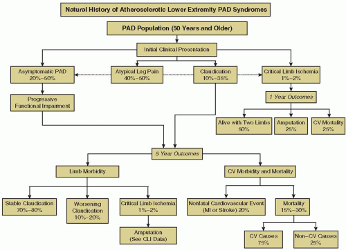
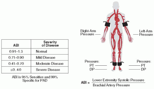
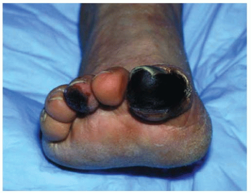
The natural history of patients with PAD varies widely depending on disease severity and progression of atherosclerosis, with symptoms ranging from intermittent claudication to critical limb ischemia (CLI) and limb loss (FIGURE 94.4). The majority of individuals with PAD have stable leg symptoms, with worsening claudication occurring in approximately 25% of patients,17 in association with diabetes, smoking, a low initial ankle-brachial index (ABI), and multiple stenotic lesions.18 CLI is the presenting symptom in 1% to 3% of PAD patients,17 and 5% to 10% of patients with previously stable PAD develop CLI over a 5-year period; risk factors being diabetes, advanced age, dyslipidemia, smoking, and an ABI of <0.5.16 In symptomatic individuals, only 1% to 3% require limb amputation over a 5-year period,17 but individuals with diabetes or those who present with CLI are at increased risk for limb loss.

Between 40% and 65% of patients with PAD are at increased risk for major adverse cardiovascular events (vascular-related death, myocardial infarction, or stroke),16,19 the annual event rate being 5% to 7% and mortality related to cardiovascular disease being the most common cause of death. Coronary artery disease accounts for 40% to 60% of patient mortality, cerebrovascular disease for 10% to 20%, and other vascular events for 10% of deaths associated with PAD.16


RISK FACTORS FOR THE DEVELOPMENT OF PAD

The risk factors for PAD are the same as those for other atherosclerotic disease processes, modifiable ones including smoking, dyslipidemia, diabetes, and hypertension. In the National Health and Nutrition Examination Survey (NHANES), 95% of patients had at least one modifiable cardiovascular risk factor and 75% of patients had at least two cardiovascular risk factors.14 Advanced age represents a nonmodifiable risk factor for PAD development.

Smoking causes several deleterious alterations in lipid metabolism, including reduced high-density lipoprotein (HDL) and increased LDL, triglyceride, and total cholesterol levels.20 In

addition, smoking directly influences atherosclerosis by causing endothelial dysfunction, inflammation, and coagulation within the vascular bed.21 Individuals who smoke are at risk for cardiovascular events, such as sudden cardiac death, nonfatal myocardial infarction, and ischemic stroke. Active smokers are at a 4.6-fold increased risk of developing PAD compared to nonsmokers,14 and smoking is associated with a threefold increased risk of CLI, with both PAD severity and progression directly correlated to the number of cigarettes consumed.


FIGURE 94.3 Prevalance of PAD in adults over 40 years by age and gender. (Reprinted from Selvin E, Erlinger TP. Prevalence of and risk factors for peripheral arterial disease in the United States: results from the National Health and Nutrition Examination Survey, 1999-2000. Circulation 2004;110:738-743, with Permission.)


FIGURE 94.4 Natural history of lower extremity PAD. Individuals may be asymptomatic or present with symptoms of leg pain or CLI. All individuals with PAD face a risk of progressive limb ischemia symptoms, increased cardiovascular ischemic event rate, and increased mortality. CV, cardiovascular; MI, myocardial infarction. (Reprintedfrom Hirsch AT, Haskal JZ, Hertzer NR, et al. ACC/AHA 2005 practice guidelines for the management of patients with peripheral arterial disease (lower extremity, renal, mesenteric, and abdominal aortic). Circulation 2006;113:e462-e654, with permission. ©2006 American Heart Association, Inc.)

Diabetes is a strong modifiable risk factor for PAD, associated with a 2.71-fold increased risk.14,22 In the Framingham registry, men were at a twofold increased risk for stroke in the presence of PAD and diabetes compared to either disease alone, and women with diabetes and claudication symptoms were at a three- to fourfold increased risk for coronary artery disease, stroke, and heart failure.22,23 The presence of diabetes signifies a more aggressive course of PAD, with diabetic patients at a 5- to 10-fold increased risk for major limb amputation compared to those without diabetes.16

Several mechanisms have been proposed to explain the aggressive nature of vascular disease observed in diabetics. Clustering of cardiovascular risk factors, such as hypertension and dyslipidemia, partly account for the increased cardiovascular event rate. Patients with diabetes frequently develop dyslipidemia secondary to insulin resistance, resulting in decreased HDL, with increased LDL and triglycerides.24 Significant deleterious effects on vascular biology also occur in the setting of insulin resistance and hyperglycemia.25 Increased oxidative stress and platelet activation occur in the setting of hyperglycemia, placing patients with diabetes at increased risk for vascular thrombotic events. Negative effects of insulin resistance on SMC proliferation and cellular apoptosis promote further atherosclerosis progression. The development of endothelial dysfunction can impair cellular glucose transport, potentially leading to further insulin resistance and worsening diabetes.26 Clinical studies support the causal relationship of hyperglycemia, insulin resistance, and accelerated atherosclerosis, as reflected by a direct correlation of cardiovascular disease with glycosylated hemoglobin levels in diabetic populations.27

Dyslipidemia: Patients with PAD demonstrate significant abnormalities in lipid metabolism; however, the associated risk appears to be lower compared to other vascular risk factors such as diabetes or smoking. In the NHANES registry, hypercholesterolemia (defined as a total cholesterol level 240 mg/dL [6.2 mmol/L]) was associated with a 1.68-fold increase in the prevalence of PAD.14 Abnormalities in total cholesterol, LDL, very low-density lipoprotein, and lipoprotein (a) levels are associated with PAD, while serum triglyceride levels are only modestly associated with PAD.28,29

Hypertension results in a 1.5- to 2-fold relative risk (RR) increase for PAD.14 Hypertension significantly alters vascular through increased endothelial dysfunction, loss of precapillary networks, and increased microvascular vasoconstriction in hypertensive animal models; all of which beget further increases in blood pressure, promoting both atherosclerosis and thrombosis.

Advanced age represents a risk factor for cardiovascular events and worse clinical outcomes in patients with established PAD. In the SMART registry of patients with symptomatic PAD, each 10-year increase in age was associated with a 1.85-fold risk increase in fatal and nonfatal vascular events.30

Genetic risk: The genetic contribution to PAD has not been clarified to the same extent as in other cardiovascular diseases. Several family studies suggest that PAD is in part related to genetic risk, estimating that genetic variation accounts for 21% to 48% of the interindividual variability in the ABI.31 Genome-wide association studies have enabled further insight into the role of common genetic variants in cardiovascular disease. The genetic epidemiology network of arteriopathy study demonstrated that PAD (identified as a reduction in the ABI) was modestly heritable in African American and white hypertensive populations. Single-nucleotide polymorphisms (SNPs) associated with PAD were observed in several loci, but no single SNP reached genome-wide significance (P ≤ 5 × 10⁻⁸).32 A common variation of the chromosome 9p21 allele has been associated with several forms of vascular disease, particularly myocardial infarction and stroke.33,34,35,36 In elderly individuals, Cluett et al.37 demonstrated a modest association between the 9p21 allele and PAD detected by ABI (odds ratio [OR] 1.29, 95% CI 1.06 to 1.56). Further studies are necessary to identify common genetic variants associated with PAD development and to clarify the role of genetic testing in risk stratification of PAD patients.


CARDIOVASCULAR RISK MODIFICATION


Antiplatelet Therapy

Platelet activation is a crucial component of vascular atherothrombosis and mediated by several compounds including thromboxane A2, adenosine diphosphate (ADP), serotonin, and norepinephrine.54 The antiplatelet therapies most extensively studied in PAD are acetylsalicylic acid (aspirin) and thienopyridines (clopidogrel and ticlopidine). Aspirin reduces platelet activation through the inhibition of cyclooxygenase, a crucial enzyme for the synthesis of thromboxane A2. Thienopyridines impair binding of ADP to the P2Y12 receptor, consequently inhibiting platelet activation through the ADP pathway.

Antiplatelet therapy is primarily utilized to reduce the risk of cardiovascular events in patients with PAD. A large meta-analysis by the Antiplatelet Trialists collaboration involving 287 studies examined the effects of antiplatelet agents on cardiovascular outcomes in patients with a history of vascular disease. In a subgroup analysis of 42 studies involving 9,214 patients with PAD, antiplatelet therapy (primarily aspirin, alone or in combination with dipyridamole) was associated with a 23% odds reduction in subsequent vascular events.55 In a recent meta-analysis of 18 randomized studies involving 5,269 patients with PAD, Berger et al.56 reported a significant reduction in nonfatal cerebrovascular events with aspirin therapy compared to placebo (RR 0.66, 95% CI 0.47 to 0.94). However, no significant differences were demonstrated for all-cause mortality, cardiovascular mortality, nonfatal myocardial infarction, or bleeding. Although numerous studies have reported significant improvements in various cardiovascular endpoints with aspirin therapy, recent evidence suggests that the overall cardiovascular benefits demonstrated with aspirin may not be applicable to very low-risk patients who have asymptomatic PAD without concomitant cardiovascular disease. A large, randomized control trial of 28,980 patients with asymptomatic PAD (diagnosed by an ABI of <0.95) failed to demonstrate a reduction in cardiovascular events in participants treated with aspirin 100 mg daily compared to placebo.57 However, the low overall mortality rate (1.9%) and mean ABI score (0.86) in the trial suggests a lower risk population compared to those evaluated in previous studies. In PAD patients undergoing revascularization, aspirin improves vessel patency after lower extremity bypass or percutaneous angioplasty. Overall, aspirin is recommended as first-line antiplatelet therapy for the majority of patients with a diagnosis of PAD.

Thienopyridines also benefit patients with PAD. The CAPRIE study showed an 8.7% RR reduction in vascular events with clopidogrel compared to aspirin therapy in patients with atherosclerotic vascular disease, including PAD.58 The greatest benefit was demonstrated within the PAD subgroup, achieving a 23.8% RR reduction in vascular events. Thienopyridines also improve symptoms related to PAD. In a randomized study of 151 PAD patients with intermittent claudication, Balsano et al.59 reported an improved ABI index, pain-free walking distance, and maximal walking distance with ticlopidine compared to placebo. Current guidelines support the use of clopidogrel as

an alternative antiplatelet agent to aspirin for cardiovascular risk reduction in patients with PAD. The development of severe neutropenia, a potentially fatal side effect, has been reported in 2.1% of patients prescribed ticlopidine, which has significantly limited its use in clinical practice.60


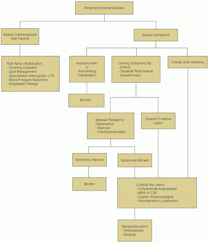
FIGURE 94.9 Overview of the management of patients with asymptomatic PAD, intermittent claudication, or CLI. (Adapted from Hiatt WR. Medical treatment of PAD and claudication. N Engl J Med 2001;344(21):1608-1621, with permission. © 2001, Massachusetts Medical Society. All rights reserved.)


Table 94.2 Treatment guidelines for cardiovascular risk reduction in patients with PAD


Treatment


Goals of Management


Smoking cessation


• Always address smoking cessation in PAD


• Both nonpharmacologic and pharmacologic methods may be utilized


Antiplatelet therapy


• Antiplatelet therapy should be considered in all PAD patients


• Aspirin (81mg daily) is the preferred antiplatelet agent


• Clopidogrel (75 mg daily) may be considered as an alternative


Lipid reduction


• LDL reduction to <2.59 mmol/L (100 mg/dL) in all PAD patients


• Consider further LDL reduction to <1.8-2 mmol/L (70-78 mg/dL)


• HMG coenzyme A reductase inhibitors (statins) are first-line therapy for treatment of dyslipidemia in PAD


Diabetes control


• Target HbA1C level of <7.0%


Blood pressure reduction


• Target blood pressure <140/90 in PAD


• Consider further reduction of blood pressure to <130/80 in patients with coexisting renal failure or diabetes


• Consider ACE inhibitor for blood pressure reduction and cardiovascular protection


PAD, peripheral arterial disease; LDL, low-density lipoprotein; HbA1C, hemoglobin A1C; ACE, angiotensin-converting enzyme.


The use of dual antiplatelet therapy to reduce secondary events in patients with established cardiovascular disease remains controversial. The CHARISMA study is the largest randomized trial which evaluated dual antiplatelet therapy with aspirin and clopidogrel in patients with known vascular disease or multiple vascular risk factors.61 Dual antiplatelet therapy did not reduce cardiovascular events compared to aspirin alone and was associated with a significant increase in bleeding events. Given the results of CHARISMA, there is currently insufficient evidence to support the routine use of dual antiplatelet therapy in patients with PAD without secondary indications (i.e., postmyocardial infarction or vascular stent implantation).


Anticoagulant Therapy

While a theoretical rationale exists for improved antithrombotic effects with the addition of an oral anticoagulation agent to antiplatelet therapy, studies examining the combination have failed to demonstrate a benefit and have shown increased bleeding. The WAVE study compared the combination of warfarin and antiplatelet therapy to antiplatelet therapy alone in patients with a history of PAD.62 Combination therapy did not reduce cardiovascular events and was associated with increased life-threatening bleeding. Combination therapy has also been studied for the prevention of graft occlusion in patients following peripheral bypass surgery with either venous or arterial grafts. In the Dutch BOA study, there were no significant differences in cardiovascular events or limb salvage with combination therapy compared to antiplatelet therapy alone in patients undergoing infrainguinal bypass.63 Although the Dutch BOA study reported fewer ischemic events with combination therapy in patients with venous grafts, other studies have not demonstrated similar results.64 Currently, the routine use of anticoagulants in addition to antiplatelet therapy is not recommended for routine use in patients with PAD to reduce cardiovascular risk or prevent graft occlusion.


Lipid-Lowering Therapy

As patients with PAD are a high-risk population for major cardiovascular events, aggressive lipid management is recommended. HMG coenzyme A reductase inhibitors (statins) are the principal agents for risk reduction in patients with vascular disease, with each reduction of 1 mmol/L of LDL with statin therapy associated with a 20% reduction in cardiovascular events.65 Large randomized control trials demonstrate profound benefits with statin therapy in patients with cardiovascular disease or vascular risk factors. The Heart Protection Study compared simvastatin 40 mg to placebo in 20,536 individuals with cardiovascular disease, including 6,748 patients with PAD.66 Simvastatin was associated with significant reduction in all-cause mortality (12.9% vs. 14.7%, P = 0.0003), primarily driven by a decrease in coronaryrelated death (5.7% vs. 6.9%, P = 0.0005). Moreover, aggressive lipid control provides further benefit compared to moderate lipid control in patients with cardiovascular disease. The Treat to New Targets study reported significant improvements in cardiovascular events with a reduction in LDL to <1.8 mmol/L (<170 mg/dL), compared to moderate LDL control in patients with coronary artery disease (RR 0.78, 95% CI 0.69 to 0.89).67 Current PAD guidelines recommend aggressive LDL reduction in PAD patients to <2.59 mmol/L (100 mg/dL).16 A number of lipid guidelines advocate for even lower LDL targets to <1.8 to 2.0 mmol/L (70 to 77 mg/dL) in patients with established vascular disease.65


Clinical studies suggest that statins may improve cardiovascular outcomes by reducing vascular inflammation, an important mediator of atherosclerosis progression. The PROVE-IT study reported significant reductions in C-reactive protein (CRP) levels with high-dose compared to low-dose statin therapy amongst 3,745 patients presenting with acute coronary syndromes who were followed over a mean period of 24 months. Patients with a reduced CRP had significantly fewer cardiovascular events compared to those with an increased CRP, regardless of LDL concentration.68 Complementary results were reported in the REVERSAL study, which examined the effects of high-dose statin therapy on atherosclerosis progression in CAD patients by coronary intravascular ultrasound.69 Participants who received high-dose statin therapy had reduced coronary atherosclerosis progression compared to those who received moderate-dose statin therapy, atheroma progression being directly related to changes in both LDL and CRP. Both PROVE-IT and REVERSAL provide support for the additional vascular benefits statins provide as potent mediators of vascular inflammation. Statin therapy also exerts positive effects on endothelial activity, likely mediated through increased endothelial nitric oxide synthase expression.70,71 Tsunekawa et al.72 demonstrated increased brachial artery flow-mediated vasodilation and plasma nitrate levels in prediabetic patients treated with statin therapy compared to placebo. Additional proposed vascular effects of statins include decreased oxidative stress through the downregulation of angiotensin II and NAD(P)H oxidase; and the stimulation of circulating endothelial progenitor cells which assist in ischemic tissue neorevascularization and repair.73 In addition to cardiovascular risk reduction, several studies have reported improvements in claudication symptoms and walking performance with statin therapy in patients with PAD.74,75,76

Individuals with PAD frequently have further disorders of lipid metabolism, such as reduced HDL and elevated triglycerides. Although epidemiologic studies have demonstrated that both HDL and triglyceride disorders are associated with increased cardiovascular events, there is limited evidence supporting targeted therapy to improve HDL or triglycerides in the current era of aggressive LDL lowering using statin therapy. Lifestyle modifications, including exercise and weight loss, have beneficial effects on both HDL and triglycerides. Niacin therapy can increase HDL up to 15% to 25%, and reduce LDL by an additional 20% in patients, and may be considered as a second agent in patients with persisting lipid abnormalities despite adequate statin therapy.65 Niacin improves surrogate markers of cardiovascular disease such as carotid artery intimal thickness, although whether this translates to a reduction in cardiovascular events in patients already receiving statin therapy remains uncertain.77 Fibric acid derivatives (fibrates) may be considered in patients with severe hypertriglyceridemia or those with persistently increased triglycerides despite statin therapy and lifestyle modification.


Blood Pressure Control

Aggressive blood pressure is recommended in all patients with vascular disease. The intersociety consensus for the management of PAD (TASC II) guidelines suggest that patients with PAD should have blood pressure controlled to <140/90, or <130/80, in the presence of diabetes or renal impairment.16 Weight reduction, limitation of alcohol consumption, and dietary changes are important lifestyle modifications which reduce blood pressure. In separate populations of overweight individuals, a weight reduction of 10 lbs resulted in a significant reduction in blood pressure ranging from 5 to 20 mm Hg.78,79,80 Maintenance of a normal body weight (BMI 18.5 to 24.9 kg/m2) should be targeted in all patients to improve blood pressure and reduce overall cardiovascular risk.80,81 Alcohol consumption should be limited to no more than 2 drinks per day in men, and no more than 1 drink per day in women.80 Dietary salt intake should be limited to 65 to 100 mmol per day in hypertensive individuals. Consumption of a diet rich in fruits, vegetables, and low-fat dairy products with limited saturated fat has been shown to lower blood pressure.80,82,83

Several classes of medications may be used alone or in combination for hypertension treatment in patients with PAD. Angiotensin-converting enzyme (ACE) inhibitors and thiazide diuretics are considered first-line therapies for hypertension management in PAD patients. Ostergren et al.84 evaluated the effects of ramipril in a subset of 3,099 patients with PAD enrolled in the HOPE study. Ramipril was associated with a 25% RR reduction in vascular events in individuals with clinical PAD, with similar benefits also demonstrated in those without clinical PAD but a reduced ABI. Long-acting calcium channel blockers and angiotensin receptor blockers (ARBs) can also be used as initial monotherapy for hypertension treatment. Despite previous concerns of worsening claudication symptoms with β-adrenergic blocking drugs, multiple studies have demonstrated their safety and effectiveness in patients with PAD.16 However, the antihypertensive effects of β-adrenergic blocking drugs are diminished in patients >60 years of age. ACE inhibitors and ARBs should not be routinely used in combination due to the increased risks of renal dysfunction and hyperkalemia.

Only gold members can continue reading. Log In or Register to continue

Stay updated, free articles. Join our Telegram channel

Jun 21, 2016 | Posted by in HEMATOLOGY | Comments Off on Medical and Surgical Treatment of Peripheral Arterial Disease

Full access? Get Clinical Tree

Get Clinical Tree app for offline access