Management of Metastatic Renal Cell Carcinoma


Management of Metastatic Renal Cell Carcinoma


▪ 45A VEGF-Targeted Therapy in the Management of Metastatic Renal Cell Carcinoma

Jorge A. Garcia

Brian I. Rini


INTRODUCTION

Prior to the identification of vascular endothelial growth factor (VEGF) as a therapeutic target in metastatic renal cell cancer (mRCC), the biologic response modifiers interferon-alfa (IFN-α) and interleukin-2 (IL-2) were the standard of care with an objective response rate (ORR) that ranged from 10% to 15%. These cytokines, however, provided only modest improvements in the median progression-free survival (PFS) and overall survival (OS) of mRCC patients compared to inactive therapy (1,2,3,4,5,6,7,8). Recent progress in understanding the biology of RCC led to the identification VEGF as a therapeutic target. VEGF is the most potent proangiogenic protein leading to increased tumor vasculature, cell proliferation, migration, and metastatic growth (9). Several studies using different strategies to inhibit the VEGF signaling pathway have demonstrated significant antitumor effects and meaningful clinical benefits. With the availability of various active agents, the management of patients with mRCC needs to be refined and thus current strategies are aimed to better identify timing of treatment, quality-of-life (QOL) issues, and the appropriate sequence or combination algorithm that can translate into maximum clinical benefit for patients.


VASCULAR ENDOTHELIAL GROWTH FACTOR AND RCC

VEGF also known as vascular permeability factor (VPF) or VEGF-A is a highly conserved, disulfide-bonded homodimeric glycoprotein and the founding member of a family of closely related cytokines with critical importance in both normal and tumor-associated angiogenesis and lymphangiogenesis (10). VEGF is a member of the platelet-derived growth factor (PDGF) superfamily of growth factors that includes VEGF-B, VEGF-C, VEGF-D, VEGF-E, and placenta growth factor (PlGF). Although originally described as a tumor-secreted protein that increased microvascular permeability to plasma proteins, further studies demonstrated its ability to mediate induction of endothelial cell division and migration (11,12), promotion of endothelial cell survival through protection from apoptosis (13), and reversal of endothelial cell senescence (14). The human VEGF gene, located on the short arm of chromosome 6, is organized as eight exons separated by seven introns and is differentially spliced to encode four major polypeptides (VEGF121, VEGF165, VEGF189, and VEGF206) (15). These isoforms have distinct physical properties relevant to their ability to bind heparin and extracellular matrix, but identical biologic activities when free in solution (16). VEGF exerts its biologic effect through interaction with transmembrane tyrosine kinase receptors that are selectively expressed by vascular endothelium. These receptors include VEGFR-1 (Flt-1) and VEGFR-2 (KDR/Flk-1) (17), selectively expressed on vascular endothelial cells; VEGFR-3 (Flt-4), expressed on lymphatic and vascular endothelium (9); and the neuropilin receptor (NRP-1), expressed on vascular endothelium and neurons (18). Upon binding of VEGF to the extracellular domain of the receptor, dimerization and autophosphorylation of both receptor tyrosine kinases occur and a cascade of downstream proteins are activated. While specific receptor functions are not well defined, VEGFR-2 (KDR/Flk-1) appears to be the main receptor responsible for microvascular permeability and endothelial cell proliferation and migration (19).


IMPORTANCE OF VEGF EXPRESSION IN RENAL CELL CANCER

Several series have demonstrated that the vast majority of patients with clear cell RCC have overexpression of VEGF in tumor tissues, as demonstrated by the level of mRNA transcripts and VEGF protein identified in RCC tumor tissue (20,21,22). Inactivation of the von Hippel-Lindau (VHL) tumorsuppressor gene which leads to deregulation of HIF almost certainly accounts for the activation of transcription genes such as VEGF and other genes relevant to RCC biology and therapy including PDGF (23), basic fibroblast growth factor (bFGF) (24), erythropoietin (25), and transforming growth factor alpha (TGF-α) (26,27). The biology of RCC has been extensively reviewed in previous chapters.


CLINICAL IMPACT OF VEGF EXPRESSION IN RENAL CELL CARCINOMA

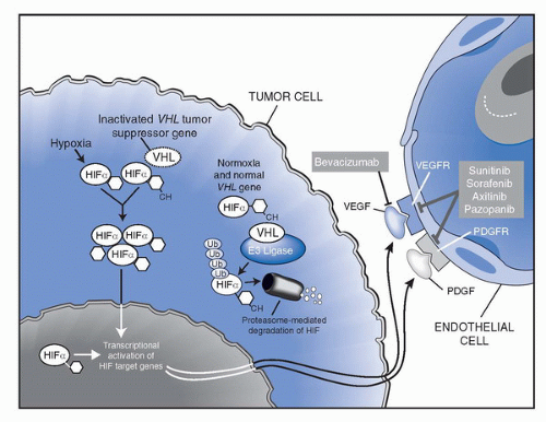
The biology of VEGF overexpression in RCC has led to the development of several novel therapeutic strategies to inhibit different steps within this proangiogenic pathway required for tumor growth and proliferation. Figure 45A.1 summarizes current targets for anti-VEGF agents in RCC. Existing data regarding the efficacy of current VEGF-targeted agents approved by the US food and drug administration (FDA) for the management of mRCC patients are summarized in Table 45A.1.


BLOCKING THE VEGF LIGAND


Bevacizumab

A recombinant human monoclonal antibody against VEGF (bevacizumab, Avastin; Genentech, South San Francisco, California) binds and neutralizes all biologically active isoforms of
VEGF (28). The clinical utility of bevacizumab in mRCC was initially investigated in a randomized phase II trial in which 116 patients with mRCC with clear cell histology were randomized to receive placebo, low-dose (3 mg/kg) bevacizumab or high-dose (10 mg/kg) bevacizumab given intravenously every 2 weeks (29). All patients had prior disease progression despite at least one systemic treatment regimen; the vast majority (93%) had received prior IL-2. Groups were balanced using established prognostic factors (3). The study was designed to detect a twofold time to disease progression (TTP) increase with either dose of bevacizumab versus placebo, with results showing a 4.8 versus 2.5 months PFS favoring the high-dose bevacizumab arm versus placebo; (p < 0.001 by log-rank test). There were four partial responses, all in the high-dose bevacizumab arm (4/39; 10% ORR). Common toxicity included grade (G) 1/2 hypertension (HTN) and proteinuria, more commonly seen in the high-dose bevacizumab arm. All toxicities were reversible with cessation of therapy.


FIGURE 45A.1. The biology of RCC leading to VEGF production. Sites of action of VEGF-targeted therapies are illustrated. Bevacizumab is a VEGF ligand-binding antibody. Sunitinib, sorafenib, axitinib, and pazopanib are small molecule inhibitors of the VEGF receptor (VEGFR) and PDGF receptor (PDGFR) tyrosine kinases.

These results represented the first proof-of-concept of the importance of angiogenesis in RCC, and highlighted the possibility that VEGF blockade may result in a low ORR but could still lead to a delay in disease progression in mRCC. That is, tumor burden reduction not meeting RECIST-defined criteria for objective response (30) may translate into a prolongation of TTP. This phenomenon has also been observed with other VEGF-targeting agents described in this chapter and underscores the limitations of our ability to assess the antitumor effect and clinical benefit of a given drug by simple tumor measurements alone.


Bevacizumab-based Combinations


Bevacizumab and Erlotinib

The combination of bevacizumab with an epidermal growth factor receptor (EGFR) inhibitor became an attractive strategy in RCC after the recognition that one of the ligands for this receptor, TGF-α is often overexpressed in this disease (31). Subsequent in vitro studies also suggested that inhibition of the EGFR pathway could lead to downregulation of VEGF expression (32,33). Considering this biologic rationale, a phase II study evaluated the addition of erlotinib, an EGFR inhibitor, to bevacizumab in mRCC patients (34). Treatment consisted of bevacizumab 10 mg/kg intravenously every 2 weeks and erlotinib 150 mg orally each day. Sixty-three untreated (68%) and cytokine-refractory (32%) patients were enrolled. Fifteen (25%) patients had objective responses. The median PFS was 11 months. Treatment was generally well tolerated; only two patients discontinued treatment because of toxicity (skin rash). G1 or 2 skin rash and diarrhea were the most frequent treatment-related toxicities.

The additive or synergistic potential of this regimen was further evaluated in a multi-institutional randomized phase II trial of bevacizumab plus placebo versus bevacizumab plus erlotinib (35). This trial demonstrated similar response rates and PFS rates for the two arms (ORR: 13.7% versus 14%, respectively, and median PFS of 8.5 versus 9.9 months, respectively; p = 0.58). Thus, it is not apparent from this trial that adding an EGFR-targeting agent increases the clinical activity of VEGF-targeting approaches. Additional trials evaluating other EGFR inhibitors have also failed to demonstrate clinical benefit in mRCC (36,37,38).


Bevacizumab and IFN-α

The addition of an antiangiogenic agent to standard cytokines has also been explored. Two multicenter international studies have evaluated the clinical activity of bevacizumab plus IFN-α versus IFN-α plus placebo or IFN-α alone in untreated mRCC patients (39,40,41,42,43).

The AVOREN study was an International phase 3 trial that randomized 649 untreated mRCC patients to receive treatment either with IFN-α (Roferon; Roche, Basel, Switzerland) plus placebo or interferon plus bevacizumab (39). Patients were balanced using established prognostic factors (44), had predominant (>50%) clear cell histology, and had undergone a previous nephrectomy. Bevacizumab 10 mg/kg or placebo was administered intravenously every 2 weeks with no dose reductions permitted. IFN-α 9 MIU was administered three times per week as a subcutaneous injection. Dose reductions to either 6 MIU or 3 MIU were allowed. The study was designed
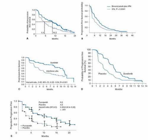
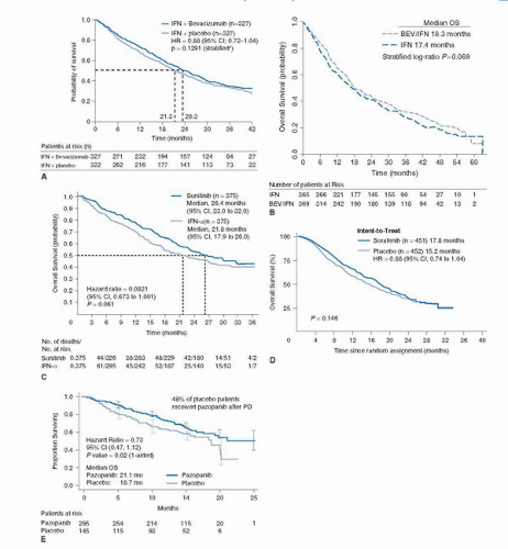
to detect an OS improvement from 13 to 17 months with PFS, ORR, and safety as secondary endpoints. Due to the change in standard of care and the availability of other active VEGF inhibitors that precluded reaching the anticipated OS endpoint, the study was amended and unblinded at the time of final PFS analysis. The median PFS observed was 10.2 months in the bevacizumab plus IFN-α group, compared with 5.4 months in the control group (HR 0.63, 95% CI 0.52-0.75; p = 0.0001) (Fig. 45A.2A). The improvement of PFS was largely confined to good- and intermediate-risk patients. (Good risk: 12.9 vs. 7.6 months; intermediate risk: 10.2 vs. 4.5 months; poor risk: 2.2 vs. 2.1 months.) A significant ORR difference was also observed in favor of the bevacizumab-treated patients (31% vs. 13%; p < 0.0001). The final median OS was 23.3 months in the bevacizumab arm compared to 21.3 for the IFN-α plus placebo-treated arm (HR 0.86, 95% CI 0.72-1.04; stratified log-rank test p = 0.1291) (43) (Fig. 45A.3A).


TABLE 45A.1 RANDOMIZED PHASE III DATA OF VEGF INHIBITORS IN MRCC


Therapy


Trial Arm


n


Setting


Patient Risk


ORR (%)a


Median PFS (months)


Median PFS by Risk Group (months)b


Median OS (months)


Bevacizumab (AVOREN)


IFN/Bevacizumab


327


Good and intermediate


31


10.2


Good risk: 12.9 vs. 7.6


23.3


Front-line


Intermediate risk: 10.2 vs. 4.5


IFN/Placebo


322


(9% poor)


13


5.4


Poor risk: 2.2 vs. 2.1


21.3


Bevacizumab (CALGB90206)


IFN/Bevacizumab


369


Good and intermediate (10% poor)


26


8.4


Good risk: 11.1 vs. 5.7


18.3


Front-line


Intermediate risk: 8.4 vs. 5.3


IFN


363


13


4.9


Poor risk: 3.3 vs. 2.6


17.4


Sunitinib


Sunitinib


375


Good and intermediate (6% poor)


47c (39)d


11


Good risk: 14.5 vs. 7.9


26.4


Front-line


Intermediate risk: 10.6 vs. 3.8


IFN


375


12c (8)d


5


Poor risk: 3.7 vs. 1.2


21.8


Sorafenib (TARGET)


Sorafenib Placebo


451
452


Cytokine-refractory


Good and intermediate


11
8


5.5
2.8


Not Available


17.8
15.2


Pazopanib


Pazopanib


290


Front-line and cytokine-refractory


Good and intermediate (3% poor)


30


9.2e (11.1)f


Not Available


21.1


Placebo


145


3


4.2e (2.8)f


18.7


a RECIST-defined. (Therasse P, Arbuck SG, Eisenhauer EA, et al. New guidelines to evaluate the response to treatment in solid tumors: European Organization for Research and Treatment of Cancer, National Cancer Institute of the United States National Cancer Institute of Canada. J Natl Cancer Inst 2000;92:205-216.)

b Memorial Sloan Kettering Cancer Center Risk Stratification. (Motzer RJ, Bacik J, Murphy BA, et al. Interferon-alfa as a comparative treatment for clinical trials of new therapies against advanced renal cell carcinoma. J Clin Oncol 2002;20:289-296.)

c Investigator assessment.

d Independent review.

e All enrolled patients.

f Treatment-naïve patients only.

gInterim analysis; prespecified design not met.


A second multicenter phase 3 trial, which was conducted in the United States and Canada through the Cancer and Leukemia Group B (CALGB 90206) (42), was nearly identical in design with the exception that it lacked a placebo infusion and did not require prior nephrectomy. This trial enrolled 732 untreated mRCC patients (369 to bevacizumab plus IFN-α and 363 to IFN-α alone). The primary endpoint of the study was to detect a 30% improvement in OS in patients randomly assigned to bevacizumab plus IFN-α compared to IFN-α monotherapy. The median PFS of the study was 8.5 months in patients who received bevacizumab plus interferon versus 5.2 months for patients who received interferon monotherapy (p < 0.0001). The hazard ratio for progression in patients who received bevacizumab plus interferon after adjusting for stratification factors was 0.71 (p < 0.0001) (Fig. 45A.2B). Moreover, among patients with measurable disease, the ORR was higher in patients who received bevacizumab plus interferon (25.5%) than in patients who received IFN-α monotherapy (13.1%; p < 0.0001). The median OS in this study was 18.3 months for bevacizumab-treated patients compared to 17.4 months for those receiving IFN-α alone (p = 0.069) (43) (Fig. 45A.3B).

The contribution of interferon to the antitumor effect of this regimen currently is unclear as neither study contained a bevacizumab monotherapy arm, precluding evaluation of the risk/benefit of the addition of cytokines. Similarly, the appropriate dose of IFN-α when given in combination
with bevacizumab remains unknown. Notwithstanding the fact that a significant percentage of patients receiving the bevacizumab-containing regimen in both phase 3 trials required dose modifications of IFN-α (41% AVOREN and 37% CALGB90206) a recent exploratory analysis of the European study would suggest that the improvement of PFS observed with the addition of the VEGF antibody to IFN-α appears to be maintained in spite of the need for IFN-α dose reductions (10.2 months with full dose vs. 12.4 months in patients who required a reduced dose of IFN-α) (41). Given the lack of dose response for interferon, it is possible that lower interferon doses in this combination can reduce toxic effects and preserve efficacy. Such a hypothesis requires prospective testing.


FIGURE 45A.2. PFS of VEGF-targeted therapy in phase III studies. Kaplan-Meier PFS probability curves by the existing VEGF inhibitors tested in a randomized phase III setting. A: Bevacizumab/INF versus IFN/placebo; B: Bevacizumab/IFN versus IFN alone; C: Sunitinib versus IFN; D: Sorafenib versus Placebo; E: Pazopanib versus Placebo.

The results of these studies have established bevacizumab-based therapy as an appropriate front-line treatment in untreated mRCC patients. Similarly, combinations of high-dose IL-2 and low-dose IL-2 plus bevacizumab are currently ongoing. These studies will further define the role of bevacizumab as front-line therapy in patients with mRCC.


FIGURE 45A.3. OS of VEGF-targeted therapy in phase III studies. Kaplan-Meier Median OS probability curves by the existing VEGF inhibitors tested in a randomized phase III setting. A: Bevacizumab/INF versus IFN/placebo; B: Bevacizumab/IFN versus IFN alone; C: Sunitinib versus IFN; D: Sorafenib versus Placebo; E: Pazopanib versus Placebo.


Bevacizumab/Small-molecule VEGF Receptor inhibitor

Despite the antitumor activity observed with bevacizumab both as monotherapy and in combination with IFN-α, not all mRCC patients respond; complete responses are rare and tumor resistance to the antibody is nearly universal. While bevacizumab leads to rapid clearance of circulating VEGF without affecting VEGF already bound to receptor, treatment with a small tyrosine kinase VEGF-receptor inhibitor can also lead to a compensatory circulating plasma VEGF increase leading to overexposure of tumors to VEGF. Thus maximal and continuous VEGF blockade with agents directed against both the VEGF ligand and receptor could result in an enhanced antitumor effect. Several phase I studies have evaluated the
combination of VEGF-R inhibitors with bevacizumab (44,45,46,47). A phase I study evaluated the maximum-tolerated dose (MTD) and safety of the combination of bevacizumab and sorafenib in 39 patients with advanced refractory malignancies (44). Among these, only three patients were mRCC. Treatment included sorafenib 200 mg orally twice daily and bevacizumab intravenously at 5 mg/kg (dose level [DL] 1 or 10 mg/kg [DL2]) every 2 weeks with a preplanned level 3 where sorafenib was escalated to 400 mg orally twice daily. These doses were below approved single-agent dose and selected because of concerns of potentiating toxicity. The most common adverse events (AEs) included HTN, hand-foot syndrome (HFS), diarrhea, transaminitis, and fatigue. A dose-limiting toxicity (DLT) of recurrent G2 HFS was noted in the first DL. The unexpected severity of toxicity seen prevented from full dose escalation suggesting that this combination may not be tolerable long term and therefore alternate sorafenib dosing schedules are currently undergoing evaluation (45). Recently, the results of two separate phase I trials evaluating the combination of bevacizumab plus sunitinib were reported (46,47). While one trial (46) evaluated the MTD and safety of the combination exclusively in mRCC (n = 26), the second trial (47) evaluated the same regimen in 38 patients with refractory solid malignancies. Among these, 16% (6/38) were mRCC. In both trials, treatment was administered in 42-day cycles, during which patients received oral sunitinib once daily from days 1 to 28 followed by 14 days off and bevacizumab intravenously every 2 weeks starting of day 0 or 1 of treatment. Despite differences in dose escalation design among the studies, the MTD of the combination was defined as 50 mg sunitinib and 10 mg/kg bevacizumab. Although antitumor effect was observed in mRCC and notably in other advanced solid tumors typically refractory to traditional chemotherapy, the common denominator for both trials was toxicity. In the exclusive RCC trial, the most frequently reported AEs (any grade [G]) for all patients included fatigue (92%), HTN (92%), proteinuria (88%), diarrhea (76%), hand-foot-skin reaction (72%), and bleeding (72%). The most frequently reported G3 to 4 AEs included HTN (60%), proteinuria (36%), and thrombocytopenia (24%). Similarly, in the broad phase I study, G3 or greater toxicity was observed in 82% of patients including most commonly fatigue (64%), HTN (51%), proteinuria (33%), thrombocytopenia (31%), and anorexia (26%). In both trials, the majority of G3 to 4 events occurred at the highest dose. A major toxicity concern was the finding of a microangiopathic hemolytic anemia (MAHA)-like clinical picture observed in five patients who have reached full dose of both sunitinib and bevacizumab. Two of these patients also developed reversible posterior leukoencephalopathy syndrome (RPLS). Although none of these patients required treatment with plamapheresis and their clinical symptoms and laboratory features reversed upon treatment discontinuation, this appears to be a severe toxicity when these two agents are combined in RCC.


SMALL-MOLECULE VEGF RECEPTOR INHIBITORS

An alternative approach to VEGF inhibition involves small-molecule tyrosine kinase inhibitors. These agents inhibit not only the VEGFR but also other receptors in the split kinase domain superfamily of receptor tyrosine kinases, including the PDGFR. PDGFR is expressed in pericytes, which serve as structural supporting cells for endothelial cells.


Sunitinib

Sunitinib (Sutent, Pfizer, Inc., New York, NY) is an orally bioavailable oxindole small-molecule tyrosine kinase inhibitor of VEGFR-2 and PDGFR-B. In vitro assays have demonstrated inhibition of VEGF-induced proliferation of endothelial cells and PDGF-induced proliferation of mouse fibroblast cells (48). Investigation in mouse xenograft models demonstrated growth inhibition of various implanted solid tumors and eradication of larger, established tumors.

Sunitinib has been investigated in two sequentially conducted single-arm multicenter phase II trials. Trial one enrolled 63 cytokine-refractory mRCC patients. The majority of patients (87%) had clear cell histology and 93% of patients had undergone cytoreductive nephrectomy. Patients were treated with 50 mg of sunitinib orally daily on a 4-weeks-on/2-weeks-off cycle. Overall, Twenty-five (40%) of 63 patients treated with sunitinib achieved a RECIST-defined PR. Median PFS was 8.7 months (95% CI 5.5, 10.7) and the median OS was 16.4 months (49). A confirmatory multi-institutional phase II trial enrolled 106 mRCC patients and required clear cell histology and prior nephrectomy (50). According to an independent third-party review, the ORR observed was 34% and the median PFS was 8.3 months. The results of these trials served as the basis for the US FDA approval of this compound in mRCC.

To evaluate the activity of sunitinib in previously untreated mRCC patients an international randomized phase III study was undertaken (51). Previously untreated mRCC patients (n = 750) with clear cell histology were randomized 1:1 to receive sunitinib 50 mg once daily, in 6-week cycles consisting of 4 weeks of treatment followed by 2 weeks without treatment or IFN-α as a subcutaneous injection three times per week on nonconsecutive days at 3 MU per dose during the first week, 6 MU per dose the second week, and 9 MU per dose thereafter. The primary endpoint was PFS (from historical control of 4.7 to 6.2 months). Secondary endpoints included ORR, OS, and safety. Health-related quality of life was also assessed with the use of the Functional Assessment of Cancer Therapy—General (FACT-G) and FACT-Kidney Symptom Index (FKSI) questionnaires (52,53). Patients were stratified according to baseline levels of LDH, ECOG performance status, and the presence or absence of nephrectomy. The ORR by investigator review was 47% in the sunitinib group (95% CI, 42%-52%) versus 12% in the IFN-α group (95% CI, 9%-16%; p < 0.001). Similarly, the median PFS by third-party independent review was 11 months versus 5 months in favor of sunitinib-treated patients corresponding to a HR of 0.42 (95% CI, 0.32-0.54; p < 0.001) (Fig. 45A.2C). After grouping patients according to MSKCC prognostic-risk criteria (3), the median PFS remain superior for patients treated with sunitinib than for those treated with IFN-image Sunitinib-treated patients had a greater median OS when compared with the IFN-α group (26.4 months; 95% CI, 23.0-32.9 months; vs. 21.8 months; 95% CI, 17.9-26.9 months, respectively; HR 0.821; 95% CI, 0.673-1.001; p = 0.051) (Fig. 45A.3C) based on the primary analysis of the unstratified log-rank test (p = 0.013 using the unstratified Wilcoxon test). By stratified log-rank test, the HR was 0.818 (95% CI, 0.669-0.999; p = 0.049). The benefit of sunitinib over IFN-α was observed in all mRCC patients regardless of their MSKCC risk classification. More than 50% of patients in both arms of this trial went to receive subsequent treatment with a VEGF-targeted agent including sunitinib, thus the lack of statistical significance observed in the prespecified OS analysis. The results of this trial have positioned sunitinib as a standard front-line therapy for mRCC patients.

The efficacy and safety of sunitinib administered to broad spectrum of mRCC patients was reported by Gore et al. (54). The study enrolled over 4,500 patients with previously treated and treatment-naive mRCC. Contrary to other existing sunitinib studies, this trial included 7% of patients with brain metastases (n = 321), 13% (n = 582) with ECOG performance status ≥2, and 13% (n = 588) with non-clear cell mRCC.
Similarly, over 30% of patients (n = 1,418) in the study were older than 65. Among all evaluable patients (3,464/4,564) the ORR observed was 17% (n = 603). Similarly, the ORR for patients with non-clear cell histology (n = 437) was 11%. The median PFS for the entire cohort was 10.9 months (95% CI 10.3-11.2) and the median OS was 18·4 months (17.4-19.2).

Although standard dose and schedule of sunitinib is effective and well tolerated, an alternative regimen of continuous once-daily dosing may offer added convenience and flexibility and might reduce the incidence and severity of AEs such as fatigue and nausea.

To test this hypothesis, the antitumor activity of continuous daily dosing of sunitinib was evaluated (56). A total of 107 cytokine-refractory mRCC were randomly assigned to receive continuous oral sunitinib at 37.5 mg daily in AM (n = 54) or PM (n = 53) dosing. More than 80% of patients have some degree of tumor burden reduction and 20% of patients achieved a confirmed RECIST-defined PR. The median PFS and OS for the entire cohort were 8.2 months (95% CI, 6.4-8.4 months) and 19.8 months (95% CI, 16.2 -24.9 months), respectively. Pharmacokinetics of this regimen appeared to be similar to those previously reported with the intermittent schedule (49,55). Therapy was well tolerated with neither dose reduction nor treatment delay required in the majority of patients. This dose and schedule appears to have comparable safety profile to the current recommended regimen of 50 mg/day administered on a 4/2 schedule (49). A large randomized trial comparing intermittent to continuous sunitinib dosing has recently completed accrual with results pending.

To study the role of sunitinib in the second-line setting for mRCC patients who have failed prior bevacizumab-based therapy a small (n = 61) phase II trial was conducted (56).

Tumor burden reduction was observed in 85% of patients including 14 patients (23%) who achieved a RECIST-defined PR. The median PFS was 30.4 weeks (95% CI, 18.3-36.7 weeks) and median OS was 47.1 weeks (95% CI, 36.9-79.4 weeks). In this study, prior response to bevacizumab did not predict for subsequent response or lack thereof to second-line sunitinib treatment. Similarly, none of the biomarkers evaluated (VEGF-A, sVEGFR-3, and PlGF) prior to initiating sunitinib treatment correlated with clinical activity. Although the precise mechanisms of response to sunitinib in bevacizumab-refractory mRCC has not been elucidated, this clinical activity supports the hypothesis that sunitinib inhibits signaling pathways involved in bevacizumab resistance, and provide support for continued targeting of the VEGF-VEGFR signaling pathway.


Sorafenib

Sorafenib (Nexavar, Bayer Pharmaceuticals, West Haven, Connecticut, and Onyx Pharmaceuticals, Emeryville, California) is an orally bioavailable bi-aryl urea Raf kinase inhibitor with demonstrated inhibition in Ras-dependent human tumor xenograft models (57). Activated Ras promotes cell proliferation through the Raf/MEK/ERK pathway by binding to and activating RAF kinase. Sorafenib has also demonstrated direct inhibition of VEGFR-2, VEGFR-3, and PDGFR-B, FMS-like tyrosine kinase 3 (Flt-3), c-Kit protein (c-Kit), and RET receptor tyrosine kinases (58, 59). Xenograft models treated with daily sorafenib have also demonstrated significant inhibition of tumor angiogenesis, as measured by anti-CD31 immunostaining (60,61).

Using a randomized discontinuation (or withdrawal) trial (RDT) design, Ratain et al. evaluated the effects of sorafenib in mRCC patients (62). This trial was initially designed to include patients with various refractory solid malignancies; however, early indications of the activity of this agent in mRCC patients permitted the investigators to focus the study on this patient population. Two-hundred and two patients with mRCC received oral sorafenib, 400 mg twice a day, and patients with stable disease after 12 weeks of treatment were randomized (double-blind) to either continue the drug or receive placebo. Patients initially received oral sorafenib 400 mg twice daily during the initial run-in period. After 12 weeks, patients with changes in bidimensional tumor measurements that were within 25% from baseline were randomly assigned to sorafenib or placebo for an additional 12 weeks, with a primary endpoint of the study of PFS after randomization. The majority of patients were low or intermediate risk, using the MSKCC prognostic factors (3), had received prior systemic therapy (84%), most commonly cytokine-based and an overwhelming majority (89%) had a prior nephrectomy. During the run-in phase of the study 73 patients had tumor shrinkage of ≥25%. Sixty-five patients with SD at 12 weeks were randomly assigned to sorafenib (n = 32) or placebo (n = 33). At 24 weeks, 50% of the sorafenib-treated patients were progression free versus 18% of the placebo-treated patients (p = 0.0077). Median PFS from randomization was significantly longer with sorafenib (24 weeks) than placebo (6 weeks; p = 0.0087).

On the basis of this study, The Treatment Approaches in Renal Cancer Global Evaluation Trial (TARGET), an international phase 3, randomized, double-blind, placebo-controlled trial of sorafenib versus placebo in cytokine-refractory mRCC was launched (63). In this trial, 903 patients with cytokine-refractory mRCC of clear cell histology were randomly assigned to receive either continuous treatment with oral sorafenib (at a dose of 400 mg twice daily) or placebo; 451 patients received sorafenib and 452 received placebo. The primary end point was to detect an OS improvement of 33.3% in favor of sorafenib treatment. All patients on trial had either favorable (52% and 50%, respectively) or intermediate-risk (48% and 49%, respectively) disease by MSKCC (3). Similarly, over 93% of patients in both arms of the study had undergone prior nephrectomy.

An independent review of the data demonstrated that while seven patients (2%) receiving sorafenib had a RECIST-defined response, 74% of sorafenib-treated patients had some degree of tumor shrinkage. The median PFS was 5.5 months in the sorafenib group and 2.8 months in the placebo group; (HR 0.44; 95% CI, 0.35-0.55; p = 0.000001). (Fig. 45A.2D). On the basis of this analysis, the data safety monitoring board (DSMB) halted the trial, leading to cross over of patients still on placebo to sorafenib and approval of sorafenib for the treatment of mRCC by the FDA. The first preplanned OS analysis was performed when almost half of placebo-treated patients had crossed over sorafenib. OS in the sorafenib group was not superior to placebo (17.8 vs. 15.2 months, respectively; HR 0.88; 95% CI, 0.74-1.04; p = 0.146.) (Fig. 45A.3D) although after censoring placebo-assigned patients at the time of cross over to sorafenib an improved survival in favor of sorafenib was observed (17.8 vs. 14.3 months, respectively; HR 0.78; 95% CI, 0.62-0.97; p = 0.0287) (64).

The Advanced Renal Cell Carcinoma Sorafenib (ARCCS) program, a nonrandomized, open-label expanded access program made sorafenib available to mRCC before regulatory approval. As reported (65), 2,504 patients with mRCC from the United States and Canada were enrolled and received sorafenib 400 mg orally twice daily. Safety and efficacy were explored overall and in subgroups of patients including those with no prior therapy (n = 1,254), non-clear cell histology (n = 202), brain metastases (n = 70), prior bevacizumab treatment (n = 290), and patients ≥70 years of age. Among patients evaluable for response (n = 1,891), 67 patients (4%) achieved a RECIST-defined PR, and 1,511 patients (80%) had SD for at least 8 weeks. The median PFS in the overall population was 24 weeks (95% CI, 22-25; censorship rate, 46%). Similarly,
the median OS in the entire cohort was 50 weeks (95% CI, 46-52; censorship rate, 63%). The efficacy and safety results were similar across the subgroups.

To investigate the clinical activity of sorafenib in the front-line setting, a multicenter, randomized, open-label, phase II trial was designed to compare the median PFS of 189 untreated mRCC patients randomly assigned to receive either sorafenib 400 mg orally twice daily or IFN-α-2a 9 million units three times weekly (66). At the time of PD, patients were allowed to continue on study in the so-called “Period 2” where those patients progressing on sorafenib were dose escalated to 600 mg orally twice daily and those on IFN-α-2a were allowed to receive sorafenib 400 mg orally twice daily within 14 days after the past IFN dose. The vast majority of patients had prior nephrectomy (94%), and all patients had clear cell histology and were either low or intermediate risk by MSKCC classification (3). There was no difference in the median PFS between both arms (5.7 months sorafenib vs. 5.6 months IFN-α-2a; p = 0.537) and similar response rates for arms were observed (5.2% sorafenib vs. 7.6% IFN-α-2a). Whereas 43/65 patients (66.2%) who progressed on sorafenib 400 mg twice daily had their dose escalated to 600 mg twice daily, all 50 patients who progressed on IFN-α-2a were switched to sorafenib 400 mg twice daily. Although no responses were observed, 41.9% of patients who had their sorafenib dose escalated to 600 mg twice daily had some degree of tumor burden reduction. Following crossover from IFN-α-2a to sorafenib, tumor burden reduction was observed in 76.2% of patients. Ten patients (20%) achieved a response (1 CR and 9 PR). The median PFS was 3.6 months for those who dose escalated sorafenib and 5.3 months for those who crossed over from IFN-α-2. Although limited by its sample size and phase 2 design, the results of this trial suggest that sorafenib is less robust a front-line agent in untreated mRCC patients.

Given the potential additive or synergistic effects of inhibiting VEGF-R in the setting of immunomodulation, two different trials evaluated the addition of sorafenib to IFN-α therapy in untreated mRCC patients. Gollob et al. (67) conducted a phase II study where 40 mRCC patients received treatment that consisted of 8-week cycles of sorafenib 400 mg orally twice daily plus IFN-α2b 10 million units subcutaneously three times a week. Although there were no restrictions with regard to histology and prior treatment of nephrectomy status, the vast majority of patients had clear cell histology (n = 35), were untreated (n = 25), and had undergone prior nephrectomy (n = 35). Among 36 pts evaluable for response, the ORR by RECIST criteria was 33% (28% PR, 5% CR). An additional 8% had ≥20% tumor shrinkage. The toxicity exceeded that of either drug alone and were mostly grade G1 and 2 fatigue (77%), anorexia (78%), rash (57%; 13% G3), diarrhea (75%), weight loss (60%), hypophosphatemia (36%; 37% G3), nausea (65%), neutropenia (26%; 25% G3), alopecia (60%), oral mucositis (48%) and HFS (10%; 10% G3). At the time of this report, the median PFS was 10 months (95% CI, 8-18 months), and median OS was not reached. A Southwest Oncology Group (SWOG) trial 0412 also evaluated this combination in 58 previously untreated mRCC patients (68). Sorafenib and IFN therapy were given at similar doses and schedule as first trial. Overall response rate was 19% (18% PR and 1% CR) and SD was observed in 39% of patients. The median PFS was 7 months (95% CI, 4-11 months) and the 6- and 12-month PFS rates were 53% and 37%, respectively. Toxicities were similar to those previously reported. Although these trials demonstrate the feasibility of combination therapy with sorafenib and cytokines in the current environment, it is unlikely that these type of combination regimens will be further developed. Currently several trials evaluating the combination of sorafenib with an oral mammalian target of rapamycin (mTOR) inhibitors are underway.


Pazopanib

Pazopanib (Votrient; GlaxoSmithKline, Research Triangle Park, North Carolina) is an oral angiogenesis inhibitor targeting VEGF-1, -2, and -3 receptors, PDGF-α and –β receptors, and c-kit (69). In vivo, pazopanib inhibited the growth of multiple human tumor xenografts in mice and bFGF- and VEGF-induced angiogenesis in two different mouse models of angiogenesis (70). After establishing the MTD and DLT of pazopanib in a phase I study of refractory solid tumors (71), Hutson et al. conducted a multicenter phase II trial to evaluate the efficacy and safety of pazopanib (800 mg orally daily) in 225 mRCC patients (72). This study was originally designed as a RDT, however, after planned interim analysis conducted after the first 60 patients completed 12 weeks of treatment demonstrated a response rate of 38%. Based on this activity and on recommendation by the independent DSMB, randomization was halted, and all continuing patients in the study were treated on an open-label basis. Patients enrolled in this study shared similar characteristics as those in other phase II RCC trials. Specifically, 91% of patients had a prior nephrectomy, 69% were treatment-naïve, and cytokines were the most common prior treatment received in remaining 31% of patients. According to MSKCC criteria, 43% of patients were favorable-, 41% intermediate-, and 2% poor-risk criteria.

The ORR observed was 35% (95% CI, 28%-41%) by independent review. This was similar regardless of previous treatment or not (37% vs. 34%, respectively). The estimated median PFS for the entire cohort was 45 weeks (95% CI, 36-59 weeks). Although the toxicity profile was similar to that seen with other small VEGF-R inhibitors, G3 AST and ALT elevation were noted in 6% and 4%, respectively, and have emerged as a somewhat unique side effect to this agent. A subsequent randomized, double-blind, placebo-controlled phase III trial that evaluated the efficacy (PFS, OS, ORR) and safety of single-agent pazopanib in treatment-naïve and cytokine-refractory mRCC patients led to the FDA approval of this compound in mRCC (73). Four-hundred thirty five patients were randomly assigned in a 2:1 ratio to receive Pazopanib (800 mg orally daily) or a matching placebo. The vast majority of patients had clear cell histology and prior nephrectomy, and 202 patients had received prior cytokine therapy. Similarly, over 90% of patients were either good or intermediate risk by MSKCC risk classification (3). Pazopanib significantly prolonged median PFS compared with placebo in the overall study population (9.2 vs. 4.2 months; HR, 0.46; 95% CI, 0.34-0.62; p < 0.0001), the treatment-naive subpopulation (11.1 vs. 2.8 months; HR, 0.40; 95% CI, 0.27-0.60; p < 0.0001), and the cytokine-pretreated subpopulation (7.4 vs. 4.2 months; HR, 0.54; 95% CI, 0.35-0.84; p < 0.001) (Fig. 45A.2E). Similarly, the interim analysis of OS result favored pazopanib over placebo (pazopanib 21.1 months vs. placebo 18.7 months; p = 0.02 [1 sided] HR = 0.73 [95% CI, 0.47-1.12]); however, these results did not cross the prespecified O’Brien-Fleming boundaries for either superiority or futility (Fig. 45A.3E). Most AEs were similar to those previously observed in the phase II study of pazopanib. The results of this phase III trial support the use of this agent as another standard of care in the management of mRCC patients entering front-line treatment.


ADDITIONAL VEGF INHBITORS WITH ACTIVITY IN RCC

Another promising agent in advanced RCC is AG013736 (axitinib; Pfizer, Inc., New York, NY), an orally bioavailable small molecule tyrosine kinase inhibitor of VEGFR-2 and PDGFR-B. In nonclinical and clinical studies, the compound
has been shown to inhibit angiogenesis, vascular permeability, and blood flow (74). Although axitinib inhibits PDGF receptors and KIT with nanomolar in vitro potencies, based on pharmacokinetic/pharmacodynamic analysis, axitinib acts primarily as a VEGFR tyrosine kinase inhibitor at the current clinical exposure. A phase II single-arm, multicenter trial of axitinib in cytokine refractory mRCC patients (n = 52) demonstrated an ORR of 44% (95% CI 30.5-58.7) (75). The median TTP was 15.7 months (8.4-23.4, range 0.03-31.5). Similar to other small molecule tyrosine kinase inhibitors, most toxicity was grade 1 or 2 and included gastrointestinal, dermatologic, fatigue, HTN, and proteinuria. To determine the activity of this agent in patients with VEGF-refractory mRCC, an additional study enrolled 62 patients who developed PD while on sorafenib treatment (76). Since the previous study indicates variable drug levels in patients (75), the axitinib dose was increased in a step-wise fashion from 5 mg twice daily to 7 mg twice daily and then to 10 mg twice daily, in the absence of predefined toxicities. Sixteen patients (25.8%) had received one additional prior therapy regimen, and 46 patients (74.2%) had been treated with at least two prior regimens. Approximately 20% of patients have received prior therapy with sunitinib. The ORR observed in the study was 22.6% and the median duration of response was 17.5 months (95% CI, 7.4 months to not estimable). Objective responses were observed regardless of the axitinib dose received. With a median follow-up of 22.7 months, median PFS for the entire study population was 7.4 months (95% CI, 6.7-11.0 months). The safety profile of axitinib in this trial was consistent with previously reported findings (74,75), although a higher incidence of HFS was noted. Currently, a randomized phase III trial evaluating this agent against sorafenib in patients with treatment-refractory disease has completed accrual with results pending.


TOXICITY OF VEGF-TARGETED THERAPY

Prior to the development of targeted agents in RCC, acute and long-term toxicities were often underappreciated as the main goal of treatment was to improve efficacy even at the expense of toxicity. VEGF inhibitors have significantly improved ORR and median PFS and OS in mRCC patients; however, most patients are required to continue on chronic therapy to maintain and maximize clinical outcome; thus, toxicity and QOL issues are more relevant than before. Existing data with VEGF inhibitors suggest that drug tolerability including type of AE, frequency, and severity is directly related to therapeutic target used (i.e., VEGF ligand vs. VEGF/PDGF-R vs. other non-VEGF targets). Available agents share similar characteristics in their ability to target VEGF; therefore, they share some common AEs such as HTN, cardiac, arterial, and venous thromboembolism, and minor bleeding events (39,42,51,63,73). Other toxicities including gastrointestinal, cutaneous, hematologic, and constitutional in nature appear to be related to the ability of some of these agents to block other non-VEGF targets such as PDGFR-b, c-KIT, Flt-3, RET (51,63,73), and RAF-1 (affecting the RAS-RAF-MEK-ERK signaling pathway) (63). While most of these AEs are National Cancer Institute Common Terminology Criteria (CTC) G1 or G2, a small but significant subset of patients (10%-20%) can develop G3 and 4 toxicities that often require dose modifications including a dose reduction, drug holiday, and even discontinuation of therapy (Table 45A.2). The most common G3 and 4 AEs observed in five different phase III trials evaluating different classes of VEGF inhibitors included HTN, declined left ventricular ejection fraction (LVEF), hand-foot-syndrome (HFS), constitutional and myelosuppression among others. While some of these AEs might not cause long-term complications, AEs such as HTN, decline in LVEF, bone marrow suppression, hypothyroidism, and the recently recognized MAHA-like syndrome and RPLS are of concern now that mRCC patients are living longer and continue receiving chronic therapy.


TABLE 45A.2 OVERVIEW OF COMMON TOXICITIES OBSERVED WITH VEGF INHIBITORS


Incidence (%)


Adverse Event


Bevacizumab/IFN (Avoren)


Sunitinib


Sorafenib


Pazopanib


Hypertension


26


24


17


40


Decline LVEF


<1


13


NR


<1


Hand-foot syndrome


NR


29


33


NR


Fatigue


33


54


29


19


Diarrhea


20


61


48


52


Stomatitis/mucositis


NR


30


5


NR


Nausea/vomiting


NR/NR


52/31


19/12


26/21


Anorexia


36


34


14


22


Skin rash


NR


24


41


NR


Hypothyroidism


NR


14


NR


NR


Neutropenia


7


34


77


7


Anemia


10


79


NR


22


Elevation AST/ALT


NR


53


NR


53


Elevation amylase/lipase


NR


35/56


NR


NR


Hypophosphatemia


NR


31


NR


34


Hyperglycemia


NR


NR


NR


41


Although there is no significant skin toxicity associated with bevacizumab (39,42), this AE is common among patients receiving therapy with sunitinib or sorafenib (51,63) and often is G1 and 2 in 17% and 40%, respectively. There is no skin rash reported with the use of pazopanib (73). Hand-foot skin reaction (HFS) characterized initially by tingling or burning with subsequent development of erythematous patches that primarily affects pressure-bearing areas on the fingers or the soles of the feet and progresses to hyperkeratosis after an average of 2 to 4 weeks is observed in 20% and 30%, respectively. Grade 3 or greater HFS observed in <6% of patients,
however, is often a cause for dose modification and even drug discontinuation. The underlying mechanism for developing HFS associated with sorafenib and sunitinib has not been determined; however, the focal occurrence of the skin lesions observed with sunitinib or sorafenib compared with the traditional HFS or palmar plantar erythro-dysesthesia (PPE) associated with classic chemotherapy suggests that blood vessels under pressure points may be more prone to damage due to the VEGF/PDGF-R inhibitory activity of these agents (77).

The frequency and severity of HTN in patients receiving a VEGF inhibitor for metastatic RCC is similar across existing phase III trials (39,42,51,63,73). Grade 1 and 2 HTN range from 13% to 36% and the incidence of G3 and 4 is in the range of 3% to 5%. HTN in patients receiving VEGF inhibitors often occurs during the first cycle of therapy and requires aggressive monitoring and management. Several retrospective series have investigated the incidence of HTN in patients receiving bevacizumab, sunitinib, or sorafenib (78,79,80,81). Data from 3,252 patients who received sorafenib in phase II trials and in an expanded access program showed all-grade HTN in 23.6% of patients and 6.5% with G3 or greater HTN (78). Due to concern for increased susceptibility for HTN due to previous nephrectomy or renal dysfunction, mRCC patients were compared with a meta-analysis of patients who received sorafenib for non-renal cell malignancies. The differences in the incidence of HTN between these patient populations were not significant with a relative risk (RR) of 1.03 (p = 0.89) for any grade HTN and RR of 1.23 (p = 0.40) for G3 or greater HTN. In these studies, the RR for developing HTN is 6.1. By comparison, a similar analysis of data from patients treated with sunitinib shows a significant increase in the incidence of HTN in mRCC patients receiving sunitinib compared with patients with a non-renal cell cancer (79). mRCC receiving sunitinib had an incidence of any grade and G3 or greater HTN of 25.9% and 8.3%, respectively, with any grade and G3 HTN of 19.6% and 5.3%, respectively, in patients with a non-renal cell cancer (p < 0.001 and p = 0.001 for any grade and G3 or greater, respectively). The RR of developing HTN while receiving sunitinib was 8.2 (95% CI 4.70-142.9). An analysis of 1,850 patients receiving bevacizumab for numerous tumor types demonstrated a RR of developing HTN of 3.0 (95% CI 2.2-4.2; p < 0.001) (80).

To date, no specific recommendations for treating VEGF-associated HTN exist, and therefore standard anti-HTN guidelines should be followed. Treatment with any of the VEGF inhibitors should be interrupted or discontinued in patients in whom HTN is not responsive to standard antihypertensive therapy. Although the etiology of this phenomenon is not well understood, it is possible that suppression of vasodilators such as nitric oxide and prostacyclin, whose production is stimulated by VEGF and through VEGFR-2, can result in an increase in peripheral vascular resistance and thus elevation in blood pressure (82,83). Despite the potential detrimental effects of HTN in patients with mRCC, this phenomenon has been proposed as a potential biomarker of activity for VEGF-targeted therapy. Two retrospective analyses have evaluated the relationship of HTN and outcome (84,85). Of interest, the median OS of mRCC patients developing HTN, defined as a DBP ≥ 90 mm Hg or SBP ≥ 140 during treatment with axitinib or sunitinib, was significantly better to those who did not have an elevated BP. Although provocative, additional prospective trials are needed to validate an elevated diastolic BP as a biomarker of activity.

Although the etiology of proteinuria that is observed with bevacizumab therapy is not well understood and HTN appears not to be a predisposing factor, this could be the result of loss of the integrity of the glomerular filtration barrier and lack of downregulation of the proteins involved in podocyte tight junction (83). The incidence of proteinuria observed in the bevacizumab-containing phase III studies is <20% and is predominantly G1 and 2 in nature (39,42). Despite its asymptomatic presentation, urine protein should be evaluated periodically in all patients receiving bevacizumab and may require temporarily holding bevacizumab if nephritic range proteinuria (>3.5 g/24 hr) is reached. Proteinuria can worsen renal function especially in those with underlying renal dysfunction and in the setting of HTN.

Cardiac toxicity is a likely underrecognized but potentially serious adverse effect of therapy with either sunitinib or sorafenib (51,63). The incidence of LVEF decline in patients receiving bevacizumab is <1% (39,42). On the contrary, up to 10% of patients receiving sunitinib can develop declines in LVEF. Similar AE is observed in <2% of patients receiving either pazopanib or sorafenib (51,73). Declines in LVEF while on sunitinib tend to improve immediately after discontinuation of the agent, although pretreatment cardiac risk factors such as HTN, coronary artery disease (CAD), arrhythmias, peripheral vascular disease, or heart failure prior to therapy can lead to irreversible myocardial damage (86,87,88,89,90). Awareness of the possible association between sunitinib and left ventricular dysfunction or symptomatic heart failure is important since the true incidence is unknown as this has not been a specific endpoint of any of the clinical trials evaluating his agent.

Something unique is the effect of VEGF-R inhibitors on thyroid tissue (91,92,93,94,95). Hypothyroidism has been reported in patients as early as 1 to 2 weeks after the initiation of sunitinib treatment, and the incidence increases progressively with duration of therapy (94). Greater than 85% of patients treated with sunitinib had abnormal results (median onset of two cycles of therapy) on one or more thyroid function tests (TFTs), including elevation of thyroid stimulating hormone (TSH) levels, decreased T3 levels, and, less commonly, decreased T4 or freethyroxine index levels (94). The hypothyroidism in patients receiving sunitinib may be preceded by a transient thyroiditis with symptoms of thyrotoxicosis occurring 10 to 30 weeks after starting sunitinib (92,93). Regular surveillance of thyroid function is warranted in patients receiving sunitinib. TSH measurements should be taken at baseline and every 2 to 3 months during treatment; any abnormal TSH value or symptoms suggestive of hypothyroidism should prompt a more thorough evaluation. The mechanism of sunitinib-associated hypothyroidism is not known. VEGF and VEGF receptors are expressed in normal thyroid tissue, although their role in regulating thyroid function has not been determined (96). Although sorafenib is also a multitarget tyrosine kinase, there are differences in the effects on thyroid function compared with sunitinib. In a series of 39 patients receiving sorafenib for RCC, only 18% had laboratory values consistent with hypothyroidism with thyroid-related symptoms in only 2 patients including 1 patient with hyperthyroidism (95). Therefore, routine monitoring of TFTs in patients on sorafenib may not be indicated. Significant thyroid dysfunction has not been observed in the clinical trials of bevacizumab and pazopanib. As with patients who develop thyroid dysfunction from any other cause, patients with hypothyroidism due to either sunitinib or sorafenib should receive standard thyroid replacement therapy.

Only gold members can continue reading. Log In or Register to continue

Stay updated, free articles. Join our Telegram channel

Jul 15, 2016 | Posted by in ONCOLOGY | Comments Off on Management of Metastatic Renal Cell Carcinoma

Full access? Get Clinical Tree
Get Clinical Tree app for offline access