Cancer patients can have acquired bleeding problems for many reasons. In this review, an approach to the evaluation and management of the bleeding patient is discussed. Specific issues including coagulation defects, thrombocytopenia, platelet dysfunction, and bleeding complications of specific hematological malignancies due to anticoagulation, are discussed.
Approach to the bleeding cancer patient
Patient Review
Patients with underlying cancer can present to the Emergency Department (ED) with bleeding related to the underlying malignancy, antineoplastic treatment, or nonmalignancy-related factors. No matter how extreme the situation, the ED physician should try to obtain a complete history about the patient including therapies that they have recently received. In additional, the physical examination can provide valuable clues. For example, the presence of multiple sites of diffuse bleeding signals the presence of coagulopathy such as disseminated intravascular coagulation (DIC). The appearance of multiple ecchymotic lesions should alert the clinician to consider purpura fulminans.
Laboratories
The first step in evaluation of any bleeding patient is to obtain a basic set of coagulation tests. The international normalized ratio (INR) activated partial thromboplastin time (aPTT), platelet count, and fibrinogen level can be obtained rapidly. Three patterns of defects can be seen in the INR and aPTT ( Box 1 ). Isolated elevations of the INR indicate a factor VII deficiency. In sick patients, low factor VII levels are also common due to third spacing and increased consumption leading to an elevated INR. A marked elevation of the INR out of proportion to the aPTT suggests vitamin K deficiency. Isolated elevation of the aPTT has many causes. Prolongation of the INR and aPTT suggests multiple defects or deficiency of factors II, V, or X, and marked prolongation of the INR and aPTT can also be seen with low levels of fibrinogen. Further coagulation tests should be ordered based on the INR and aPTT to better define the defect if the reason for the coagulation deficiency is not apparent from the history. Ideally, these tests should be done in conjunction with a hematology consultant.
Elevated prothrombin time, normal aPTT
Factor VII deficiency
Vitamin K deficiency
Warfarin
Sepsis
Normal prothrombin time, elevated aPTT
Isolated factor deficiency (VIII, IX, XI, XII, contact pathway proteins)
Specific factor inhibitor
Heparin
Lupus inhibitor
Elevated prothrombin time, elevated aPTT
Multiple coagulation factor deficiencies
Liver disease
Disseminated intravascular coagulation (DIC)
Isolated factor X, V or II deficiency
Factor V inhibitors
High hematocrits (>60% spurious)
High heparin levels
Severe vitamin K deficiency
Low fibrinogen (<50 mg/dL)
Dysfibrinogemia
Dilutional
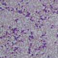
If the platelet count is low, examination of the blood smear by the laboratory technician is essential to make sure that the artifact of platelet clumping (pseudothrombocytopenia) is not present. Although many processes can cause a moderately low platelet count, the differential diagnosis for isolated profound thrombocytopenia (<10,000/μL) is generally limited to immune thrombocytopenia, drug-induced thrombocytopenia, or post—transfusion purpura.
Excessive bleeding has been reported with plasma fibrinogen levels less than 50 mg/dL. The end points of the INR and aPTT are timed to the formation of the fibrin clot. If plasma levels of fibrinogen decrease to less than 80 mg/dL, the clot may be small and not detected by the machine, resulting in a prolonged INR and aPTT. Low fibrinogen levels reflect severe liver disease, consumptive coagulopathy, or dilution by infusion of massive amounts of resuscitative fluids.
Some bleeding disorders, such as platelet function defects or increases in fibrinolysis, cannot be detected by routine laboratory tests. Performing rapid tests to assess platelet function remains controversial. Bleeding times are difficult to perform in the ED and are not predictive of bleeding risk. The PFA—100 is a rapid automatic test for platelet function that is likely to replace the bleeding time, but there are no data on the use of this rapid test to guide therapy for acute bleeding in the ED. It is also difficult to assess for excessive fibrinolysis. The euglobulin clot lysis time is a screen for fibrinolysis but it is not standardized and can be difficult to obtain. Thromboelastography is a unique point-of-care laboratory test that examines whole blood thrombus formation and lysis, but it is not widely available and requires experience in interpretation.
Massive transfusion therapy
Acute bleeding in cancer patients can require large amounts of transfusion products. Early data showed high mortality rates with transfusion of more than 20 units of blood. With modern blood bank techniques and improved laboratory testing, survival rates of 43% to 70% are achieved in patients transfused with more than 50 units of blood.
The approach to massive transfusions is to measure the five basic laboratory tests outlined in Box 2 These tests reflect the fundamental parameters essential for blood volume and hemostasis. Current recommended replacement therapy is based on the results of these tests and the clinical situation of the patient. With rapid transfusion devices, a unit of product can be given in minutes. After infusing the initial blood products, the basic laboratories test should be repeated to guide additional therapy. The transfusion threshold for a low hematocrit depends on the stability of the patient. If the hematocrit is less than 30% and the patient is bleeding or hemodynamically unstable, packed red cells should be transfused. Stable patients can tolerate a lower hematocrit; an aggressive transfusion policy may even be detrimental.
The five basic tests of hemostasis
Hematocrit
Platelet count
Prothrombin time (PT-INR)
Activated partial thromboplastin time (aPTT)
Fibrinogen level
Management guidelines
- 1.
Platelets <50 to 75,000/μL: give 1 plateletpheresis concentrate or 6 to 8 packs of single donor platelets
- 2.
Fibrinogen <125 mg/dL: give 10 units of cryoprecipitate
- 3.
Hematocrit less than 30%: give red cells
- 4.
Prothrombin time INR >2.0 and aPTT abnormal: give 2 to 4 units of fresh frozen plasma (FFP).
If the patient is actively bleeding, has florid DIC, or has received platelet aggregation inhibitors, then keeping the platelet count above 50,000/μL is reasonable because this results in less microvascular bleeding. The conventional dose of platelets is six to eight platelet concentrates or one plateletpheresis unit.
For a fibrinogen level of less than 100 mg/dL, transfusion of 10 units of cryoprecipitate will increase the plasma fibrinogen level by approximately 100 mg/dL. In certain clinical situations, such as acute promyelocytic leukemia, severe fibrinolysis can occur, and the need for large amounts of cryoprecipitate should be anticipated.
In patients with an INR greater than two and an abnormal aPTT, two to four units of fresh frozen plasma (FFP) can be given. For an aPTT greater than 1.5 times normal, two to four units of plasma should be given. Patients with marked abnormalities such as an aPTT twice normal may require aggressive therapy with at least 15 to 30 mL/kg (4–8 units for an average adult) of plasma.
Occasionally, empirical transfusion therapy for the severely bleeding patient is required. Platelet products should be given first because they will also provide plasma replacement. In patients also likely to have DIC (eg, leukemia), administration of 10 units of cryoprecipitate is indicated. For patients who are likely to receive 10 or more units of blood, early use of FFP may help preserve coagulation. One approach is to thaw the plasma and give four units whenever six or more units of uncrossmatched blood are given to a patient with massive bleeding. Recent studies suggest that matching 1:1 units of RBC and FFP may improve outcomes.
Complications of Transfusions
Hypothermia commonly complicates the care of massively transfused patients and can worsen bleeding. Hypothermia impairs platelet function, decreases the efficiency of coagulation reactions, and enhances fibrinolysis. Unwarmed packed red cells can lower the body temperature by 0.25°C. Hypothermia can be prevented by transfusion of blood through blood warmers. Electrolyte abnormalities are unusual even after massive transfusion. Platelet concentrates and plasma contains citrate, which can chelate calcium. However, the citrate is rapidly metabolized and clinically significant hypocalcemia is rare. Although empirical calcium replacement is often recommended, one study suggests that its use is associated with a worse outcome. If hypocalcemia is a clinical concern, then ionized calcium levels should be monitored to guide therapy.
Although potassium leaks out of stored red cells, even older units of blood only contain a total of 8 mEq/L of potassium, so hyperkalemia is not usually a concern. Stored blood is acidic with a pH of 6.5 to 6.9, but acidosis attributed solely to transfused blood is rare. Empirical bicarbonate replacement has been associated with severe alkalosis and is not recommended.
Recombinant Factor VIIa
Recombinant factor VIIa (rVIIa) was originally developed as a bypass agent to support hemostasis in hemophiliacs with inhibitors. Recently, there has been an explosion of information concerning rVIIa use for a wide array of bleeding disorders. Increasingly, rVIIa is being used as a universal hemostatic agent for patients with uncontrolled bleeding from any mechanism. Multiple case reports have demonstrated successful use of rVIIa for bleeding in cardiac surgery patients, obstetric bleeding, reversal of anticoagulation, and trauma. Unfortunately, a paucity of prospective data exists to put these anecdotes into recommended clinical practice.
A general approach for use of rVIIa for acute bleeding would be to first ensure that a reasonable attempt has been made to correct coagulation status, and that there is a defined, survivable condition. Although dosing recommendations vary, a reasonable dose of rVIIa for most situations is 90 μg/kg. If hemostasis is not obtained within 30 minutes, there is little use in giving a second dose. If the patient improves and then rebleeds after 2 to 3 hours, another dose can be given.
Although hypothermia has a detrimental effect on blood coagulation, this does not seem to affect the function of rVIIa. In vitro studies show no decrease in the effect of rVIIa on thrombin generation at 33°C and this finding is supported by clinical data. In contrast, low pH does seem to have a negative effect. In vitro, the effect of rVIIa was reduced by 90% when the pH was lowered from 7.4 to 7.0, which is also supported by clinical data showing that trauma patients who did not respond to rVIIa were more likely to have pH less than 7.2 compared with responders. This finding supports the idea of aggressive resuscitation before resorting to rVIIa.
In theory, rVIIa-induced coagulation activation would be expected to result in DIC or overwhelming thrombosis, but thrombotic complications are rare. In 700,000 doses given to hemophiliacs, only 16 thrombotic complications were seen. However, if used in older patients, especially those with vascular risk factors, the risk of arterial thrombosis seems to increase. In the trials for intracranial hemorrhage, the thrombosis rate after use of rVIIa was 5% to 9%. This risk may be augmented by the known prothrombotic state of cancer. Until more safety data are available, the risk/benefit ratio should be considered before using rVIIa in cancer patients, especially in those with a history of vascular disease such as strokes or myocardial infarction.
Coagulation factor-related bleeding
Acquired Von Willebrand Disease
Acquired von Willebrand disease (VWD) occurs in lymphomas, myeloproliferative syndromes, myeloma, and monoclonal gammopathies. The most common presentations are diffuse oozing from surgical sites, the nose, or gastrointestinal bleeding. Patients with acquired VWD can present as type 1 (decreased protein) or type 2 (abnormal protein) disease.
Patients with acquired VWD have variable responses to therapy. Desmopressin is effective in many patients with acquired VWD types 1 and 2 but, consistent with the antibody-mediated destruction, the magnitude and duration of the effect is often reduced. For bleeding patients, high doses of the von Willebrand concentrate Humate—P are indicated. For patients with strong inhibitors that factor concentrates cannot overcome or with life-threatening bleeding, rVIIa may prove useful.
Acquired Factor VIII Inhibitors
Factor VIII deficiency due to autoantibodies is the most frequent acquired coagulation factor deficiency complication in older cancer patients. Unlike classic hemophiliacs, these patients often have ecchymoses covering large areas of their body, and can have massive muscle and soft tissue bleeding. Patients will have prolonged aPTTs, a positive test for an inhibitor, and a low factor VIII level. For severe or life-threatening bleeding, recombinant VIIa is the treatment of choice. The dose is 90 μg/kg repeated every 2 to 3 hours until bleeding has stopped.
Disseminated Intravascular Coagulation
DIC is the clinical manifestation of inappropriate thrombin activation. The activation of thrombin leads to (1) fibrinogen conversion to fibrin, (2) platelet activation and consumption, (3) factors V and VIII activation, (4) protein C activation (and degradation of factors Va and VIIIa), (5) endothelial cell activation, and (6) fibrinolysis.
Patients with DIC can present in 1 of 4 patterns:
- 1)
Asymptomatic: patients can present with laboratory evidence of DIC but no bleeding or thrombosis (often seen in patients with sepsis or cancer). However, with further progression of the underlying disease, these patients can rapidly become symptomatic.
- 2)
Bleeding: bleeding is due to a combination of factor depletion, platelet dysfunction, thrombocytopenia, and excessive fibrinolysis. These patients may present with diffuse bleeding from multiple sites.
- 3)
Thrombosis: despite the general activation of the coagulation process, thrombosis is unusual in most patients with acute DIC. An exception to this is cancer patients in whom thrombosis can be the major complicating factor. Most often the thrombosis is venous, but arterial thrombosis and nonbacterial thrombotic endocarditis have been reported.
- 4)
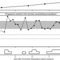
Purpura fulminans, which is DIC in association with symmetric limb ecchymosis and necrosis of the skin, is seen in two situations. Primary purpura fulminans is most often seen after a viral infection such as varicella in an immunocompromised child. In these patients the purpura fulminans starts with a painful red area on an extremity that rapidly progresses to a black ischemic area. Secondary purpura fulminans is most often associated with meningococcemia infections, but can be seen in any patient with overwhelming infection. Postsplenectomy sepsis syndrome patients and cancer patients are also at risk for purpura fulminans. Patients present with signs of sepsis and the skin lesions often involve the extremities and may lead to amputation.
The best way to treat DIC is to treat the underlying cause ( Box 3 ). However, factors must be replaced if depletion occurs and bleeding ensues. Management should be guided using the five basic tests of coagulation (see Box 2 ).
Adenocarcinoma
Amniotic fluid embolism
Burns
Intravascular hemolysis
Infections
Leukemia
Penetrating brain injury
Placental abruption
Retained fetal death in utero
Shock
Snake bites
Trauma
Heparin therapy is reserved for the patient who has large vessel thrombosis as a component of their DIC. Specific heparin levels should be used instead of the aPTT to monitor anticoagulation. Cancer patients with chronic DIC will require long-term heparin therapy.
Platelet number and function
Immune Thrombocytopenia
Immune thrombocytopenia (ITP) is a common condition affecting about 1:20,000 individuals, most often young women. ITP most commonly complicates the care of patients with chronic lymphocytotic leukemia but can be seen with Hodgkin and non-Hodgkin lymphoma. ITP is due to antibodies binding to platelet proteins, most often to the platelet receptor GP IIb/IIIa. Patients often present with signs of bleeding and petechiae. Life-threatening bleeding is unusual and the physical examination is only remarkable for stigmata of bleeding such as petechiae. ITP can occur anytime in the course of the hematological neoplasm, ranging from predating the diagnosis to occurring when the patient is years in remission.
There is no specific laboratory test that “rules-in” ITP but rather it is a diagnosis of exclusion. Extremely low platelet counts with a normal blood smear and a negative history can be diagnostic of ITP. The patient should be questioned carefully about drug exposure including over-the-counter medicines, natural remedies, or recreational drugs. Therapy in ITP should be guided by the patient’s signs of bleeding and platelet counts. Overall, patients tolerate thrombocytopenia well. It is unusual to have life-threatening bleeding with platelet counts more than 1 to 5000/μL unless other sites of bleeding are present, such as a gastric ulcer. The primary therapy for ITP is pulse dexamethasone 40 mg/d for 4 days. In patients with severe thrombocytopenia (counts less than 10,000/μL) or active bleeding, one of two treatments can be tried for rapid induction of a response. Either intravenous immune globulin at 1 g/kg repeated in 24 hours or intravenous anti-D antibody at 75 μg/kg single dose can induce a response in more than 80% of patients in 24 to 48 hours.
Drug-induced Thrombocytopenia
Patients with drug-induced thrombocytopenia present with low platelet counts 1 to 3 weeks after starting a new medication. In patients with a possible drug-induced thrombocytopenia, the primary therapy is to stop the suspect drug. If there are multiple new medications, the best approach is to stop any drug that has been strongly associated with thrombocytopenia ( Box 4 ). Immune globulin, corticosteroids, or intravenous anti—D have been suggested as useful in drug-related thrombocytopenia. However, because most of these thrombocytopenic patients recover when the agent is cleared from the body, this therapy is probably not necessary and avoids exposing the patients to the adverse events associated with further therapy.
Anti-arrhythmic drugs
Procainamide
Quinidine
Anti GP IIb/IIIa agents
Abciximab
Eptifibatide
Tirofiban
Antimicrobial
Amphotericin B
Rifampin
Trimethoprim-sulfamethoxazole
Vancomycin
H2-blockers
Cimetidine
Ranitidine
Acetaminophen
Amrirone
Carbamazepine
Efalizumab
Gold
Heparin
Hydrochlorothiazide
Nonsteroidal antiinflammatory agents
Quinine
Post-transfusion Purpura
In patients with post-transfusion purpura (PTP), the onset of severe thrombocytopenia (<10,000/μL), often with severe bleeding, will occur 1 to 2 weeks after receiving blood products. These patients often lack platelet antigen PLA1. For unknown reasons, exposure to the antigens from the transfusion leads to rapid destruction of the patient’s own platelets. The diagnostic clue is severe thrombocytopenia in a patient, typically female, who has received a red cell or platelet blood product in the past 7 to 10 days. Treatment consists of intravenous immunoglobulin and plasmapheresis to remove the offending antibody. If patients with a history of PTP require further transfusions, only PLA1 negative platelets should be given.
Bleeding in the Platelet Refractory Patient
Many patients with cancer, particularly hematological cancers, become resistant to transfused platelets. Bleeding in patients who are refractory to platelet transfusion presents a difficult clinical problem ( Box 5 ). If patients are shown to have HLA antibodies, HLA-matched platelets can be trnsfused. Unfortunately, matched platelet transfusions may not be available and do not work in 20% to 70% of these patients. Even then, as many as 25% of patients have antiplatelet antibodies in which HLA-matched products will be ineffective. Platelet cross-matching can be performed to find compatible units for these patients, but this may not always be successful and may not be available in a timely manner. Use of antifibrinolytic agents such as ϵ-aminocaproic acid or tranexamic acid may decrease the incidence of bleeding. “Platelet drips” consisting of infusing either a platelet concentrate per hour or 1 plateletpheresis unit every 6 hours may be given as a continuous infusion. For life-threatening bleeding, rVIIa may be of use. For platelet refractory patients with arterial bleeding, the use of angiographic delivery of platelets has been reported to be successful in stopping bleeding.
- 1.
Evaluate for other causes of thrombocytopenia (heparin-induced thrombocytopenia [HIT], drugs).
- 2.
Consider a platelet “drip” (1 unit of platelets given over 4–6 h).
- 3.
Consider antifibrinolytic therapy:
- A.
ϵ-Aminocaproic acid 1 g/h intravenously, or
- B.
Tranexamic acid 10 mg/kg every 8 hours.
- A.
Heparin-induced Thrombocytopenia
Heparin-induced thrombocytopenia (HIT) occurs due to the formation of antibodies directed against the complex of heparin bonded to platelet factor 4, which results in a platelet and monocyte activation and thrombosis. HIT is more common in cancer patients and has a higher rate of thrombotic complications. The frequency of HIT is 1% to 5% if unfractionated heparin is used but less than 1% with low molecular weight heparin. HIT should be suspected if there is a sudden onset of thrombocytopenia with either at least a 50% drop in the platelet count from baseline or the platelet count falls to less than 100,000/μL in a patient receiving heparin in any form. HIT usually occurs 4 days after starting heparin but may occur suddenly in patients with recent (less than 3 months) exposure. A feature of heparin-induced thrombocytopenia that is often overlooked is recurrent thrombosis in a patient receiving heparin despite a normal platelet count. Patients with HIT can present to the ED up to 2 weeks after their hospital stay (and heparin exposure) with new thrombosis and thrombocytopenia.
The diagnosis of HIT can be challenging in the cancer patient who has multiple reasons for being thrombocytopenic. In this situation, the laboratory assay for HIT may be helpful. Two general forms of HIT tests exist. One is a platelet aggregation assay whereby patient plasma, donor platelets, and heparin are added. If added heparin induces platelet aggregation, the test is considered positive. One caveat is that early in the HIT disease process the test can be negative but then turns positive 24 hours later as the antibody titer increases. There is also an ELISA assay for the presumed pathogenic antiplatelet factor 4 antibodies, which is sensitive but not specific. Positive ELISA results must be interpreted in the clinical context of the patient.
The first step in therapy for HIT consists of stopping all heparin. Low molecular weight heparins cross-react with the HIT antibodies and therefore these agents are also contraindicated. Institution of warfarin therapy alone has been associated with an increased risk of thromboses. For immediate antithrombotic therapy for HIT patients, three new antithrombotic agents are available ( Box 6 ). Patients with HIT should also be carefully screened for any thrombosis including examination by lower extremity Doppler ultrasound because of the propensity to have silent thrombosis.


