MAGNETIC RESONANCE IMAGING OF THE THYROID
MRI depicts the interactions that occur between the hydrogen atoms (protons) in different tissues in the patient’s body and an externally applied magnetic field (radiowaves of a specific frequency). Different properties (relaxation times) of the hydrogen atoms, termed T1 and T2, can be selectively emphasized. Because the hydrogen atoms of different tissues have different T1 and T2 properties, a computer-assisted analysis of T1-weighted and T2-weighted signals is used to differentiate the thyroid gland from skeletal muscle, blood vessels, or regional lymph nodes. Normal thyroid tissue tends to be slightly more intense than muscle on a T1-weighted image, and tumors frequently appear even more intense. The intravenous administration of noniodinated contrast agents, such as gadolinium-DTPA (pentetic acid), may further enhance the characterization of tissue and organs, as may suppressing the signal that is derived from fat (short tau inversion, or STIR).
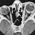
The test is uncomfortable; claustrophobic reactions occur, and considerable noise is inherent to the technique. Generally, the patient’s entire body must be inserted into a large cylinder. Advances such as the use of special surface electromagnets over the neck provide impressive images and, thus, may increase the usefulness of MRI for thyroid evaluation. The equipment is costly and in great competitive demand for other types of examinations. The guidelines for use were discussed earlier in Sectional Thyroid Imaging. Figure 35-9 shows the evaluation of a suspicious ultrasonographic finding, and Figure 35-12 demonstrates the use of MRI to show the extent of a large obstructive goiter and to help plan a surgical approach. Occasionally, in this setting, sectional imaging may help the physician to choose between medical management and surgery. In Figure 35-13, an MRI shows the size and extent of postsurgery cancer. Figure 35-14 demonstrates the use of MRI to examine enlarged upper cervical lymph nodes in a patient with thyroid cancer. Figure 35-15
shows an MRI that helped the physician to diagnose a complex clinical problem. Several studies have suggested new directions for future research, including correlation of images with the biochemistry and histopathology of tissue.21,22 Qualitative and quantitative similarities of the proton response of thyroid tissue in the neck and chest have been demonstrated,23 and differences in these characteristics in malignant and benign thyroid tissues have been studied in vitro.24
shows an MRI that helped the physician to diagnose a complex clinical problem. Several studies have suggested new directions for future research, including correlation of images with the biochemistry and histopathology of tissue.21,22 Qualitative and quantitative similarities of the proton response of thyroid tissue in the neck and chest have been demonstrated,23 and differences in these characteristics in malignant and benign thyroid tissues have been studied in vitro.24
Stay updated, free articles. Join our Telegram channel

Full access? Get Clinical Tree


