VII.G.001 Acute Lymphocytic Leukemia
VII.G.001
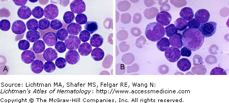
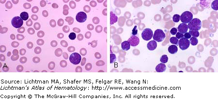
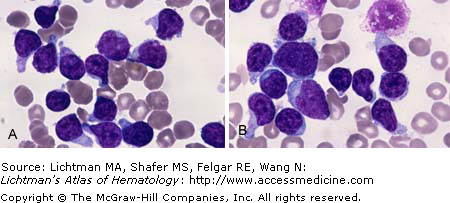
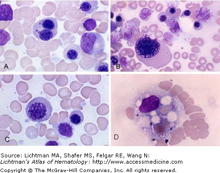
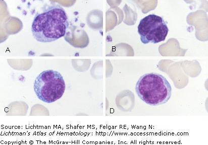
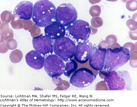
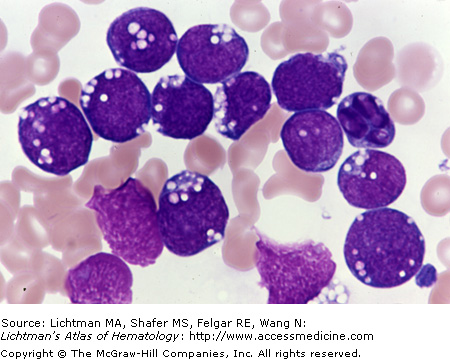
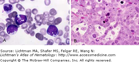
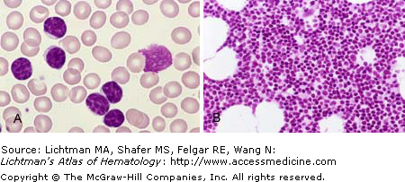
Acute lymphocytic leukemia. Marrow films. (A) Leukemic lymphoblasts ranging from small to medium in size with a very high nuclear to cytoplasmic ratio and condensed chromatin and inconspicuous nucleoli. (B) Leukemic lymphoblasts ranging from small to medium size with a very high nuclear to cytoplasmic ratio and condensed chromatin and inconspicuous nucleoli. Note large lymphoblast with azure granules. The latter may be seen in this disorder and can be the predominant cell type.
VII.G.002 Acute Lymphocytic Leukemia
VII.G.002
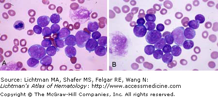
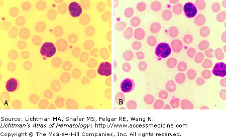
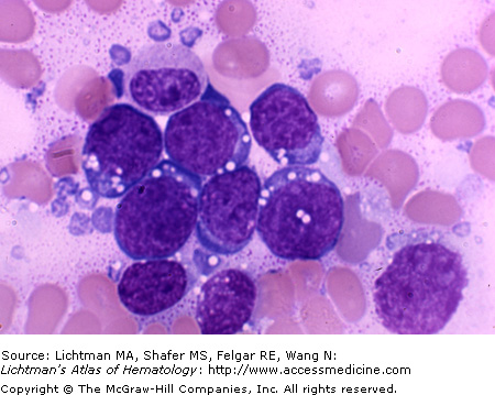
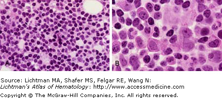
Acute lymphocytic leukemia. Marrow films. (A) Leukemic lymphoblasts ranging from small to large size with a very high nuclear to cytoplasmic ratio and condensed chromatin but conspicuous nucleoli in some cases multiple. Note scalloped nuclear margin of the larger lymphoblasts. (B) Leukemic lymphoblasts ranging from small to large size with a very high nuclear to cytoplasmic ratio and condensed chromatin and very conspicuous nucleoli. Note small lymphoblasts with pseudonucleoli, which is cytoplasm trapped in the center of the nucleus. In the cell at the top left, this phenomenon gives the appearance of a doughnut.
VII.G.003 Acute Lymphocytic Leukemia
VII.G.003
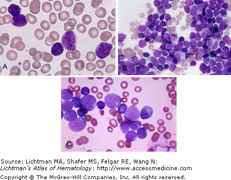
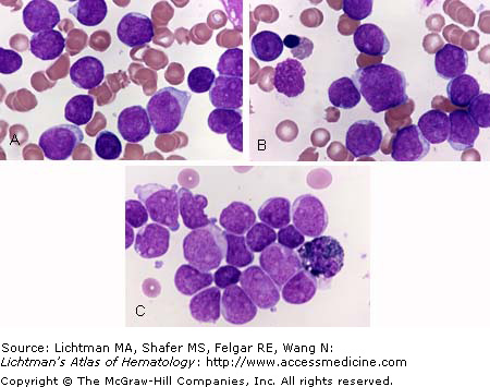
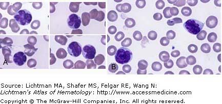
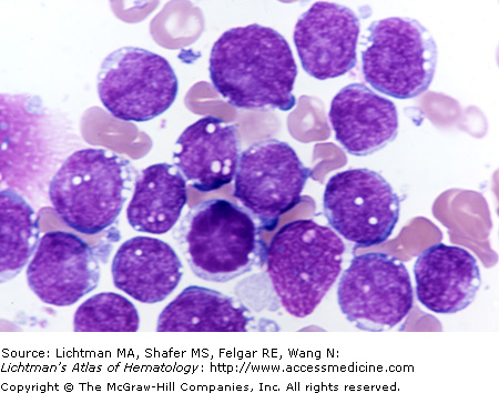
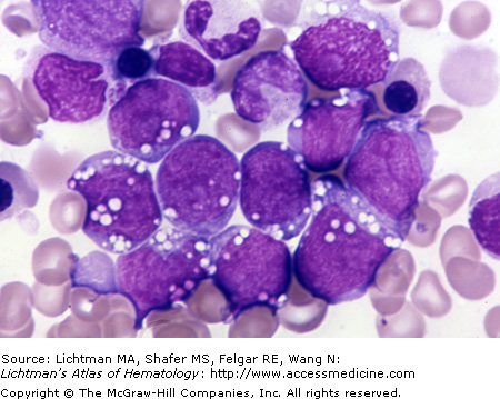
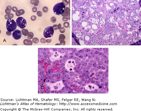
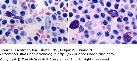
Acute lymphocytic leukemia. (A) Blood film. Leukemic lymphoblasts ranging from small to large size with a very high nuclear to cytoplasmic ratio. The small blasts have condensed chromatin and usually inconspicuous nucleoli. The large blast has a finer chromatin pattern and overt nucleoli. (B) Marrow film. Leukemic lymphoblasts ranging from small to large size. The medium sized and large blasts have a finer chromatin pattern and overt nucleoli. The small blasts have condensed chromatin and usually less conspicuous nucleoli. (C) Marrow film. Higher power. Leukemic lymphoblasts ranging from small to large size. The medium sized and large blasts have a finer chromatin pattern and overt nucleoli. The small blasts have condensed chromatin and less conspicuous nucleoli.
VII.G.004 Acute Lymphocytic Leukemia
VII.G.004
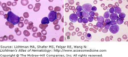
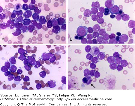
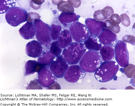
Acute lymphocytic leukemia. (A) Blood film. Leukemic lymphoblasts ranging from small to large size with a very high nuclear to cytoplasmic ratio. These blasts have condensed chromatin and nucleoli are evident in the large- and mid-sized blasts. (B) Marrow film. Marrow replaced with lymphoblasts ranging in size from small to large. The smaller blasts have condensed chromatin and the larger blasts have finer chromatin patterns and more conspicuous nucleoli. A large orthochromatic erythroblast with atypical nuclear chromatin pattern and nuclear fragment is present (megaloblastoid cell).
VII.G.005 Acute Lymphocytic Leukemia, Peroxidase Stain
VII.G.005
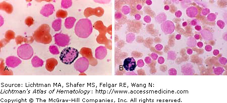
Acute lymphocytic leukemia. (A) Blood film. Leukemic lymphoblasts ranging from small to medium size with a very high nuclear to cytoplasmic ratio, condensed chromatin, and inconspicuous nucleoli. (B) Marrow film. Peroxidase stain. A promyelocyte is stained intensely positive for peroxidase (black reaction product). Leukemic lymphoblasts do not react with peroxidase.
VII.G.006 Acute Lymphocytic Leukemia

VII.G.006
Acute lymphocytic leukemia. (A) Blood film. Leukemic lymphoblasts ranging from medium to large size with a very high nuclear to cytoplasmic ratio. The blasts have intermediate condensed chromatin and conspicuous nucleoli. (B) Marrow film. Leukemic lymphoblasts ranging from small to large size. The medium-sized and larger blasts have an overt nucleoli. The small blasts have condensed chromatin and usually less conspicuous nucleoli. (C) Marrow film. Higher power. Leukemic lymphoblasts ranging from small to large size. The medium-sized and large blasts have a finer chromatin pattern and overt nucleoli. The small blasts have condensed chromatin and less conspicuous nucleoli.
VII.G.007 Acute Lymphocytic Leukemia, Periodic Acid Schiff Stain
VII.G.007
Acute lymphocytic leukemia. Periodic acid Schiff stain. (A) Blood film. Blood film. Leukemic lymphoblasts ranging from small to large size with a very high nuclear:cytoplasmic ratio. The small blasts have condensed chromatin and usually inconspicuous nucleoli. The large blasts have a finer chromatin pattern and overt nucleoli. Note vacuoles in several blast cells. (B) Marrow film. Periodic acid Schiff stain. Leukemic lymphoblasts react with reddish-purple clumps, sometimes more rectangular in character and referred to as “block” positivity (as opposed to the fine granular staining that can be seen in some myeloblasts and other cells).
VII.G.008 Acute Lymphocytic Leukemia
VII.G.008
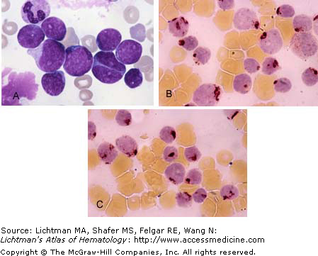
Acute lymphocytic leukemia. (A) Blood film. Leukemic lymphoblasts ranging from medium to large size with a very high nuclear to cytoplasmic ratio, condensed chromatin, and conspicuous nucleoli in the larger cells. (B) Marrow film. Leukemic lymphoblasts ranging from medium to large size with a very high nuclear to cytoplasmic ratio, condensed chromatin, and conspicuous nucleoli in the larger cells. (C) Marrow film. Peroxidase stain. A myelocyte is stained intensely positive for peroxidase (cytoplasmic black reaction product). Leukemic lymphoblasts do not react with peroxidase.
VII.G.009 Acute Lymphocytic Leukemia
VII.G.009
Acute lymphocytic leukemia. Peroxidase stain. Marrow films. (A) Small to large leukemic lymphoblasts do not react with peroxidase. One myelocyte reacts intensely (cytoplasmic black reaction product). (B) Mostly small and several large leukemic lymphoblasts. A metamyelocyte is stained intensely positive for peroxidase (cytoplasmic black reaction product). Leukemic lymphoblasts do not react with peroxidase.
VII.G.010 Acute Lymphocytic Leukemia, Acid Phosphatase Stain
VII.G.010
Acute lymphocytic leukemia. Precursor T-cell type. (A) Marrow film. Medium to large leukemic lymphoblasts with high nuclear:cytoplasmic ratio and overt nucleoli. One lymphoblast is binucleate with each nucleus containing a nucleolus. (B) and (C) Marrow films. Acid phosphatase stains. T lymphoblasts react with this stain by forming focal large dark purple cytoplasmic clumps.
VII.G.011 Acute Lymphocytic Leukemia, Vacuolated Blast Cells
VII.G.012 Acute Lymphocytic Leukemia
VII.G.013 Acute Lymphocytic Leukemia
VII.G.013
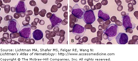
Acute lymphocytic leukemia. This case was Ph-chromosome and CD10 positive. (A) and (B) Blood films. Principally large- and medium-sized lymphoblasts with variation in nuclear cytoplasmic ratio from high to lower. One cell in (B) has cytoplasmic azure granules, occasionally seen in lymphoblasts.
VII.G.014 Acute Lymphocytic Leukemia, Erythroblast Dysmorphia
VII.G.014
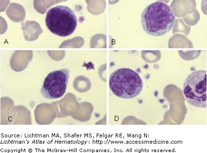
Acute lymphocytic leukemia. Abnormalities of erythroblasts are common in acute lymphoblastic leukemia at diagnosis or during treatment. In the former case the intense and profound lymphoproliferation probably depletes essential components for DNA synthesis in erythroblasts and during treatment several agents, especially methotrexate and cytarabine, if used, induce pseudomegaloblastic changes. (A)-(D) Marrow film. (A) Binucleate erythroblast with nuclear fragments. (B) Gigantic erythroblast. (C) Giant erythroblast (megaloblastoid). (D) Macrophage activation is common and intense cytophagocytosis may be present during therapy. Macrophage with several ingested erythrocytes.
VII.G.015 Acute Lymphocytic Leukemia, Radiation-Related Nuclear Dysmorphia
VII.G.015
Acute lymphocytic leukemia. (A) and (B) Leukemic lymphoblasts from a patient in the fall out zone of the Chernobyl nuclear power plant disaster near Pripyat, Ukraine in April 1986. The fallout principally affected the Ukraine, Belarus, and Russia, although it spread throughout many European countries. This case occurred in June 1991 in the fallout area in the Ukraine. No further information is available. The leukemic lymphoblasts had a high prevalence of contorted nuclei with nuclear lobulations radiating out from the center. This shape has been referred to as “Reider cells,” although the latter were first described in normal lymphocytes exposed to Na2EDTA in vitro.
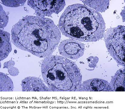
VII.G.016 Acute Leukemia Lymphoblasts. Transmission Electron Micrograph
VII.G.017 Acute Lymphoblastic Leukemia, Precursor B-Cell Type
VII.G.017
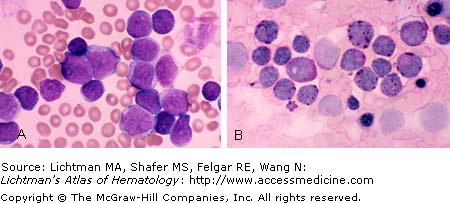
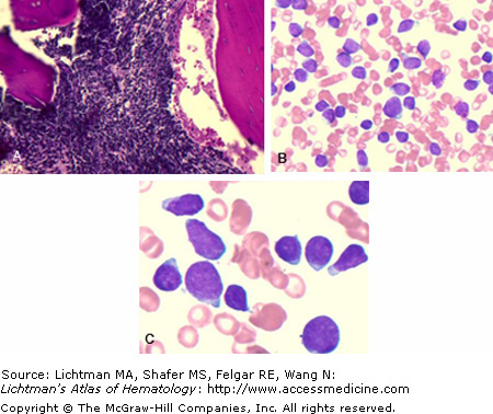
Acute lymphoblastic leukemia. Precursor B-cell type. Two-year-old infant with leukocytosis, anemia, and thrombocytopenia. Blood blast count was 43,896/μl. (A) Marrow biopsy. Markedly hypercellular with >90% lymphoblast. (B and C) Marrow film-lower power and high power. Characteristic lymphoblasts.
VII.G.018 Acute Lymphoblastic Leukemia, Precursor B-Cell Type, Flow Cytometry
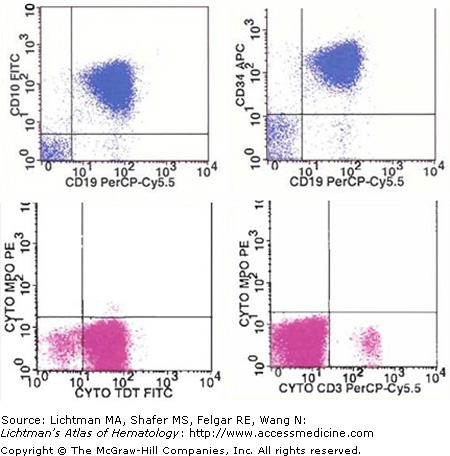
VII.G.018
Acute lymphoblastic leukemia. Precursor B-cell type. Flow cytometry. (Upper left) CD10 and CD19 positive leukemic cells. (Upper right) CD19 and CD34 positive cells. (Lower left) TDT positive, myeloperoxidase (MPO) negative cells. (Lower right) CD3 negative cells. TDT, HLA-Dr, surface kappa and lambda chains, and myeloid markers were negative.
VII.G.019 Acute Lymphoblastic Leukemia, Childhood, Phenotypes
VII.G.019
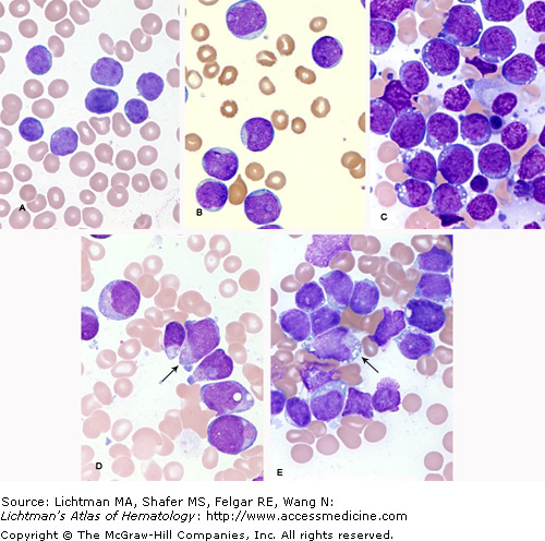
Acute lymphoblastic leukemia of childhood. Marrow films. Phenotypic variations. (A) Smaller sized blasts with a very high nuclear:cytoplasmic ratio, and indistinct or unapparent nucleoli. (B) Larger sized blasts with lower nuclear:cytoplasmic ratio and overt large nucleoli, sometimes multiple. (C) Burkitt type lymphoblasts. Deeply blue cytoplasm with lipid vacuoles ranging from few to many per cell. Overt nucleoli (D and E). Granular variant with small (D) to giant (E) granules in blast cells.
VII.G.020 Adult T-Cell Leukemia-Lymphoma (HTLV-1)
VII.G.021 Adult T-Cell Leukemia-Lymphoma (HTLV-1)

VII.G.022 Adult T-Cell Leukemia-Lymphoma (ATLL)
VII.G.022
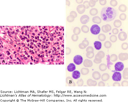
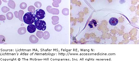
Adult T-cell leukemia-lymphoma (ATLL). (A) Lymph node biopsy. Architecture effaced by replacement with abnormal CD4+/CD25+ mature T-cell infiltrate. (B) Blood film. Classic “flower” or cloverleaf cells. By morphology alone, ATLL can be mistaken for other T-cell lymphomas. ATLL can have skin involvement and be morphologically indistinguishable on skin biopsy from cutaneous T-cell lymphoma (CTCL). Some of the distinguishing features from cutaneous T-cell lymphoma include: expression of the IL-2 receptor, as assessed by CD25 staining, history of HTLV-1 exposure, and presence of hypercalcemia. These latter three findings are characteristic of ATLL, but not CTCL.
VII.G.023 Adult T-Cell Leukemia-Lymphoma (HTLV-1)
VII.G.024 Adult T-Cell Leukemia-Lymphoma (HTLV-1)
VII.G.025 Burkitt Cell Leukemia
VII.G.026 Burkitt Cell Leukemia
VII.G.027 Burkitt Cell Leukemia
VII.G.028 Burkitt Cell Leukemia
VII.G.029 Burkitt Cell Leukemia
VII.G.030 Burkitt Cell Leukemia
VII.G.031 Burkitt Cell Leukemia
VII.G.032 Burkitt Cell Leukemia
VII.G.032
Burkitt cell leukemia. (A) Marrow film. Several lymphoblasts with high nuclear:cytoplasmic ratio, nucleoli, and characteristic vacuoles. (B) Marrow section. Marrow replaced by lymphoblasts with interspersed macrophages with ingested cellular debris. (C) Marrow section. Higher power. Marrow replaced by lymphoblasts with interspersed macrophages with ingested cellular debris.
VII.G.033 Burkitt Cell Leukemia, Oil Red O Stain
VII.G.034 Chronic Lymphocytic Leukemia
VII.G.034
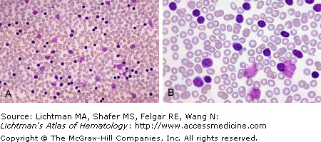
Chronic lymphocytic leukemia. Blood films. (A) Low power. Marked increase in small lymphocytes. Smudge cells (lymphocyte nuclear remnant) characteristic of the tendency of lymphocytes to be disrupted by the shear forces during the preparation of the blood film. Occasional nuclear abnormalities evident. (B) Higher power. Marked increase in small lymphocytes. Smudge cells (lymphocyte nuclear remnants).
VII.G.035 Chronic Lymphocytic Leukemia
VII.G.035
Chronic lymphocytic leukemia. Blood films. (A) Blood film. Marked increase in small lymphocytes. Smudge cells (lymphocyte nuclear remnant) characteristic of the tendency of lymphocytes to be disrupted by the shear forces during the preparation of the blood film. (B) Marrow film. Marrow replaced by diffuse infiltrate of small leukemic lymphocytes.
VII.G.036 Chronic Lymphocytic Leukemia
VII.G.037 Chronic Lymphocytic Leukemia
VII.G.037
Related posts:

Stay updated, free articles. Join our Telegram channel

Full access? Get Clinical Tree


