Breast cancer-related lymphedema remains a feared complication following breast cancer treatment (1) because it is a chronic process that cannot reliably be prevented. Dramatic modifications to surgical approaches, including less radical breast surgery and widespread adoption of sentinel lymph node biopsy (SLNB) as the standard of care for axillary staging, have contributed to significant decreases in the incidence of lymphedema. However, despite these changes, there remains approximately a 20% risk of lymphedema after axillary dissection (ALND) and a 0% to 7% risk after SLNB. Persistent risk and patient worry result in almost uniform adoption of lifestyle modifications (1). Unfortunately, it is unclear if these modifications reduce the risk for lymphedema, and it is further unclear how these changes impact overall quality of life (QOL).
Women affected by lymphedema have historically had an overall poorer QOL (2). Lymphedema can contribute to musculoskeletal pain and reductions in shoulder range of motion, limiting performance of activities of daily living. Unattended severely lymphedematous limbs can lead to elephantiasis and, in a few cases, to Stewart-Treves Syndrome, a rare but deadly angiosarcoma arising from the lymphedematous tissues. Despite these limitations, most patients with lymphedema do not consider themselves as disabled (3). They do, however, suffer from decreased body image and loss of self-esteem, but further refinement in assessment of all QOL measures has been lacking. Emerging interest supports standardization of QOL assessment in lymphedema patients as a recent meta-analysis demonstrated significant heterogeneity with 17 different instruments used (only two of which were specific to lymphedema patients) in 39 studies to assess health-related QOL outcomes in breast cancer-related lymphedema patients (4). The authors encourage future studies to use high quality lymphedema-specific patient-reported outcome instruments such as the Upper Limb Lymphedema 27 (ULL 27), which had the strongest psychometric properties.
The medical community has devoted little time and attention to lymphedema research and the resources necessary for successful lymphedema treatment. The financial burden placed on patients is frequently a source of anxiety. While Medicare supports consultation and treatment by trained lymphedema specialists, treatment coverage is frequently limited to diagnosis, acute intervention, and establishment of the treatment plan with visits and services covered only as patients demonstrate improvement. Interestingly, federal guidelines exist supporting the coverage of postmastectomy bras and prostheses; however, coverage for lymphedema compression garments and short stretch bandages, both mainstays for in-home maintenance therapy, varies among private insurers and Medicare. Advocating for equality in patient rights, the National Lymphedema Network (NLN) supports the Lymphedema Treatment Act (5, 6), a bill before Congress that would help establish and standardize treatment coverage, patient education, development of self-treatment plans, and, ultimately, would reduce healthcare costs. As of July 2011, this bill was referred to the House of Representatives subcommittee on Health and has not been discussed again.
ANATOMY AND PATHOPHYSIOLOGY
The lymphatic system is composed of lymphatic capillaries, transporting vessels, and lymph nodes. The lymphatic system has a low oncotic pressure, allowing diffusion of protein-rich interstitial fluid into lymphatic vessels, which transport it to the venous system. In the upper extremities, the superficial lymphatic system is composed of valveless capillaries located at the dermal-subcutaneous level that communicate directly with collecting lymphatic vessels coursing through subcutaneous tissues with superficial veins. These collecting vessels, or secondary lymphatics, drain into tertiary lymphatics, which progressively network and ascend to the axilla. On the left, these lymphatic vessels join other thoracic and intercostal lymphatic channels, draining into the thoracic duct, which empties into the left subclavian vein. A smaller right lymphatic duct drains the right upper extremity and neck and enters the right subclavian vein. Secondary and tertiary lymphatic vessels have valves to aid in the unidirectional propulsion of lymph fluid. Lymphatic flow is encouraged by active skeletal muscle contraction, causing intermittent compression of the subcutaneous compartment, and by nearby arterial pulsations. The lymph nodes act as points of filtration throughout the lymphatic drainage process and serve a primarily immunologic function. The vessels of the deep lymphatic system run beneath the muscular fascia near neurovascular bundles. Little communication exists between the superficial and deep systems, and lymphedema generally spares the deep component.
The etiology of lymphedema is incompletely understood, but it likely results from (1) lymphatic obstruction due to obliteration of the lymphatic pathways or removal of the lymph nodes, (2) mechanical insufficiency due to faulty lymphatic pumping or malfunction of the lymphatic valves, or (3) loss of lymphatic vessel integrity. The quantity of fluid within the interstitial space is determined by the delicate balance of hydrostatic and oncotic pressures between the vascular capillaries and the interstitial space. More than 90% of fluid within the interstitial space is removed by the venous capillaries; what remains is normally returned to the vascular system by lymphatics. The combination of the negative oncotic pressure of the lymphatic vessels and their indistinct, virtually nonexistent basement membranes allows larger proteins and macromolecules, such as bacteria and cellular debris, to passively diffuse from the interstitium into the lymphatic system. When the lymphatic system is dysfunctional, fluid transport is disrupted, and interstitial protein accumulates, increasing its oncotic pressure. This draws more fluid into the interstitium. Excessive accumulation of interstitial fluid due to impaired lymphatic transport is called lymphedema.
The cycle of lymphedema self-perpetuates as increased lymphatic fluid volume causes stretch in lymphatic vessels, leading to incompetent valves and further failure of lymph transport. Additionally, the stagnant bacteria ignites a chronic inflammatory cascade, recruiting macrophages and neutrophils to the interstitium for wound healing, and leading to collagen deposition and fibrosis, hindering lymphatic contraction. Furthermore, the severity of edema can be exacerbated by episodes of lymphangitis, chronic inflammation, or recurrent cellulitis. It is hoped that the recent development of lymphatic endothelial markers such as LYVE-1, Prox1, and podoplanin will help the study of lymphangiogenesis and regeneration.
CLINICAL EVALUATION OF UPPER EXTREMITY LYMPHEDEMA
To evaluate for lymphedema, the patient sits with arms outstretched and then flexed and rested on the hips. The clinician should pay close attention to subtle differences in symmetry including loss of bony prominences, especially at the olecranon process, styloid process of the ulnar head, and over the extensor tendons of the hand. Other subjective signs of swelling include imprints from tight-fitting shirt sleeves, watches, or jewelry. The physical exam also includes evaluation of skin turgor, firmness, and the presence of pitting or nonpitting edema. Lymphedematous changes may involve the entire upper extremity but can also be isolated to the hand in 61%, the lower arm in 55%, and the upper arm in 72% of patients (7). Table 40-1 lists the clinical stages of lymphedema.
TABLE 40-1 Clinical Stages of Lymphedema
Stage
Clinical Findings
0
Subclinical lymphedema
Impaired lymphatic transport
Swelling not visible by gross evaluation
Can be latent for months to years
I
Visibly swollen limb
Pitting edema
Edema may resolve without treatment
II
Limb is visibly swollen but swelling is nonpitting
The diagnosis of lymphedema is confirmed by physical exam and a combination of subjective and objective measures (Table 40-2). Unfortunately, the most challenging problems in accurately determining the incidence of lymphedema remain poor standardization in defining lymphedema and the lack of robust data with long-term follow-up. Many studies diagnose lymphedema using subjective measures such as patient questionnaires or survey instruments to directly and indirectly assess symptoms of arm or breast swelling, tightness, tenderness, or edema (7, 8, 9 and 10). Little correlation between subjective assessment tools and arm measurement changes constituting lymphedema exist; however, it could be argued that it is the patient’s perception of lymphedema, not the presence or absence of objective measurement changes, that negatively impacts QOL and contributes to adoption of risk reducing behaviors. A recent study found that, in patients having SLNB, perceptions of lymphedema appear to decline significantly over the first year; however, among ALND patients, perceptions of lymphedema increase between 6 and 12 months after surgery (1). At 5 years’ follow-up, a large prospective study found perceived rates of lymphedema to be less than measured after SLNB (3% vs. 5%), while patients undergoing ALND perceived more swelling than was measured (27% vs. 16%) (11). More recently, a retrospective study with 10 years of follow-up documented subjective lymphedema in 10% of SLNB patients and 33% of ALND patients (12). This study did not obtain baseline assessments. Well-documented sensory changes occurring after axillary surgery and the inconsistent practice of sparing the intercostal brachial nerve likely explain the differences between perceptions and measurements of lymphedema (12).
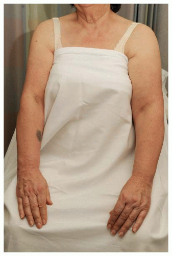
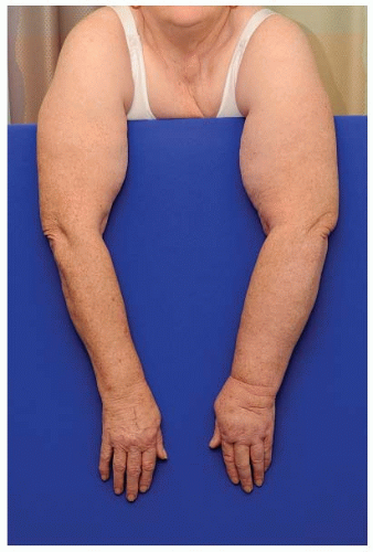
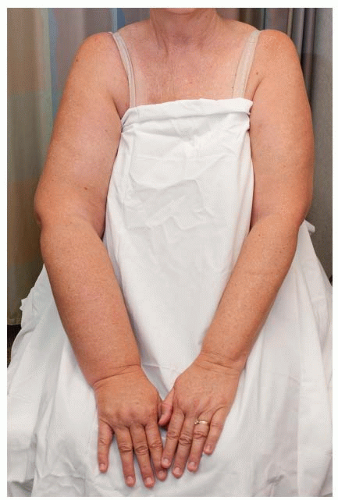
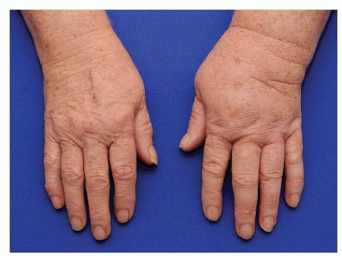
The objective measures of lymphedema that quantify volume differences and volume displacement using water remain the gold standard in assessing lymphedema despite multiple limitations (see Table 40-2). Circumferential arm measurements with a non-elastic tape measure remain the most commonly reported method for objectively assessing lymphedema. Implementation in clinical practice is relatively straightforward; however, a few guidelines should be followed. First, baseline measurements of the ipsilateral and contralateral arm are essential to control for normal variations between the dominant and nondominant arms at baseline and for any weight gain during follow-up. Second, to minimize intra-rater and inter-rater circumferential arm measurement variability, patients should be measured by the same healthcare professional at all visits and ideally measured multiple times at each point to ensure the most accurate results. Unfortunately, the number of anatomic locations and the number of measurements obtained vary between studies. Some investigators measure at only two points while others obtain 10 to 15 measurements at 3 or 4 cm increments from the nail bed to the axillary fold and then calculate the arm volume according to the volume of a frustrum or truncated cone. When compared to water displacement, multiple measures used to calculate arm volume and patient self-report had the highest specificities (90% and 89%, respectively), while measurement of arm circumferences at 2 points alone had the lowest specificity (73%) (13). Finally, the measurement change constituting lymphedema is not standardized; some consider lymphedema a 2-cm increase in circumference (9), while others consider a volume increase of less than 10% as minimal lymphedema, 10% to 20% moderate, and greater than 20% severe when compared to the baseline (14). The clinician should decide the diagnostic thresholds prior to commencement of screening to ensure consistency in measurements. Figures 40-1, 40-2, 40-3 and 40-4 demonstrate mild, moderate, severe, and isolated hand lymphedema. Regardless of the implementation strategy, arm measurements cannot determine the actual volume of extra lymphatic fluid.
TABLE 40-2 Advantages and Disadvantages of Common Lymphedema Diagnostic Tools
Method
Advantages
Limitations
Patient report (subjective assessment)
• Patient perception of symptoms and their impact on function
• Detection of prodromal symptoms of heaviness or subtle arm changes undetected by objective measures
• Recall bias of risk factors
• Unclear influence of postoperative sensation sequelae
• No standardization in subjective assessment or QOL tools
Water displacement
• Gold standard
• Volume calculation of entire limb
• Cumbersome
• Infection control limitations (water must be changed between each patient use)
• Cannot isolate location of lymphedema (i.e., to hand, forearm, or upper arm)
Circumferential tape measurements
• Portable
• Easy to learn
• Noninvasive
• Cost efficient
• Relatively quick to perform
• Intra-rater and inter-rater variability
• Nonstandardized process (i.e., inconsistent measurement intervals and diagnostic thresholds)
• Requires baseline and bilateral arm measurements
Perometry
• Standardized process
• Multiple measurement intervals
• Reproducible
• Sensitive
• Not portable
• Expensive equipment
• Cannot reliably measure the upper arm or hand
Bioimpedance spectroscopy (BIS)
• Measures only extracellular fluid compartment
• May detect subtle volume changes of <150 mL (prodromal or Stage 0 lymphedema)
• Portable
• Cost effective
• Standardized
• FDA approved
• Variable reimbursement
• Recommended at baseline and every 3 mo after surgery
Perometry or opto-electric volumetry uses a perometer to emit infrared light beams and to measure changes in the beam angles caused by the shadows of the limb. The frame moves at 3-mm increments along the length of the limb, obtaining circular cross-sectional measures, and then calculates overall total limb volume. The advantages and limitations are listed in Table 40-2. When perometry is used, clinicians consider a change of 3% over baseline measurements to be diagnostic for lymphedema (15).
Another noninvasive measurement option is bioimpedance spectroscopy (BIS), previously known as multifrequency bioelectrical impedance (Table 40-2). BIS uses resistance to electrical current to compare the composition of extracellular fluid compartments within the body and specifically between the affected and unaffected limbs (16). An increase in extracellular fluid results in a decrease in impedance of the affected limb. The most popular device available is the L-Dex marketed by Impedimed. Exploratory studies suggest an increase of 10 L-Dex units from the baseline or a value outside of the normal range may aid in the clinical assessment of unilateral arm lymphedema when compared to matched normal controls (17). Smoot et al. compared BIS to circumferential arm measures and found BIS to have the highest Area Under the Curve (AUC) value of 0.88, while using the 2-cm diagnostic cutoff option had an AUC of approximately only 0.60 (18). BIS may be best suited to identify the prodromal early stage of lymphedema as circumferential arm measures or volume calculations may fail to capture subclinical fluid accumulations of less than 150 mL (14). The device is portable and easy to use; however, reimbursement for the procedure varies by state across private insurers and Medicare. Taking all measurement options into account, the NLN recommends circumferential tape measurements made with a flexible non-elastic tape measure at a minimum of six anatomical locations per arm, infrared perometry, or BIS (5); the National Accreditation Program for Breast Centers (NAPBC) strongly recommends the use of BIS or perometry, given their high correlation of results in the assessment of stage 0 lymphedema. The dilemma in diagnosing lymphedema remains that measurement changes alone may not find all patients who are suffering from clinically significant lymphedema and may overdiagnose those who are unaffected by their measurement changes. Imaging techniques including lymphoscintigraphy, CT, and MRI are occasionally discussed to image a lymphedematous limb; however, these imaging techniques are predominantly limited to research, not clinical use.
FIGURE 40-1 Mild lymphedema in the left arm.
FIGURE 40-2 Moderate lymphedema in the left arm.
FIGURE 40-3 Severe lymphedema in the right arm.
FIGURE 40-4 Moderate lymphedema in the left hand.
Early Detection and Progression
An emerging body of literature supports the early detection of breast cancer-related lymphedema. These data cite improvements in functional outcome, resolution of prodromal symptoms, and decreased cost as reasons to support aggressive early detection practices (19). Stout presented data (15) supporting early detection and found intervention at this early stage can reduce or resolve the progression of lymphedema. This study prospectively followed 196 women, measuring arm volumes by perometer at baseline and every three months postoperatively. They controlled interventions, prescribing compression sleeves for 4 weeks to all women with a 3% change from baseline in perometer measurements. After intervention, the investigators found a mean arm volume decrease of 58% that was maintained for nearly 5 months after the compression sleeve was discontinued. Torres-Lacomba also highlights the importance of early intervention and conducted a prospective randomized trial to assess the role of early physical therapy, including manual lymphatic drainage, scar massage, and progressive active shoulder range of motion, on the incidence of lymphedema. After one year of follow-up, those in the intervention group demonstrated significantly less lymphedema (7% vs. 25%, p = .01) (20).
Luckily, contemporary estimates find that, among those with lymphedema, the majority have only a mild form (7). However, women with mild lymphedema are more than three times more likely to develop moderate or severe lymphedema compared to women with no lymphedema. Bar et al. reinforces these findings, documenting that 48% of mild lymphedema patients progressed to more severe lymphedema by 5 years follow-up (21). Risk factors for the progression of lymphedema included age more than 65 at diagnosis, morbid obesity, and regional nodal irradiation including posterior axillary boost (22). While studies agree on the importance of early identification and intervention, there is less agreement on what type of intervention should be pursued. Regardless, prospective evaluation and intervention for lymphedema is associated with significant cost savings with a recent study finding the cost to manage early-stage breast cancer-related lymphedema to be $636 annually, while the cost to manage late-stage lymphedema (traditional model) is $3,124 (23).
Risk Factors
Many retrospective studies have reported risk factors for lymphedema, including the extent of axillary surgery, mastectomy, obesity, patient age, radiation, and infection or injury in the ispilateral upper extremity. The strength of association between these treatment and epidemiologic risk factors and lymphedema is inconsistent across studies (24). A meta-analysis reviewed lymphedema risk factors from 98 studies and found a significantly increased incidence of lymphedema after mastectomy compared to lumpectomy (RR, 1.42; CI, 1.15-1.76), ALND compared to no dissection (RR, 3.47; CI, 2.34-5.15), ALND compared to SLNB (RR, 3.07; CI, 2.20-4.29), radiation versus no radiation therapy (RR, 1.92; CI, 1.61-2.28), and for positive versus negative axillary lymph nodes (RR, 1.54; CI, 1.32-1.80). While these data represent a comprehensive, contemporary review of potential risk factors, it should be acknowledged that the 98 studies used 11 different definitions for lymphedema and follow-up ranged between 1 month and 30 years. Finally, the influence of infection and injury must be tempered, as most accounts documenting these occurrences are obtained by patient recall and are therefore subject to significant bias as those affected by lymphedema are more likely to recall infection or injury (1).
The number of nodes removed or the extent of axillary surgery is the most commonly cited risk for lymphedema. However, the relationship between the number of lymph nodes removed and lymphedema risk is unclear, as some retrospective studies find no correlation and others find an increasing risk with more lymph nodes removed (25, 26, 27, 28 and 29). The prospective randomized trials establishing SLNB as the standard of care for axillary staging support the theory that lymphedema is proportional to the number of nodes removed. They also document the small but definitive risk of lymphedema after SLNB. Although follow-up ranges from 6 to 60 months, these prospective randomized trials comparing SLNB and ALND find SLNB reduces rates of lymphedema to 0% to 7% after SLNB compared to 12-16% after ALND (30, 31 and 32).
Only gold members can continue reading. Log In or Register to continue
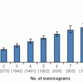 Breast Cancer Screening
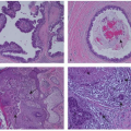 Ductal Carcinoma In Situ and Other Intraductal Lesions: Pathology, Immunohistochemistry, and Molecular Alterations
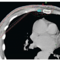 Postmastectomy Radiation Therapy
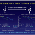 Preoperative Endocrine Therapy for Operable Breast Cancer
 Management Summary for the Care of Patients with Metastatic Breast Cancer


