Late Effects of Childhood Cancer and its Treatment


Late Effects of Childhood Cancer and its Treatment


Wendy Landier

Saro H. Armenian

Anna T. Meadows

Smita Bhatia


Currently, an estimated 15,780 new cases of cancer are diagnosed, and an estimated 1,960 deaths from cancer occur annually among children and adolescents age birth to 19 years.1 An estimated 379,112 survivors of childhood and adolescent cancer were alive in the United States as of January 1, 2010, and this number is estimated to increase to over 500,000 by 2020.2 The top three cancers among childhood cancer survivors are acute lymphoblastic leukemia (ALL), brain tumors, and Hodgkin lymphoma (HL). Most (70%) survivors of childhood and adolescent cancer are aged 20 years or older. Approximately 1 in every 530 young adults between the ages of 20 and 39 years is a childhood cancer survivor.1 It is therefore likely that most pediatric and primary care practices will be involved in the follow-up of these survivors.

Unlike adults, the growing child tolerates the acute side effects of therapy relatively well. However, the use of cancer therapy at an early age can produce complications that may not become apparent until years later as the child matures—hence the term “late effect” for late-occurring or chronic outcome, either physical or psychological, which persists or develops beyond 5 years from the diagnosis of cancer. Table 48.1 summarizes the more commonly occurring late effects.


BURDEN OF MORBIDITY

The burden of morbidity is described by quantifying the chronic medical problems experienced by this population3; the incidence of at least one chronic health condition approaches 75% among childhood cancer survivors at 30 years from cancer diagnosis.4 Furthermore, approximately 40% experience a late effect that is severe/life-threatening/disabling or fatal.4 Individuals identified to be at highest risk include those treated for HL or brain tumors, and those exposed to chest radiation and anthracyclines. More importantly, the prevalence of adverse health outcomes increases as the cohort ages. This was demonstrated in a recent study where the estimated cumulative prevalence was 95.5% for any chronic health condition and was 80.5% for a serious/disabling/life-threatening chronic condition by age 45 years.5 Recipients of hematopoietic stem cell transplantation (HSCT) during childhood are at an especially increased risk of developing severe/life-threatening/disabling conditions; the risk is nearly fourfold that of childhood cancer survivors treated with conventional therapy.6 Childhood cancer survivors are also at an increased risk of hospitalization.7,8 Female gender, an existing chronic health condition and/or a second malignant neoplasm (SMN), and prior treatment with radiation are associated with an increased risk of nonobstetrical hospitalization.

These studies demonstrate quite conclusively that the implications of cure are not trivial, and that the burden of morbidity carried by childhood cancer survivors is quite substantial. Furthermore, these data support a critical need for continuing follow-up of childhood cancer survivors into adult life. There exists a need for increased awareness of this burden of morbidity by both the survivors and their health care providers to develop and institute appropriate surveillance strategies.


STANDARDIZED RECOMMENDATIONS FOR FOLLOW-UP OF CHILDHOOD CANCER

The Institute of Medicine has recognized the need for a systematic plan for lifelong surveillance that incorporates risks based on therapeutic exposures, genetic predisposition, health-related behaviors, and comorbid health conditions.12 In response to this, the Children’s Oncology Group (COG) developed risk-based, exposure-related guidelines (Long-Term Follow-Up Guidelines for Survivors of Childhood, Adolescent, and Young Adult Cancers)13 specifically designed for follow-up care of patients who have completed treatment for pediatric malignancies. Specially tailored patient education materials, known as “Health Links” accompany the guidelines, offering detailed information on guideline-specific topics to enhance health promotion in this population with specialized health care needs. The Guidelines and the Health Links can be downloaded from www.survivorshipguidelines.org,14 and screening recommendations for commonly occurring late effects are summarized in Table 48.1.


TABLE 48.1 Exposure-Based Screening Recommendations for Commonly Occurring Late Effects in Childhood Cancer Survivors


Adverse Outcome


Therapeutic Exposures Associated with Increased Risk


Factors Associated with Highest Risk


Recommended Screening


Adverse psychosocial effects (mental health disorders, risky behaviors, psychosocial disability due to pain, fatigue, limitations in health care and insurance access)


Any cancer experience


CNS tumor; cranial irradiation; hearing loss; premorbid learning or emotional difficulties; older age at diagnosis


Psychosocial assessment with attention to: Educational and/or vocational progress, social withdrawal, anxiety, depression, posttraumatic stress, suicidal ideation, health care and insurance access


Yearly


Hearing loss


Cranial irradiation, platinum-based chemotherapy


Younger age (<4 y) at treatment, increasing radiation and chemotherapy dose


Complete audiologic evaluation


Baseline at entry to LTFU and as clinically indicated for patients who received platinum; every 5 y for patients who received radiation.


Cataracts


Cranial irradiation, total body irradiation, corticosteroids


Higher radiation dose, combination of steroids and radiation, single daily fraction


Eye exam (visual acuity, funduscopic exam)


Yearly


Radiation only:


Evaluation by ophthalmologist


Yearly if radiation dose was >30 Gy; every 3 y if radiation dose was <30 Gy


Dental abnormalities


Cranial irradiation, any chemotherapy prior to permanent dentition


Younger age at treatment


Dental exam and cleaning


Every 6 mo


Neurocognitive deficits


Cranial irradiation, intrathecal methotrexate, high-dose methotrexate and cytarabine


Female sex, younger age (<5 y) at treatment, cranial irradiation, intrathecal methotrexate


Review of educational and/or vocational progress


Yearly


Neuropsychological evaluation


Baseline at entry to LTFU; repeat as clinically indicated


Obesity


Cranial irradiation; neurosurgery involving the hypothalamic-pituitary axis


Younger age at treatment (<8 y), female sex, cranial irradiation dose >20 Gy


Height, weight, BMI


Yearly


Growth hormone deficiency


Cranial irradiation


Radiation dose >18 Gy


Targeted history and physical examination including height, weight, BMI and Tanner staging


Every 6 mo until growth is completed, then yearly


Precocious puberty


Cranial irradiation


Female sex, younger age at treatment, radiation dose >18 Gy


Height, weight, Tanner staging


Yearly until sexually mature


Hypothyroidism


Radiation to the thyroid gland (neck, mantle, etc.)


Increasing dose, female sex, age at treatment


Free T4, TSH


Yearly


Cardiomyopathy/congestive heart failure


Atherosclerotic heart disease, myocardial infarction, valvular disease


Anthracyclines, chest and spinal irradiation


Chest and spinal irradiation


High cumulative doses (>500 mg/m2), females, younger than 5 y at treatment, mediastinal irradiation


Echocardiogram


Every 1 to 5 y as indicated based on age at treatment, anthracycline dose, and history of radiation with potential impact to the heart


Electrocardiogram


Baseline at entry to LTFU; repeat as clinically indicated


Radiation only:


Fasting blood glucose or HgA1C and lipid profile


Every 2 y; if abnormal, refer for ongoing management


Pulmonary fibrosis/interstitial pneumonitis


Bleomycin, chest or whole lung irradiation, carmustine, lomustine, busulfan


Younger age at treatment, bleomycin dose >400 U/m2


Pulmonary function tests


Baseline at entry to LTFU; repeat as clinically indicated


Hepatic dysfunction


Methotrexate, mercaptopurine, thioguanine, irradiation involving the liver


Previous veno-occlusive disease of the liver, chronic viral hepatitis


ALT, AST, total bilirubin


Baseline at entry to LTFU; repeat as clinically indicated


Renal dysfunction (glomerular and/or tubular)


Platinum-based therapy, ifosfamide, high-dose methotrexate, abdominal irradiation, surgery


High-dose chemotherapy, younger age, abdominal radiation, and chemotherapy


Blood pressure, urinalysis


Yearly


Serum BUN, creatinine, and electrolytes including calcium, phosphorus, magnesium


Baseline at entry to LTFU; repeat as clinically indicated


Bladder complications


Alkylating agents, abdominal irradiation, surgery


Use of high-dose alkylating agents without bladder uroprophylaxis, abdominal radiation


Targeted history, urinalysis


Yearly


Hypogonadism (acute or premature ovarian failure in females)


Alkylating agents, craniospinal irradiation, abdomino-pelvic irradiation, gonadal irradiation


Treatment during peripubertal or postpubertal period in girls, higher cumulative doses of alkylators; gonadal irradiation


Pubertal onset, tempo, Tanner staging


Yearly until sexually mature


Females: Serum FSH, LH, estradiol


Baseline at age 13, repeat as clinically indicated


Males: Serum testosterone


Baseline at age 14, repeat as clinically indicated; ideally obtain in the morning


Infertility


Alkylating agents, craniospinal irradiation, abdomino-pelvic irradiation, gonadal irradiation


Males sex; higher doses of alkylators; gonadal irradiation; total body irradiation


Females:


Targeted history and physical examination


Yearly


Males: Semen analysis


At request of sexually mature patient


Males: FSH


If unable to obtain semen analysis


Short stature; musculoskeletal growth problems


Cranial irradiation, corticosteroids, total body irradiation


Younger age at treatment, cranial radiation dose >18 Gy, unfractionated (10 Gy) total body irradiation


Standing and sitting height


Yearly until growth completed


Scoliosis/kyphosis


Radiation involving the chest, abdomen, or spine; thoracic surgery; neurosurgery-spine


Younger age at irradiation, higher radiation doses; hemithoracic, abdominal, or spinal surgery


Spine exam for scoliosis and kyphosis


Yearly until growth completed; may need more frequent assessment during puberty


Reduced bone mineral density


Corticosteroids, craniospinal irradiation, gonadal irradiation, total body irradiation


Associated hypothyroidism, hypogonadism, growth hormone deficiency


Bone density evaluation (DEXA or quantitative CT)


Baseline at entry to LTFU; repeat as clinically indicated


Avascular necrosis


Corticosteroids, high-dose radiation to any bone


Dexamethasone, adolescence, female sex


Targeted history and physical examination


Yearly


Life-threatening infection


Splenectomy, radiation impacting the spleen (>40 Gy), chronic active graft-versus-host disease


Anatomic asplenia; higher radiation doses to the spleen; ongoing immunosuppression; hypogammaglobulinemia


Blood culture


When febrile, temperature > 101°F (>38.3°C)


Chronic Hepatitis C Virus (HCV) infection and HCV-related sequelae


Transfusions before 1993


Living in hyperendemic area


Hepatitis C antibody


Once if treated prior to 1993 (date may vary for international patients)


Hepatitis C PCR


Once in patients with positive Hepatitis C antibody


Therapy-related myelodysplasia, therapy-related acute myeloid leukemia


Alkylating agents, epipodophyllotoxins, anthracyclines


Increasing dose of chemotherapeutic agents, older age at therapeutic exposure, autologous hematopoietic cell transplant


Targeted history/physical examination


Yearly


Skin cancer (basal cell, squamous cell, melanoma)


Radiation (any field)


Orthovoltage radiation (prior to 1970)—delivery of greater dose to skin, additional excessive exposure to sun, tanning booths


Physical examination


Yearly


Secondary brain tumor


Cranial irradiation


Increasing dose, younger age at treatment


Targeted history and neurologic examination


Yearly


Thyroid cancer


Radiation to the thyroid gland (neck, mantle, etc.)


Increasing dose up to 29 Gy, female sex, younger age at radiation


Physical examination


Yearly


Breast cancer


Chest irradiation


Increasing dose, female sex, longer time since radiation


Females:


Clinical breast exam


Yearly beginning at puberty until age 25, then every 6 mo


Mammogram and breast MRI


Yearly for patients who received >20 Gy beginning 8 y after radiation or at age 25, whichever occurs last. For patients who received 10-19 Gy, clinician should discuss benefits and risks/harms of screening with patient; if decision made to screen, then follow recommendations for >20 Gy


Colorectal cancer


Abdominal/pelvic irradiation Spinal irradiation


Higher radiation dose to bowel; higher daily dose fraction; combined with chemotherapy (especially alkylators)


Colonoscopy


Every 5 y (minimum) for patients who received >30 Gy, beginning 10 y after radiation or at age 35 y, whichever occurs last; more frequently if indicated based on colonoscopy results. Monitoring of patients who received total body irradiation without additional radiation potentially impacting the colon/rectum should be determined on an individual basis.


ALT, alanine aminotransferase; AST, aspartate aminotransferase; BMI, body mass index; BUN, blood urea nitrogen; CT, computed tomography; DEXA, dual x-ray absorptiometry; FSH, follicle-stimulating hormone; Gy, Gray; HgA1C, hemoglobin A1C; HIV, human immunodeficiency virus; IV, intravenous; LH, luteinizing hormone; LTFU, long-term follow-up; MRI, magnetic resonance imaging; N/A, not applicable; PCR, polymerase chain reaction; T4, thyroxine; TSH, thyroid-stimulating hormone.


Screening recommendations are based on the Children’s Oncology Group Long-Term Follow-Up Guidelines, Version 4.0, available in their entirety at www.survivorshipguidelines.org.


A web-based version of the COG Long-Term Follow-Up Guidelines, known as Passport For CareSM (PFC), generates individualized follow-up recommendations for screening, evaluation, and survivor education, which are customized to the needs of each patient based on their disease and treatment. The PFC, developed by investigators at the Baylor College of Medicine and Texas Children’s Cancer Center, in collaboration with the COG,15 is a national resource that serves as the model for meeting the needs of cancer survivors. In addition to the COG Long-Term Follow-Up Guidelines, several other guidelines16,17,18 have been developed to address survivorship care. These guideline groups have formed a worldwide collaboration, aimed at international harmonization of long-term follow-up guidelines for childhood cancer survivors.19

Regardless of the setting for follow-up, the first step in any evaluation is to develop an outline of the patient’s medical history, including a treatment summary, and other elements listed in Table 48.2. Once completed, the treatment summary allows the survivor or their health care provider to determine recommended follow-up care according to long-term follow-up guidelines. Before the survivor graduates from a pediatric oncologist’s care, this treatment summary and list of potential long-term complications with associated screening recommendations should be reviewed with the family and, in the case of adolescents or young adults, with the patient. Correspondence between the pediatric oncologist and subsequent caretakers should address these same issues.

In the sections below, we review some of the known and emerging late effects in survivors of childhood cancer, and the relationship between these late effects and specific therapeutic exposures, so as to suggest reasonable starting points for evaluation of specific long-term problems using the screening recommendations from the COG Long-Term Follow-Up Guidelines. Therapeutic agents commonly used to treat specific childhood cancer types are summarized in Table 48.3.


TABLE 48.2 Key Elements of a Comprehensive Treatment Summary


Key Elements


Details


Demographics


Name, sex, date of birth, treating institution, treatment team


Diagnosis


Initial diagnosis type, date, stage, site(s) relapses(s) with date(s) and site(s), if applicable


Subsequent malignant neoplasms with type and site(s), if applicable


Therapeutic exposures


Chemotherapy


List of all agents received including route of administration


Include cumulative doses (per m2) for alkylators, anthracyclines, and bleomycin


If available, include cumulative doses for all agents


If patient received IV methotrexate or IV cytarabine, indicate whether any doses were in the intermediate/high dose range (>1,000 mg/m2)


If patient received carboplatin, indicate whether any dose was myeloablative and whether survivor was <age 1 at time of diagnosis


Radiation


Dates, type, fields (laterality), total dose, no. of fractions/dose per fraction


Surgical procedures


Type(s), date(s)


Hematopoietic cell transplant


Type(s), date(s), GVHD prophylaxis/treatment


Completion of therapy


Date


GVHD, graft-versus-host disease; IV, intravenous.


TABLE 48.3 Agents Commonly Used to Treat Specific Childhood Cancer Types


Tumor Type


Commonly Used Agents


Class


Acute lymphoblastic leukemia


Lymphoblastic lymphoma


Vincristine


Vinca alkaloids


Prednisone


Dexamethasone


Corticosteroids


Asparaginase


Enzymes


6-mercaptopurine


6-thioguanine


Methotrexate (low and high-dose IV, IT)


Cytarabine (IV, IT)


Nelarabine


Antimetabolites


Doxorubicin


Daunorubicin


Anthracyclines


Cyclophosphamide


Alkylators


Imatinib, sorafenib


Tyrosine kinase inhibitors


Cranial irradiation (historical)


Spinal irradiation (historical)


Radiation


Acute myeloid leukemia


Cytarabine (high-dose IV, IT)


Antimetabolites


Daunorubicin


Anthracyclines


Etoposide


Epipodophyllotoxins


Asparaginase


Enzymes


6-thioguanine


Antimetabolites


Dexamethasone


Corticosteroids


All-trans-retinoic acid (ATRA)


Retinoids


Hematopoietic stem cell transplant (HSCT)


Transplant


Busulfan (HSCT conditioning)


Cyclophosphamide (HSCT conditioning)


Alkylators


Total body irradiation (HSCT conditioning)


Radiation


Hodgkin lymphoma


Nitrogen mustard


Cyclophosphamide


Procarbazine


Dacarbazine (DTIC)


Alkylators


Vincristine


Vinblastine


Vinca alkaloids


Prednisone


Corticosteroids


Doxorubicin


Anthracyclines


Bleomycin


Antitumor antibiotics


Oophoropexy


Surgery


Mantle irradiation


Inverted Y irradiation


Involved field irradiation


Radiation


Non-Hodgkin lymphoma


Cyclophosphamide


Ifosfamide


Alkylators


Doxorubicin


Anthracyclines


Methotrexate (high dose)


Antimetabolites


Prednisone


Corticosteroids


Vincristine


Vinca alkaloids


Asparaginase


Enzymes


Etoposide


Epipodophyllotoxins


Solid tumors


Cyclophosphamide


Ifosfamide


Melphalan


Thiotepa


Dacarbazine (DTIC)


Alkylators


Cisplatin


Carboplatin


Platinum analogs


Doxorubicin


Epirubicin


Anthracyclines


Etoposide


Teniposide


Epipodophyllotoxins


Methotrexate (high dose) 5-FU


Antimetabolites


Irinotecan


Topotecan


Plant alkaloids


(Topoisomerase I Inhibitors)


Paclitaxel


Docetaxel


Taxanes


13-cis-retinoic acid


Retinoids


Imatinib


Sorafenib


Tyrosine kinase inhibitors


Vincristine


Vinblastine


Vinorelbine


Vinca alkaloids


Bleomycin


Actinomycin D


Antitumor antibiotics


Nephrectomy (renal tumors)


Surgery


Cystectomy (pelvic tumors)


Surgery


Amputation (musculoskeletal tumors)


Limb salvage (musculoskeletal tumors


Surgery


MIBG (metaiodobenzylguanidine) Iodine-131


Systemic radiation


Whole lung irradiation


Radiation


Local irradiation (to tumor bed)


Radiation


Central nervous system tumors


Cisplatin


Carboplatin


Platinum analogs


Cyclophosphamide


Procarbazine


CCNU


Temozolomide


Alkylators


Vincristine


Vinblastine (germ cell tumors)


Vinca alkaloids


Prednisone


Dexamethasone


Corticosteroids


Etoposide


Epipodophyllotoxins


6-Thioguanine


Antimetabolites


Bleomycin (germ cell tumors)


Actinomycin D (optic pathway tumors)


Antitumor antibiotics


Neurosurgery—brain


Neurosurgery—spine


Surgery


Cranial irradiation


Spinal irradiation


Radiation


IV, intravenous; IT, intrathecal.


NEUROCOGNITIVE SEQUELAE

Childhood cancer survivors are at increased risk for neurocognitive impairment. Cranial radiation has long been associated with neurocognitive late effects,39,40 although antimetabolite chemotherapy and corticosteroids have also been implicated as potential contributors to neurocognitive impairment.41 Neurocognitive deficits usually become evident within 1 to 2 years following cranial radiation and are progressive in nature. The decline over time is typically reflective of the child’s failure to acquire new abilities or information at a rate similar to peers, rather than because of a progressive loss of skills and knowledge. Affected children experience information-processing deficits resulting in academic difficulties, and are prone to problems with receptive and expressive language, attention span, and visual and perceptual motor skills. Neurocognitive function in long-term survivors of childhood cancer appears particularly vulnerable to the effects of fatigue and sleep disruption.42

Brain tumor survivors are at significant risk for impairment in neurocognitive functioning, particularly if they have received cranial radiation at a young age, had a VP shunt placed, suffered a cerebrovascular incident, or are left with hearing or motor impairments.43,44 Younger children may experience significant drops in intelligence quotient (IQ) scores, with irradiation-orchemotherapy-induced destruction in normal white matter over time, partially explaining intellectual and academic achievement deficits in these survivors.43,44 Neurocognitive difficulties include slow processing speed, inattention, and memory impairment, and deficits in verbal skills, visual spatial skills, psychomotor speed, and learning.44,45,46,47,48,49 These neurocognitive impairments adversely affect education, employment, income, and marital status.45 Patients with medulloblastoma exposed to lower doses of whole-brain irradiation (23.5 Gy) are at a lower risk of neurocognitive
impairment, when compared with those receiving higher doses of radiation.50

Survivors of childhood ALL are at risk for neurocognitive problems, generally characterized by reduced attention, processing speed, executive function, and global intellectual function.51,52 Treatment of ALL patients with cranial radiation at a dose of 24 Gy is associated with cognitive deficits.53 Additional risk factors include increased treatment intensity, younger age at treatment exposure, and female sex.54,55 Although neurocognitive problems are clearly linked to cranial radiation, there is evidence of subtle long-term neurocognitive deficits in survivors of childhood ALL after treatment with chemotherapy alone.56,57 These deficits are restricted to attention, executive function, and complex fine-motor functioning; global intellectual function is relatively preserved. Younger patients and females are at higher risk for these deficits.51,56,58,59,60 The neurocognitive impairment observed in survivors of childhood ALL persists into adulthood; this impairment is associated with reduced educational attainment and unemployment,51,61 independent living, and health care use.62

The observed interindividual variability in the risk of neurocognitive impairment suggests the role for genetic susceptibility to neurocognitive impairment. Preliminary evidence supports an association between A2756G polymorphism in methionine synthase (MS) and reduced attentiveness/response speed in childhood ALL survivors.63,64 Polymorphisms in MS are associated with hyperhomocysteinemia.65 Excess homocysteine increases risk for vascular abnormalities, including stroke.66,67 Survivors with MS polymorphisms who were treated with methotrexate, which could increase homocysteine levels, may be at increased risk for these abnormalities. There is also evidence for an association between glutathione S-transferase (GST) and increased performance variability and reduced attentiveness, in survivors of childhood ALL and medulloblastoma.68 GSTs are enzymes involved in sequestering reactive oxygen species,69 and polymorphisms in the GST gene may interfere with the ability to respond to oxidative stress. An association between monoamine oxidase (T1460CA) and increased attention variability has also been reported. MAOA is involved in serotonin and norepinephrine catabolism, and low activity of this gene has been associated with increased norepinephrine and overactivation of the sympathetic nervous system. This process may result in increased anxiety and/or physiologic stress, which are associated with attention problems.70,71 The association with polymorphisms in MAOA may suggest a predisposition for attention problems in a subset of survivors, one that may not be related to specific chemotherapy agents. Finally, parent-reported attention problems are more common in children with the Cys112Arg polymorphism in apolipoprotein E4.64 APOE is involved in lipoprotein metabolism,72,73 and the E4 polymorphism is a risk factor for dementia.74 APOE-4 has also been associated with age-related myelin breakdown and risk of neurocognitive impairment after traumatic brain injury.75


Recommendations for Screening and Follow-Up

A baseline neuropsychological evaluation is recommended for patients who received therapy that may impact neurocognitive function. This should be repeated as clinically indicated and at key transition points (transitioning from grade school to middle/junior high school); an annual assessment of their vocational or educational progress should also be monitored.


CARDIOVASCULAR FUNCTION

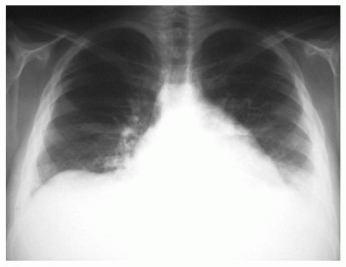
The anthracyclines doxorubicin and daunomycin are well-known causes of cardiomyopathy and clinical congestive heart failure (CHF) (Fig. 48.1). Anthracyclines have a wide range of clinical activity against pediatric cancers, and it is estimated that as many as 60% of childhood cancer survivors have been treated with anthracyclines, making it one of the more common therapeutic exposures.76 Anthracycline cardiotoxicity is thought to be related to direct myocardial injury due to formation of free radicals. Over time, the left ventricular wall thins, leading to increased myocardial stress and decreased contractility.76 Progressive cardiomyopathy can occur early within the first year of treatment or can be delayed, being diagnosed years following completion of therapy; the risk of disease is dose-dependent.76 The incidence of CHF is less than 5% with cumulative anthracyclines exposure of <300 mg/m2; the incidence approaches 20% at doses between 300 and 600 mg/m2, and exceeds 35% for doses >600 mg/m2.77,78,79 The risk of anthracycline-related CHF is modified by younger age (<5 years) at exposure and concomitant mediastinal irradiation.76 CHF is associated with poor prognosis; 5-year overall survival rates are reported to be less than 50%.80,81 Advances in noninvasive cardiac imaging have allowed investigators to identify a growing population of survivors who may be at risk for late-occurring CHF, setting the stage for pharmacologic interventions to prevent progression to clinical CHF.82,83


Figure 48.1 Anthracycline-related congestive heart failure (dose-response relation).

It is increasingly evident that well-recognized clinical and therapeutic risk factors may not fully explain the wide interindividual variability in susceptibility to therapy-related CHF. Significant cardiotoxicity has been reported at cumulative doses of less than 250 mg/m2 in some patients,84 whereas doses that exceed 1000 mg/m2 have been tolerated without long-term sequelae by some. Investigators have begun examining the role of genetic susceptibility in the development of therapy-related CHF. Using a biologically plausible candidate gene approach, studies have identified genetic polymorphisms involved in metabolism of anthracyclines (CBR, ABCC2, SLC28A3), iron homeostasis (HFE), antioxidant defense (RAC2, NCF4, CYBA), and the myocardial response to the drug (HAS3), which could place survivors at increased risk for therapy-related CHF.84,85,86,87,88,89 Many of these genomic variables, when fully established, could be important in facilitating the implementation of targeted primary prevention strategies.

Mediastinal radiation has been implicated in the development of constrictive pericarditis, cardiomyopathy, valvular heart disease, coronary artery disease, and conduction abnormalities.76 Exposure to mediastinal radiation is associated with valvular fibrosis or insufficiency in 40% to 60% of HL survivors, whereas conduction defects are present in as many as 75%.90 Although clinically evident CHF is rare following mediastinal radiation alone, when present, it is primarily in the form of diastolic dysfunction, as opposed to systolic disease seen following anthracycline exposure.76,90

Coronary artery disease (CAD) has been reported following radiation to the mediastinum, with a cumulative risk of 21% at 20 years.76 The morphologic changes in radiation-induced vascular
disease are similar to those observed in spontaneous atherosclerosis. Coronary ostia and left anterior descending artery are frequently involved. The exact mechanism by which radiation produces atherosclerosis is not well understood, but it is likely that endothelial injury secondary to radiation initiates the process. However, significant radiation-associated CAD rarely occurs in the absence of other cardiovascular risk factors such as dyslipidemia, hypertension, and obesity.76

The pericardium is one of the most commonly affected structures of the heart after radiotherapy.76 Patients can present with chronic pericardial effusions, constrictive pericarditis, or sometimes with chronic effusions in association with pancarditis. Although total-heart radiation at a dose of 40 Gy appears to be the usual threshold, pericarditis has been reported following doses as low as 15 Gy, even in the absence of radiomimetic chemotherapy.76

Prevention of cardiotoxicity is currently being explored. Analogs of doxorubicin and daunomycin and liposomal anthracyclines, with a potential for decreased cardiotoxicity while retaining equivalent antitumor activity have been studied.91,92,93 Cardio-protectants such as dexrazoxane have been shown to minimize cardiac injury and remodeling shortly after anthracycline administration without compromising its antitumor efficacy.91,94 However, long-term data on efficacy of dexrazoxane is lacking, and certain subgroups, particularly children who have the greatest potential number of life years following cancer therapy, remain understudied.91 The role of pharmacologic intervention for prevention of CHF in asymptomatic survivors with left ventricular dysfunction is less well studied. A randomized placebo-controlled study using angiotensin-converting enzyme (ACE) inhibitors demonstrated that while ACE inhibitors did not prevent decline in ventricular function, they were able to provide some respite in the form of afterload reduction.95

Finally, studies conducted in non-oncology populations support the benefits of interventions to reduce modifiable risk factors, such as obesity, smoking, hypertension, diabetes, and dyslipidemia.96,97 Childhood cancer survivors are at a higher risk of developing hypertension, diabetes, and dyslipidemia, when compared to the general population.98,99 In fact, survivors who have hypertension or diabetes in addition to past exposure to anthracyclines and/or radiation are at an especially high risk of cardiovascular disease.100 While there have been no studies conducted to demonstrate a reduction in cardiovascular events after risk factor modification in cancer survivors, findings from studies in non-oncology populations suggest that routine screening for these risk factors may be beneficial, setting the stage for possible interventions to mitigate adverse cardiovascular outcomes.


Recommendations for Screening and Follow-Up

Patients exposed to anthracyclines need ongoing monitoring for late-onset cardiomyopathy using physical examination and serial noninvasive testing (echocardiogram).101 The frequency of echocardiograms can range from yearly to every 5 years, depending on cumulative anthracycline dose, age at exposure, and treatment with mediastinal radiation. Aerobic exercise is generally safe and should be encouraged for most patients. However, intensive isometric activities (i.e., heavy weight lifting, wrestling) should be avoided. Pregnant women previously treated with anthracyclines should be closely monitored, as changes in volume during the third trimester could add significant stress to a potentially compromised myocardium. In addition to monitoring for cardiomyopathy, survivors who received radiation involving the heart field also need monitoring for potential early-onset atherosclerosis. Heart-healthy lifestyles should be encouraged for all survivors, including implementation of a regular exercise program, dietary recommendations, as well as screening for dyslipidemia, hypertension, and diabetes. Additional specific recommendations for monitoring, based on age and therapeutic exposure, are delineated within the COG Long-Term Follow-Up Guidelines.14


PULMONARY FUNCTION

Lungs are particularly susceptible to radiation-induced injury. Radiation-related complications such as pulmonary fibrosis and pneumonitis are most often seen in patients with malignant diseases of the chest, notably HL and solid tumors, with pulmonary metastases such as in Wilms tumor and Ewing sarcoma. These changes can be detected months to years after completion of therapy, and are most prevalent in individuals with a history of pneumonitis as an acute complication of therapy. The incidence and severity of radiation-associated lung damage is related to the total dose, fractionation of that dose, type of radiation, total volume of lung irradiated, and age at exposure.102

Radiation-related pulmonary injury is likely mediated by cytokines, which stimulate septal fibroblasts, increasing collagen production, resulting in pulmonary fibrosis.102 The basis for respiratory damage in young children appears to be different from that in the adult or adolescent, and is likely because of resulting chest wall hypoplasia and compromised lung parenchymal growth. Studies in children who received whole lung radiation to treat metastatic disease consistently demonstrate deficits in total lung capacity (TLC), forced vital capacity (FVC), and diffusion capacity of the lung (DLCO), consistent with a proportionate interference with growth of the lungs and chest wall.103,104 While the incidence of radiation-induced late pulmonary toxicity has dramatically decreased in the past decade secondary to refined radiation therapy techniques, these patients remain at risk for declining pulmonary function years after completion of their cancer treatment.102,103,105

Certain chemotherapeutic agents can also compromise pulmonary function. Bleomycin toxicity is the prototype for chemotherapy-related lung injury. Clinically apparent bleomycin pneumonopathy (interstitial pneumonitis and pulmonary fibrosis) is more frequent in older adolescents and adults. The chronic lung toxicity usually follows persistence or progression of abnormalities, which develop within 3 months of completion of therapy. Dose-dependent toxicity occurs at treatment doses greater than 400 units, and can be exacerbated by concurrent or previous radiation therapy to the mediastinum.105 Above this dose, 10% of adult patients experience fibrosis102; data are limited for such high drug doses in children because few are exposed. Other chemotherapeutic agents such as cyclophosphamide, carmustine (BCNU), lomustine (CCNU), busulfan, and melphalan have also been associated with acute and late-onset pulmonary toxicity.102 However, most reports describing long-term pulmonary toxicity due to these agents have included doses that are no longer used in conventional cancer therapy.102

Pulmonary complications are a leading cause of morbidity and mortality in long-term HSCT survivors, where both restrictive and obstructive lung disease can significantly complicate the medical management of these survivors.106,107 The intensity of therapy in patients undergoing HSCT and the additive effects of previous therapies magnify the risk. Patients who received un-fractionated, single dose, TBI as part of their conditioning regimen have the highest risk for late pulmonary complications. Fractionated TBI has reduced the risk for many of these complications.107 Obliterative bronchiolitis (BO), a chronic, irreversible, obstructive lung disease can be diagnosed months to years after transplantation.108 Chest radiographs show hyperinflation, and chest CT scans demonstrate parenchymal hypoattenuation and air trapping. The treatment of BO remains a challenge; many patients with BO succumb to their pulmonary disease.108


Recommendations for Screening and Follow-Up

Monitoring for pulmonary dysfunction in childhood cancer survivors includes assessment of symptoms such as chronic cough or dyspnea on annual follow-up. Risks of smoking and exposure to secondhand smoke should be discussed with all patients. The
best approach to chronic pulmonary toxicity of anticancer therapy is preventive and includes respecting cumulative dosage restrictions of bleomycin and alkylators, limiting radiation dosage and port sizes, and avoidance of primary or secondhand smoke. Pulmonary function tests are recommended as a baseline upon entry into long-term follow-up for patients at risk, repeated as clinically indicated in symptomatic patients and in those with subclinical abnormalities identified on screening evaluation. Repeat evaluation should also be considered for at-risk patients prior to general anesthesia. Influenza and pneumococcal vaccines are encouraged in survivors at risk for pulmonary compromise.


GENITOURINARY ABNORMALITIES


Renal

Long-term renal complications in childhood cancer survivors can occur as a result of chemotherapy (cisplatin, carboplatin, ifosfamide, and methotrexate), radiation, or surgery. Chemotherapy-induced nephrotoxicity can manifest as acute irreversible renal failure, slow progressive chronic renal failure, or renal tubular dysfunction.109 Clinical manifestations of renal injury include hypertension, proteinuria, and varying degrees of renal insufficiency.109,110 Radiation nephropathy can present with many of the clinical symptoms seen in chemotherapy-related nephrotoxicity, whereas survivors who have undergone nephrectomy are at risk for additional complication such as hyperfiltration injury.109

Ifosfamide nephrotoxicity typically manifests as proximal tubular dysfunction, and, less often, as decreased glomerular filtration rate (GFR).111,112 It is estimated that approximately 30% of ifosfamide-treated children develop a persistent nephropathy, and 5% have clinically significant Fanconi renal syndrome (hypokalemia, hypophosphatemia, glucosuria, proteinuria, renal tubular acidosis, and rickets).113 Risk factors for chronic nephrotoxicity include higher cumulative dose (≥60 gm/m2), young age (<4 years) at treatment, concurrent or previous platinum-based therapy, irradiation involving the kidneys at a dose exceeding 15 Gy, and unilateral nephrectomy.112,113,114

Cisplatin can damage the glomerulus and distal renal tubules, potentially causing diminished GFR and electrolyte wasting, most commonly involving magnesium, calcium, potassium, and sodium.109 Hypomagnesemia may persist in some patients, and can be severe enough to require magnesium supplementation.115 It is estimated that up to 50% of patients remain hypomagnesemic, and the risk is greatest for those exposed to other nephrotoxic agents such as ifosfamide.109

Irradiation to the kidney can result in radiation nephritis or nephropathy after a latent period of 3 to 12 months. Doses in excess of 20 Gy can result in significant nephropathy.109 The prevalence of renal insufficiency approached 10% in one study of 5-year Wilms tumor survivors.114


Recommendations for Screening and Follow-Up

Surveillance in at-risk survivors should include monitoring of serum creatinine, blood urea nitrogen, and serum chemistries at baseline at entry into long-term follow-up; and should be repeated as clinically indicated. Urinalysis and measurement of blood pressure should be performed at baseline and annually thereafter. Ongoing management may include electrolyte replacement, treatment of hypertension, and avoidance of further nephrotoxic agents. In children with a history of radiation to the renal artery who develop hypertension, radiologic studies for stenosis of the renal artery should be ordered and, if stenosis is present, surgical correction should be undertaken. Patients with a history of nephrectomy should be counseled regarding the importance of protecting the remaining single kidney. These patients should be cautioned to avoid nephrotoxic agents (e.g., ibuprofen, aminoglycosides) when possible; to maintain normal weight; to use seatbelts properly (i.e., to wear lap belts around the hips, not the waist), and to seek early intervention for urinary tract infections. Additionally, mononephric survivors should be counseled regarding participation in sports and physical activity, stressing the importance of physical fitness in maintaining cardiovascular health, and taking into consideration current kidney health (position, size, function). The unlikely risk of renal injury related to sports participation should be considered in discussing the risk/benefit ratio of sports and physical activity with the survivor and his or her family.116,117,118


Bladder

Well-recognized complications involving the bladder include hemorrhagic cystitis, bladder fibrosis, and neurogenic bladder. Hemorrhagic cystitis (HC) is a condition in which irritation of the lining of the bladder leads to exposure of the submucosal blood vessels and bleeding.119,120 Treatment with alkylating agents such as cyclophosphamide, ifosfamide, and radiation has been implicated in the development of HC.119,120 Patients who receive both chemotherapy and radiation are at highest risk.119 Patients may report urinary urgency, frequency, dysuria, and suprapubic pain. Although HC typically occurs during therapy, it may become a chronic recurring problem after completion of therapy. The incidence of HC has been reported to be 15% in Ewing sarcoma patients treated with cyclophosphamide.120 Uroprophylaxis with 2-mercaptoethane (mesna) in conjunction with cyclophosphamide administration has decreased the incidence of HC; mesna administration is now the standard of care for protocols utilizing high-dose cyclophosphamide and ifosfamide.120

Radiation to the pelvis or bladder can result in fibrosis and scarring, with resultant decreased bladder capacity and predisposition to urinary tract infections.120 Changes in bladder function with diminished bladder compliance are noted within 6 to 9 months after irradiation and correlate with radiation dose. Patients exposed to radiation dose greater than 45 Gy to the whole bladder are at highest risk for late fibrosis and contractures.121

Surgery involving the lower genitourinary tract has the potential to impair normal function of the bladder and normal voiding mechanisms. Myogenic and neurogenic impairment may occur as a result of bladder or peripheral nerve injury during surgery to remove portions of the bladder or adjacent pelvic tumors.122,123 In addition, neural innervation to the bladder can be compromised if there is injury to the central nervous system (CNS) or at the spinal cord level, resulting in inability to void and/or incontinence.120 In patients with bladder rhabdomyosarcoma, partial cystectomy can affect bladder function by reducing bladder volume. The extent of resection generally predicts severity of bladder function compromise.120,123

Only gold members can continue reading. Log In or Register to continue

Stay updated, free articles. Join our Telegram channel

Aug 25, 2016 | Posted by in ONCOLOGY | Comments Off on Late Effects of Childhood Cancer and its Treatment

Full access? Get Clinical Tree

Get Clinical Tree app for offline access