Laboratory Markers of Activated Coagulation and Its Regulation


Laboratory Markers of Activated Coagulation and Its Regulation


Kenneth A. Bauer

Helen A. Ireland


The development of blood tests to predict thrombotic events, or to confirm or exclude the presence of thrombosis, has been a goal of scientists working in the field for many years. The hemostatic mechanism comprises platelets, vessel wall components, and procoagulant, inhibitory, and fibrinolytic proteins. Advances in our knowledge of the biochemistry of these processes have facilitated the development of sensitive and specific assays that are able to detect platelet activation, generation of coagulation enzymes, products of intravascular fibrin formation or dissolution, and activated inhibitory proteins in addition to the inactive coagulation factors (zymogens) and their inhibitors. This chapter focuses on measures of activated markers of hemostasis and their relationship to thrombotic disease. While many of these assays have not found a place in clinical practice, their use as research tools has furthered our understanding of the hemostatic mechanism and its perturbation in many disease states.

Due to the short half-life of most of the hemostatic enzymes, it has not been possible to measure directly the levels of most of the active forms in vivo. Many of the enzymes are neutralized rapidly by naturally occurring protease inhibitors or bound to cellular receptors in the locale in which they are generated. Faced with these obstacles, immunochemical assays have been developed for peptides that are liberated with the activation of coagulation zymogens or for enzyme-inhibitor complexes. The assays that have been developed, along with the reported in vivo half-lives for some of the species, are listed in Table 58.1 and described in detail in a later section of this chapter. Most of these assays were initially developed in research laboratories, but some are now available commercially in kit form using either polyclonal or monoclonal antibodies.

The assay formats for these markers are generally of two types. The first approach that has been used is a competitive radioimmunoassay procedure. This requires an antibody population that recognizes antigenic determinants on the activation fragment or enzyme-inhibitor complex that are hidden in the parent zymogen or inhibitor. The need for highly specific antibodies to the marker of interest could sometimes be obviated by devising sample processing procedures that efficiently remove the cross-reacting species in plasma before assay. The use of the radioimmunoassay has, however, waned due to the requirement of radioactive tracers. The second approach, used more commonly today, is the enzyme-linked immunosorbent assay. In most instances, immunoglobulin G (IgG) or F(ab′) two fragments directed toward epitopes on one region of the marker are bound to wells of plastic microtiter plates to “capture” the antigen from the plasma specimen. After washing away unbound species, a second IgG or F(ab′) two population recognizing epitopes in a different domain of the marker molecule is added to the wells. These antibodies are coupled to an enzyme capable of cleaving a suitable colorimetric substrate, allowing the development of a titration curve.

Successful execution of clinical investigations seeking to establish a relation between markers of coagulation, fibrinolysis, and thromboembolic disease requires, first, that the selected assays are specific for the products of interest and possess sufficient sensitivity. Second, the tests must be standardized so that the assays perform in a reproducible manner. Third, care must be taken to ensure that technical factors do not introduce in vitro artifacts that can significantly alter the immunoassay results (e.g., drawing of blood samples through indwelling intravenous catheters). These include venipuncture quality, choice of anticoagulant, sample processing procedures (if required), and plasma storage conditions.


ASSAYS FOR COAGULATION ACTIVATION

Many of the protein components of the hemostatic mechanism are inactive zymogens that are converted to their active forms, serine proteases. For coagulation system enzymes to be generated at any significant rate, a zymogen, a cofactor, and a converting enzyme must form a multimolecular complex on a natural surface. These transformations are suppressed if the converting enzyme is inhibited, the protein cofactor is destroyed, or the surface receptors that are essential for the assembly of the macromolecular complex are sequestered. For an overview of the blood coagulation mechanism, see Chapter 8.


Assays for Contact System Activation

Contact system activation can be monitored by immunoassays for factor XIIa, factor XIIa-C1 inhibitor complex, kallikrein-C1 inhibitor, and kallikrein-α2-macroglobulin. Factor XI activation can be measured as factor XIa and factor XIa-factor XIa inhibitor (α1-antitrypsin, antithrombin, C1 inhibitor, and α2-antiplasmin) complexes.1,2,3,6,7


Assays for Intrinsic and Extrinsic Pathway Activation

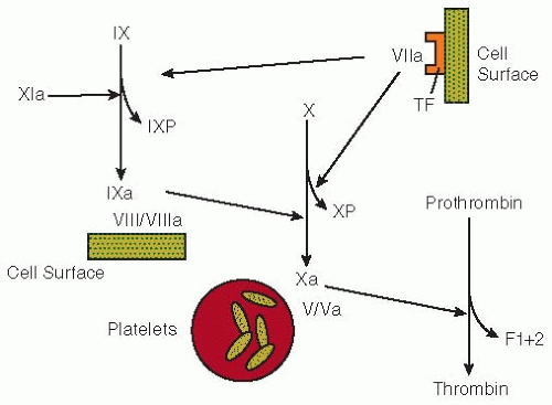
The activation of factor IX can be monitored by measuring the levels of the factor IX activation peptide10,47 or factor IXa-antithrombin complexes.11 These assays reflect the action of factor XIa, the factor VII-tissue factor complex, or both, on factor IX (FIGURE 58.1). Factor X activation mediated by the extrinsic or intrinsic pathways can be monitored by measuring the factor X activation peptide.12


Table 58.1 Immunochemical and enzymatic markers of coagulation and fibrinolytic system activation


Biochemical Step


Marker (Reference)


T1/2 (Reference)


Coagulation


Factor XII-factor XIIa


Factor XIIa1


Factor XIIa-C1 inhibitor complex2,3


Prekallikrein-kallikrein


Kallikrein-C1 inhibitor complex3,4


Kallikrein-α2-macroglobulin complex5


Factor XI-factor XIa


Factor XIa-XIa inhibitor complexes6,7


Factor VII-factor VIIa


Activated factor VII8,9


Factor IX-factor IXa


Factor IX activation peptide10


15 min


Factor IX-antithrombin complex11


30 min


Factor X-factor Xa


Factor X activation peptide12


30 min


Factor X-antithrombin complex13


Prothrombin-thrombin


Prothrombin activation fragment F1+215,16,17,18


90 min14


TAT complex19,21,22


15 min20,23


Protein C-activated protein C


Protein C activation peptide24


5 min


Activated protein C-protein C inhibitor complex26,27


40 min25


Activated protein C-α1-proteinase inhibitor (antitrypsin)27


140 min25


Fibrinogen-fibrin


Fibrinopeptide A28,29,30


3-5 min29


Fibrinopeptide B31,32


Fibrinolysis


Plasminogen-plasmin


Plasmin-α2-antiplasmin complex22,33,34,35,36,37,38


Plasmin-α2-macroglobulin complexes34


Plasmin action on fibrin I


Bβ1-42 fragment39,40


Plasmin action on fibrin II


Bβ15-42 fragment41


Plasmin action on fibrinogen/fibrin


Fibrinogen (fibrin) degradation products42,43


Plasmin action on cross-linked fibrin


D-dimer44,45


8 h46


An assay for factor VIIa uses a clotting assay utilizing recombinant tissue factor that has undergone a C-terminal truncation.8,9 This tissue factor mutant maintains cofactor activity toward factor VIIa but does not support factor VII activation.


Assays for Measuring In Vivo Thrombin Generation

Prothrombin activation fragment F1+2 (F1+2): Thrombin generation takes place at an appreciable rate under physiologic conditions only in the presence of factor Xa, factor Va, calcium ions, and activated platelets. During this process, the aminoterminus of the prothrombin molecule is released as F1+2 (see FIGURES 58.1 and 58.2).

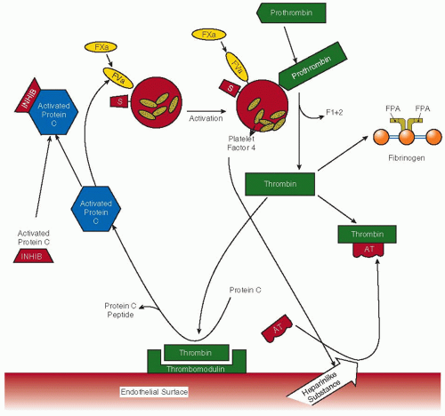
Thrombin-antithrombin complex measurement: Thrombin and other serine proteases within the coagulation pathway can be rapidly inhibited by circulating antithrombin, in a mechanism potentiated by endogenous heparan sulfate. Thrombin-antithrombin (TAT) complex can be measured in plasma and used as a marker of thrombin generation19,21,48 (FIGURE 58.2).


Assays for Thrombin Activity

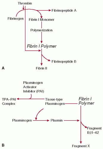
Once evolved, the serine protease, thrombin, converts fibrinogen into fibrin, releasing fibrinopeptides A and B from the aminoterminal regions of the Aα and Bβ chains of fibrinogen, respectively49 (see FIGURE 58.3A). The rate of fibrinopeptide A release from fibrinogen is considerably faster than that of fibrinopeptide B.31,49,50 Cleavage of fibrinopeptide A from fibrinogen generates an intermediate known as fibrin I that can polymerize but crosslinks poorly.51,52 Further proteolysis of fibrin I can occur through the action of either thrombin or plasmin. If thrombin action predominates, fibrinopeptide B is released, and fibrin II is formed. Immunoassays for fibrinopeptide A28,30,53,54,55 and fibrinopeptide B31,32 provide an index of thrombin action on fibrinogen.


ASSAYS FOR INHIBITOR COMPLEXES AND ACTIVATED INHIBITORS OF COAGULATION


Protein C Pathway

Thrombin activity and generation can also be inhibited rapidly by activation of the protein C pathway. Thrombin binds to
thrombomodulin on vascular endothelial cells and once bound can no longer participate in procoagulant mechanisms, either to activate platelets or to cleave fibrinogen. Endothelial protein C receptor, which lies adjacent to thrombomodulin within the caveolae region of the endothelial membrane, augments this mechanism by capture of protein C from the circulation, particularly in large vessels, and presents protein C to the thrombin-thrombomodulin complex. Thrombin-thrombomodulin activates protein C, which then inactivates factor Va and factor VIIIa by cleavage, thereby also regulating thrombin generation. Assays have been developed to monitor this pathway (FIGURE 58.2). Immunoassays to measure protein C activation include measurements of the protein C activation peptide,24 an immunoactivity assay for activated protein C,56,57,58 and activated protein C-inhibitor complexes.26,27,59


FIGURE 58.1 Pathways of coagulation activation. The activation of factor IX by factor XIa or the factor VII-tissue factor (TF) mechanism liberates the factor IX activation peptide (IXP). The conversion of factor X to factor Xa by the factor IXa-factor VIII/VIIIa-cell surface complex releases the factor X activation peptide (XP). The generation of thrombin from prothrombin is mediated by factor Xa in the presence of factor Va and activated platelets. During this process, the F1+2 fragment is released.


ASSAYS FOR ACTIVATED FIBRINOLYSIS AND ITS INHIBITION

Tissue plasminogen activator (tPA), produced by endothelial cells, converts circulating plasminogen to plasmin, the enzyme responsible for degradation of the fibrin clot. This process can be suppressed by complex formation between tPA and circulating plasminogen activator inhibitor 1 (PAI-1).


Fibrinopeptides Released by Plasmin

Plasmin action on fibrin I releases Bβ1-42 from the aminoterminus of the Bβ chain and cleaves the carboxyl terminus of the Aα chains, thereby generating fragment X60 (FIGURE 58.3B). In contrast, plasmin proteolysis of fibrin II results in the release of Bβ15-42 rather than Bβ1-42 because fibrinopeptide B has already been removed by the action of thrombin. Plasma levels of Bβ1-4239,40 and Bβ15-42,41 peptides released by the proteolytic action of plasmin on fibrinogen and fibrin, respectively, have been used as indices of in vivo plasmin activity. The antisera used in these assays show significant, but not absolute, specificity for the various fibrinopeptides relative to fibrinogen. Consequently, cross-reacting fibrinogen must be removed from plasma samples before analysis, using techniques that do not alter the levels of the peptides.29,61

The initial assay for Bβ1-42 was indirect and involved measurement of fibrinopeptide B immunoreactivity before and after thrombin addition to fibrinogen-depleted plasma. The resultant increase in immunoreactivity, termed thrombin-increasable fibrinopeptide B, reflects the presence of Bβ1-42, a fibrinopeptide B-containing fragment.62 Assays were subsequently developed that directly measured the levels of Bβ1-42 with either a polyclonal40 or a monoclonal antibody.39 These antisera are specific for Bβ1-42 and do not cross-react with fibrinopeptide B or Bβ15-42. An alternative approach is to use an antiserum raised against Bβ15-42 that cross-reacts completely with Bβ1-42 because it recognizes the carboxyl-terminal region of these peptides.63 However, the Bβ1-42/Bβ15-42 assay may underestimate the circulating levels of Bβ1-42 because release of the carboxyl-terminal arginine residue by circulating carboxypeptidases decreases the immunoreactivity of the peptide when measured with a carboxyl-terminal-directed antisera.40

Using a monoclonal antibody, a specific assay for Bβ15-42 has been developed41 that provides a marker of plasmin action on fibrin II. However, two factors limit the utility of this test. First, because the antibody exhibits some cross-reactivity with Bβ1-42, the assay may not be a specific marker of fibrin proteolysis in patients with marked fibrinogenolysis.64 Second, only low levels of Bβ15-42 are generated during thrombolysis,65,66 probably because the Bβ42-43 bond of fibrin II is not cleaved as readily by plasmin as is the same bond of fibrinogen or fibrin I. Exploiting this phenomenon, the fibrin specificity of thrombolytic agents has been increased by linking them to monoclonal antibodies against the Bβ15-42 domain of fibrin, thereby targeting them to fibrin.67,68


Assays for Large Fragments of Fibrinogen and Fibrin

The levels of fibrinogen or fibrin degradation products (FDPs) can be measured using a variety of immunologic techniques42,43 or by use of the staphylococcal clumping reaction.69 Most of the antisera used in these assays cross-react with fibrinogen, and the tests therefore must be performed on serum samples. These assays, like those for fragment E,70,71 a degradation product found in most high molecular weight derivatives produced by plasmin proteolysis of fibrinogen or fibrin, do not differentiate between fibrin and fibrinogen degradation products. Furthermore, serum FDP measurements can be problematic72 because incomplete removal of fibrinogen in the formation of serum falsely elevates FDP levels, whereas adsorption of degradation products to the clot results in a spurious reduction in FDP values.73 These limitations have prompted the development of antibodies against neoantigenic determinants on plasmin-derived fibrinogen or fibrin fragments that do not cross-react with fibrinogen. Both polyclonal and monoclonal antibodies against neoantigens on fragments D and E74,75,76,77 have been developed for this purpose. Although some clinical testing has been done with these assays,78,79 they have largely been replaced by assays for D-dimer.

Plasmin-induced lysis of cross-linked fibrin results in the formation of a variety of degradation products, D-dimer being
the smallest. This fibrin derivative is comprised of two fragment D moieties covalently linked by their γ chains.80,81 D-dimer levels in whole blood can be measured by red cell agglutination, whereas levels in plasma can be quantified using an enzyme immunoassay (EIA) or latex bead agglutination. All assays use monoclonal antibodies that recognize neoepitopes on D-dimer that are not expressed on the D-domains of non-cross-linked fibrinogen or fibrin.44,45 Although agglutination assays can be done more rapidly than the EIA, latex agglutination assays are less sensitive and provide only semiquantitative results.


FIGURE 58.2 Regulation of thrombin generation by the natural anticoagulant mechanisms of the endothelium. Factor Xa, factor Va, protein S, prothrombin activation fragment F1+2, fibrinopeptide A, antithrombin, and activated protein C inhibitor(s) are designated as FXa, FVa, S, F1+2, FPA, AT, and activated protein C inhibitor, respectively. Thrombin may be inactivated by forming 1:1 stoichiometric complexes with its major physiologic inhibitor, antithrombin, thereby resulting in the formation of TAT complexes. Activated protein C can be neutralized by inhibitors of activated protein C (e.g., protein C inhibitor, α1-proteinase inhibitor, and α2-macroglobulin), which results in the generation of activated protein C inhibitor complexes.

The performance of each D-dimer assay differs, depending on the specificity of the monoclonal antibodies used in the test, variability in the cutoff value selected to identify positive results, and characteristics of the patient populations in which the test was evaluated.82,83,84 Most commercial D-dimer assays use authentic D-dimer as the reference standard, others use fibrinogen. When a fibrinogen standard is used, results are reported as fibrinogen equivalent units, with two of these units approximately equivalent to a single D-dimer. The use of different standards makes it difficult to compare study results because the units for D-dimer measurement are rarely specified.

The concept that D-dimer is derived only from the breakdown of cross-linked fibrin in thrombi has been challenged, because it was observed that the levels of D-dimer in patients undergoing coronary thrombolysis are higher than those that would be predicted given the volume of the coronary
thrombus.85 In addition, D-dimer levels can be elevated in the absence of concomitant evidence of angiographic reperfusion after thrombolysis.85 There are two potential explanations for these findings. The first is that a flaw in the design of D-dimer assays can limit its specificity and lead to higher than expected plasma levels.64,86 Although a monoclonal antibody is used to “capture” the D-dimer in plasma, a panspecific antibody is sometimes used to identify the bound antigen. Cross-reactivity of this “tag” antibody with FDPs would lead to an overestimate of D-dimer levels in the setting of thrombolytic therapy. Rather than reflecting clot lysis, a second possibility is that the elevated D-dimer levels after thrombolytic therapy may reflect large amounts of circulating soluble cross-linked fibrin,64 thereby explaining the increased D-dimer levels that occur in the absence of angiographic evidence of reperfusion.64


FIGURE 58.3 The actions of thrombin (A) and plasmin (B) on fibrin I polymer. A: The conversion of fibrinogen to fibrin II polymer by thrombin. B: The action of plasmin on fibrin I polymer. The fibrinolytic capacity of human plasma is determined primarily by the activity of tissue-type plasminogen activator (tPA) and its inhibitor (plasminogen activator inhibitor 1 [PAI-1]), which are released from vascular endothelial cells. tPA is able to bind to fibrin I polymer, which allows plasminogen to be activated to plasmin at an increased rate.

As an alternative to D-dimer, plasma levels of soluble fibrin monomer, a complex of fibrin monomer with fibrinogen, can be measured as an index of thrombin action on fibrinogen. Early assays used protamine sulfate to precipitate soluble fibrin monomers,87 but newer tests use monoclonal antibodies against fibrin-specific epitopes on the α or γ chains, or examine the cofactor activity of fibrin monomer in a tPA-induced plasminogen activation assay.88 The two types of assays yield different results, highlighting the need for standardization. Theoretically, assays for soluble fibrin monomer may be superior to D-dimer assays if patients with thrombosis have reduced fibrinolytic activity, as has been suggested by some investigators.89


Assays for Plasmin-α2-Antiplasmin

α2-antiplasmin, the major plasma inhibitor of plasmin, forms a 1:1 stoichiometric complex with the enzyme.90,91,92,93,94 The reaction between plasmin and α2-antiplasmin is extremely rapid and the enzyme-inhibitor complex is stable and devoid of enzyme activity.33,95,96 Assays that measure the plasma levels of plasmin-α2-antiplasmin complexes have been developed and can be used as an index of in vivo plasmin generation.22,33,34,35,36,37,38 Increased levels of complex have been detected in patients with intravascular coagulation,97 consistent with the activation of fibrinolysis that occurs in this disorder.


APPLICATIONS OF ASSAYS


Influence of Physiologic Factors and Organ Dysfunction

A number of variables can result in overlap of assay results between individuals with a pathologic condition or altered physiologic status and health. Although values further from the mean of the normal distribution are more likely to be abnormal, appropriate interpretation requires an appreciation of the factors that can influence measurements.

The normal aging process alters coagulation activation in a predictable fashion.18,98,99 With advancing age from 45 to 70 years, an increasing number of patients who are otherwise healthy exhibit elevated levels of F1+2. This reflects increased thrombin generation because clearance of radiolabeled F1+2 is unchanged.98 The levels of the factor IX and X activation peptides also increase with advancing age.10 Significant, but somewhat less striking, positive correlations have been observed between increasing age and the levels of fibrinopeptide A and protein C activation peptide.98 In a study of 25 healthy Italian centenarians, coagulation and fibrinolysis measurements were compared with two control groups of healthy adults ranging from 18 to 50 years and 51 to 69 years.99 Older controls generally had slightly higher values of several measurements than younger controls. Centenarians had striking signs of heightened coagulation enzyme activity as assessed by plasma measurements of factor VIIa (P < 0.01 compared with either control group) or the activation peptides of factor IX, factor X, and prothrombin and TAT complexes (P < 0.001 for all). Heightened coagulation enzyme activity was accompanied by enhanced formation of fibrin (high fibrinopeptide A and D-dimer, P < 0.001) and secondary fibrinolysis (plasmin-antiplasmin complex, P < 0.001).

It has been reported that strenuous exercise in the form of long-distance running leads to increased TAT complex levels without elevations in the levels of fibrinopeptide A.100 Cigarette smoking has been associated with elevations in activation markers of blood coagulation.101


Certain medications that are not known to have a direct effect on blood coagulation can alter the activity of the coagulation mechanism. Caine et al.102 showed that administration of conjugated equine estrogen daily to menopausal women for 3 months increased the levels of F1+2 in a dose-dependent manner. The activity of fibrinopeptide A also increased, and levels of protein S and antithrombin were decreased as compared to placebo treatment. In women who had undergone oophorectomy, Kroon et al.103 confirmed that 0.625 mg of conjugated equine estrogen, as well as 0.05 transdermal 17β-estradiol daily for 6 weeks increased levels of F1+2. Scarabin et al.104 found that 2 mg of estrogen valerate daily with cyclic progesterone increased F1+2 levels and decreased antithrombin activity, whereas transdermal estrogen (2.5 mg 17β-estradiol daily with cyclic progesterone) did not. The treatment of patients with coronary heart disease with gemfibrozil, a drug used to lower serum cholesterol and triglyceride concentrations, reduces F1+2 levels by approximately 25%.105

In healthy volunteers, postprandial elevations in the levels of factors VIIa and IX activation peptide have been demonstrated, but no changes were observed in the levels of factor XIIa or indices of thrombin generation.106,107,108 The roles of factors XII, XI, and IX in factor VII activation were evaluated by investigating patients with isolated deficiencies of these factors after a fatty meal.106,108 Factor VIIa levels increased postprandially in patients with factor XII deficiency but did not change in those with factor IX deficiency. In patients with factor XI deficiency, Miller et al.106 found that factor VIIa levels rose postprandially, whereas Silveira et al.108 did not observe significant alterations. From these studies, it can be concluded that factor IX plays a critical role in the events linking postprandial lipemia to factor VII activation, but factor XII does not. This implies that factor XII is not involved in the activation of factor VII by lipolysis of triglyceride-rich lipoproteins.

Dysfunction in normal physiologic clearance mechanisms can also result in substantial elevations in the levels of activation peptides. For F1+2, this has been demonstrated in patients with chronic renal failure on dialysis.109,110 Caution is required, therefore, in interpreting an elevated level of a marker as evidence of heightened coagulation or fibrinolytic activity in disorders associated with renal (e.g., thrombotic thrombocytopenic purpura, systemic lupus erythematosus, nephrotic syndrome, renal transplant rejection) or hepatic dysfunction. In a prospective epidemiologic study of healthy middle-aged males, the Second Northwick Park Heart Study (NPHSII) measured the levels of F1+2, fibrinopeptide A, factor IX activation peptide, factor X activation peptide, factor VIIa, and factor XIIa. The factor IX activation peptide was the only coagulation activation marker found to correlate with creatinine.111

To determine the influence of inflammation on levels of activated markers of coagulation, correlation analysis was done between C-reactive protein (CRP) and factor XIIa, factor VIIa, factor IX activation peptide, factor X activation peptide, F1+2, and FPA in healthy individuals within NPHSII.111 Only weak significant correlations (Pearson correlation 0.15, n > 1,135) were observed for each analyte with CRP, suggesting that inflammation does not contribute a great deal to levels of coagulation activation markers. The Pearson correlation between CRP and fibrinogen in the same individuals was 0.45, suggesting a large influence of inflammation upon fibrinogen. Stronger correlations were, however, observed between the coagulation activation markers downstream from FVIIa-tissue factor. Factor IX activation peptide and factor X activation peptide were highly correlated (0.47, n = 1,335), while the correlation coefficients between factor X activation peptide and prothrombin F1+2 or factor VIIa were 0.23 and 0.21, respectively.


Disseminated Intravascular Coagulation

A number of factors can trigger disseminated intravascular coagulation (DIC), and increased coagulation system activation and secondary fibrinolysis are cardinal features of the disorder. The aforementioned assays should therefore be sensitive markers of acute or chronic DIC syndromes, and elevations in levels of factor X activation peptide,12 protein C activation markers,27,59,112 F1+2,16,24 TAT complex,16,113,114,115 fibrinopeptide A,24,29,30,53,55,116 fibrinopeptide B,31,32,62 and Bβ1-4262 have been observed in such patients. However, the sensitivity and specificity of these assays for the diagnosis of DIC have yet to be determined, and the diagnosis of DIC can usually be made with simpler laboratory tests.

Only gold members can continue reading. Log In or Register to continue

Stay updated, free articles. Join our Telegram channel

Jun 21, 2016 | Posted by in HEMATOLOGY | Comments Off on Laboratory Markers of Activated Coagulation and Its Regulation

Full access? Get Clinical Tree

Get Clinical Tree app for offline access