Ischemic Stroke: Acute Thrombolytic Therapy, Antithrombotic Approaches, and General Management


Ischemic Stroke: Acute Thrombolytic Therapy, Antithrombotic Approaches, and General Management


Gregory J. Del Zoppo

Jeffrey L. Saver


PREVENTION AND TREATMENT OF ACUTE STROKE

Stroke is a syndrome of fixed or transient neurologic deficits that result from atherothrombotic events, thromboembolism, subarachnoid hemorrhage, intracerebral hemorrhage, lacunae, and other cerebrovascular causes (Table 93.1).1,2 Atherothrombotic stroke refers to cerebral arterial occlusions from either in situ arterial thrombosis or artery-to-artery emboli originating in intracranial arteries or the internal carotid artery (ICA), the aortic arch, and the vertebral-basilar (VB) artery system.

Platelet-fibrin thrombi arise on arteriosclerotic lesions in the extracranial portion of the ICA (i.e., at the flow divider) or in the aortic arch and can embolize downstream, predominantly into the middle cerebral artery (MCA) and anterior cerebral arterial territories. A similar process can send emboli from atheromata at the subclavian-vertebral artery junctions into the basilar artery. Emboli from the heart originate from left ventricular mural thrombi formed during myocardial ischemia (infarction) (MI) or other injury; from atrial thrombi formed in association with (nonvalvular) atrial fibrillation (AF); from valvular injury; from prosthetic valves; or, in association with patent foramen ovale and atrial septal aneurysm. Less common than atherothrombotic stroke, thrombosis of small, penetrating cerebral arteries leads to lipohyalinosis and lacunae formation.3 Studies employing angiography within 6 hours of onset of carotid artery territory ischemic stroke have shown a high frequency of atherothrombotic or thromboembolic arterial occlusions in the symptomatic territory (FIGURE 93.1).4,5,6,7,8,9,10


NATURAL HISTORY OF CEREBRAL ATHEROTHROMBOTIC AND THROMBOEMBOLIC DISEASE

Transient ischemic attacks (TIAs) are often accompanied by subsequent stroke or other cardiovascular disease. Completed stroke has been reported in 40% to 75% of individuals with one or more TIAs,11,12 with a prevalence of approximately 30% per year.11,12 Among individuals with a premonitory TIA, approximately 50% may have a stroke within the 1st year.13 The early risk of stroke is high, occurring in 3% to 5% of patients within 48 hours after TIA and in 8% to 14% within 3 months.14 Stroke-related death and cardiac death occur in 28% and 37% of TIA individuals, respectively, indicating that cardiovascular mortality is significant in the population of patients with stroke.13 More recent evaluations, which have included data from placebo groups in large trials of antithrombotic efficacy, suggest a lower mortality. For example, the combined outcome events of stroke, MI, and death occurred in 14% of individuals treated with placebo for more than 2 years in the United Kingdom transient ischemic attack (UK-TIA) Study Group Trial.15 Approximately 64% of individuals with TIAs showed evidence of cerebral infarction in their initial computed tomography (CT) scan.16

Patients presenting with an ischemic stroke are at risk for recurrence, often within the same vascular territory. The 5-year cumulative incidence of secondary stroke was 42% among men in one prospective follow-up study. A separate study noted a 32% 7-year cumulative incidence for recurrent stroke.17 Again, the highest recurrence of stroke is within the 1st year following the initial event.

Stroke-related mortality during the first 7 days after ictus was examined prospectively by Silver et al.18 Cerebral edema from large, hemispheric ischemic lesions led to transtentorial herniation and death in 78% of 46 patients who died in that interval. This is consistent with the number of fatal events (82%) in one retrospective pathology study.19 Generally, however, mortality is not considered to be the primary outcome in current acute stroke intervention trials because of its low incidence and multifactorial basis.

Improvements in neurologic outcome are commonly observed among patients who survive a stroke. Both neurologic presentation and outcome depend on stroke subtype.20 Patients with lacunar strokes fare better than those patients with thromboembolic events. The conditions under which stroke occurs can alter outcome. It has been recently appreciated, both clinically and experimentally, that female patients have a better 1-year survival and lower infarct volume than male patients, although the reasons are unclear.21,22,23 Experience from two statin intervention trials for prevention of MI demonstrated a significant reduction in stroke incidence in patients treated with the 3-hydroxy-3-methylglutaryl-coenzyme A reductase inhibitors.24,25,26,27,28 Although, epidemiologic studies have not established cholesterol levels as a risk factor for ischemic stroke,29 in both situations, the mechanisms of protection are unknown.


STROKE AS A SYSTEMIC DISORDER

Stroke is a systemic disorder, and as such, in the acute and early postictal phase, a number of conditions can influence stroke outcome. These require acute and intensive medical attention. Systemic oxygen desaturation exacerbates brain injury that originates from focal cerebral ischemia. One-third of ischemic stroke patients suffer aspiration acutely,30 while pneumonia
complicates acute stroke in 5% to 12% of patients.31 All stroke patients should be monitored for hypoxia, with a goal of maintaining O2 saturation levels of >95%.


Table 93.1 Etiology of focal cerebral ischemia


Source


Frequency (%)


Ischemic Stroke


Atherothrombotic events


40-57


Thromboembolism


16-23


Lacunae


14


Hemorrhagic Stroke


Intracerebral hemorrhage


4-18


Subarachnoid hemorrhage


10-19


Focal ischemia impairs autoregulation in the injured territory so that regional cerebral blood flow (rCBF) fluctuates with mean arterial pressure.32,33,34 Management of postischemic hypertension is problematic, and the approaches have been open to debate.35 In select patients carefully managed in intensive care, induced hypertension can improve cerebral perfusion and may favorably influence the clinical course.36 It is recommended that antihypertensive treatment be withheld in the first few hours after ischemic stroke onset, unless the systolic blood pressure is above 220 mm Hg or the mean arterial pressure >130 mm Hg on two consecutive measurements at least 5 minutes apart.37,38 This approach is recommended unless the patient has a concurrent condition that independently requires moderation of blood pressure (e.g., acute MI, aortic dissection, or early hemorrhagic transformation of cerebral infarct). Notable, to minimize the risk of cerebral hemorrhagic transformation, is the requirement that in the first 24 hours after thrombolytic treatment the systolic blood pressure should be maintained at <180 mm Hg and diastolic blood pressure <105 mm Hg.


FIGURE 93.1 Series of angiography-based studies from separate trials demonstrating the frequency of carotid artery territory obstruction accompanying symptomatic ischemic stroke. Data points 1 to 6 are derived from citations4,5,7,8,9,10 in order.

Careful fluid management is mandatory in patients with acute cerebral ischemia. This is usually accomplished with intravenous administration of normal saline with infusion rates of 75 to 125 mL/h to maintain intravascular volume.37

Hyperglycemia is the most common metabolic derangement in acute ischemic stroke, and elevated blood glucose at admission in acute ischemic stroke correlates with poor outcome.39 However, it has not been demonstrated whether this association reflects a direct adverse effect of hyperglycemia upon outcome, or an epiphenomenon of severe initial brain injury producing a secondary rise in blood sugar. In patients receiving intravenous thrombolysis, acute hyperglycemia is a risk factor for hemorrhagic transformation.40 It is suggested that severe hyperglycemia should be treated promptly in acute stroke, aiming to maintain a glucose level of <200 mg/dL.41,42

Hyperthermia on admission is an independent risk factor for poor outcome from acute ischemic stroke, and increased core temperature markedly worsens ischemic and cerebral injury in animal models.43 It is sometimes uncertain whether hyperthermia is a consequence of severe stroke, rather than a promoter of increased injury. Induced hypothermia is beneficial as a neuroprotective treatment in human global brain ischemia after cardiac arrest and has been a promising strategy in focal ischemic stroke. Interventional trials are underway to determine whether systemic or selective cerebral hypothermic treatment can improve stroke outcome. Fever should be investigated for a source of infection and treated, and the elevated temperature reduced promptly with antipyretics and cooling blankets if necessary.


HEMOSTASIS AND CEREBROVASCULAR ISCHEMIA

Thrombosis and thromboembolism play a central role in focal cerebral ischemia, as suggested by clinical, angiographic, and laboratory observations. The findings of migrating thromboemboli in the retinal artery and refractile bodies in patients with focal cerebral ischemia44,45,46 and of thrombi in cortical arteries during cerebral ischemia and on affected carotid arteries support a pathogenic role for these thrombotic events.47,48

Select acute angiographic studies have documented cerebrovascular occlusions in individuals with focal ischemia. In three prospective studies, symptomatic occlusion of a brain-supplying artery within the carotid territory was documented in 81% of patients within 8 hours of symptom onset.9 Separate angiographic studies have shown arterial occlusions in 59% of patients at 24 hours and in 41% at 1 week after symptom onset in patients with focal cerebral ischemia.4,5,7,8,9 Furthermore, large-artery events in the carotid territory are primarily thrombotic or embolic in origin. VB ischemia results from in situ thrombosis on atheromata in the basilar or vertebral arteries or from emboli from more proximal sources.49 Indeed, atherosclerosis of brain-supplying arteries is a thrombophilic state (FIGURE 93.2).

There are differences among races with regard to thrombosis predisposition. For instance, among white populations, cardiogenic thromboemboli or extracranial arterial sources are common, whereas in situ intracranial atherosclerosis with supervening thrombosis is more frequent in the Japanese population.50 Thrombi derived from the ICA or brain-supplying arteries have mixed architectural features of erythrocytes and fibrin, rather than one or the other, with fibrin/platelet deposits interspersed with linear collections of monocytes and
polymorphonuclear leukocytes and confined erythrocyte-rich regions.51 It has been suggested that red cell and mixed thrombi frequently produce hyperdense middle cerebral artery (HMCA) signs sometimes observed on CT and hypointense artery signs on magnetic resonance imaging (MRI).52


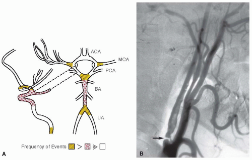
FIGURE 93.2 Atherosclerosis as a source of thrombotic and embolic obstruction of arterial circuits in the central nervous system (CNS). A: Predilection sites for atheromata in brain-supplying arteries. B: Example of severe (99%) stenosis of the ICA in patient with TIAs (arrow). ACA, anterior cerebral artery; MCA, middle cerebral artery, PCA, posterior cerebral artery; BA, basilar artery; UA, uncal artery.

Indirect evidence for the involvement of thrombi in cerebrovascular ischemia derives from observations of platelet, coagulation, and fibrinolytic system activation in patients with thrombotic stroke or TIAs.53,54,55,56,57 Platelet activation, spontaneous platelet aggregation, and circulating platelet aggregates have been reported in patients with recent atherothrombotic and thromboembolic cerebral ischemia.53,56,57,58,59,60,61,62,63 Experimental studies confirm the deposition of fibrin and activated platelets in microvessels of the ischemic regions shortly after MCA occlusion.64,65,66,67,68,69 These observations implicate the local activation of hemostasis in the ischemic microvascular bed.


HEMORRHAGIC TRANSFORMATION IN CEREBRAL ISCHEMIA

Hemorrhage accounts for approximately 10% to 15% of all strokes.1,6,7,9,70,71,72,73,74,75 Normal platelet function appears necessary to maintain the integrity of the cerebrovascular beds and to prevent clinically detectable hemorrhage. Antithrombotic agents increase hemorrhagic risk in the ischemic territory.76 Increases in the incidence of symptomatic intracerebral hemorrhage were observed with aspirin (acetylsalicylic acid [ASA]) in the UK-TIA Study, the International Stroke Trial (IST), and the European Stroke Prevention Study-2 (ESPS-2) trial.72,73,77 An increased risk of intracerebral hemorrhage is associated with advanced age (>75 years), the intensity of anticoagulation, and the concomitant use of antithrombotics.76

Hemorrhage within the ischemic lesion occurs normally during thromboembolic stroke and is classified as hemorrhagic infarction (HI), parenchymal hematoma (PH), or both (FIGURE 93.3). HI refers to petechial or confluent petechial hemorrhage in the area of ischemic injury, typically involving cortical or basal ganglia gray matter.78,79,80,81 HI occurs in 50% to 70% of individuals in postmortem studies,78,79,80 in 10% to 43% of nonanticoagulated individuals with acute cerebral infarction in CT scan-based studies,82,83 in 37.5% of patients with cardiogenic cerebral embolism, but in only 1.9% of patients with carotid territory thrombosis.84 Petechial hemorrhage can result from ischemia-related degradation of the microvessel basal lamina matrix components.85,86

PH is a homogeneous mass of blood (coagulum) that can displace brain tissue. It is most often the cause of symptomatic hemorrhage. Many reports of PH in patients with cerebral embolism are associated with anticoagulant treatment.87,88 In addition, PH can result from the rupture of small penetrating arteries (FIGURE 93.4).


THE PENUMBRA

Astrup and colleagues depicted the development of the core of ischemic injury destined for tissue destruction (infarction), as a circumferential region, or “penumbra,” of metabolically metastable tissue that has the potential for full recovery, following occlusion of a brain-supplying artery.89,90,91,92,93 This depiction has greatly influenced the concept of ischemic stroke following occlusion of brain-supplying arteries and the attempts to enhance tissue recovery clinically in the acute phase. This
concept encompasses characteristic electrophysiologic changes, biochemical and molecular alterations, microvessel responses, metabolic changes, and regional differences in tissue perfusion and H2O diffusion as displayed by imaging studies (e.g., the DEFUSE study).93,94,95 By positron emission tomography, the infarction corresponds to rCBF below 12 mL/100 g/min and a cerebral metabolic rate for oxygen (CMRO2) below 65 µm/100 g/min.96 The penumbra has been defined as rCBF decreased to 12 to 22 mL/100 g/min, CMRO2 above 65 µm/100 g/min, and an oxygen extraction fraction of 50% to 90%.96 Experimental data from vascular and molecular modeling studies as well as recent imaging work indicate that the penumbra is dynamic and
demonstrates that in the early minutes and hours after ischemia onset the core contains pockets of injury surrounded by “minipenumbras.”93 It has been hypothesized that failure to resolve these mini-penumbras into viable tissue leads ultimately to homogenous injured tissue (FIGURE 93.5). The cell and microvessel events in large part relate to local microvascular flow and may underlie recoverability of the ischemic territory-at-risk when flow is reestablished. Normal collateral arterial circuits protect cerebral cortical tissue and may contribute to the reversibility of the ischemic penumbra. Importantly, acute reperfusion of the occluded artery (arteries) is taken to decrease the penumbra and recover normally functioning tissue.


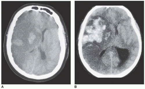
FIGURE 93.3 Hemorrhagic transformation. A: HI typified by regions of confluent petechiae 24 hours after MCA occlusion. Note midline shift due to tissue swelling. B: Parenchymal hemorrhage marked by coagulum displacing midline structures and compressing ventricle. (Image in panel A: From von Kummer R, Bozzao L, Manelf C. Early CT diagnosis of hemispheric infarction. Berlin, Germany: Springer Verlag, 1995:1, with permission.)


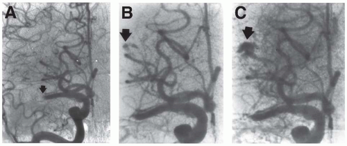
FIGURE 93.4 Evolution of parenchymal hemorrhage following intravenous infusion of recombinant tissue-type plasminogen activator (rtPA). Arrows indicate evolving hemorrhage. A: Obstruction of proximal middle cerebral artery (MCA) (M1 segment). B,C: During systemic rtPA infusion, following partial recanalization of the MCA, progressive extravasation from a distal lenticulostriatal artery was noted. A large PH with ventricular extension resulted (see FIGURE 93.3). (From the collection of M. Pessin, with permission.)


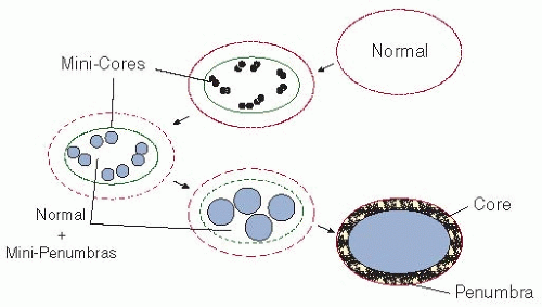
FIGURE 93.5 A proposed construct of the “penumbra” that takes into account the heterogeneous evolution of injury within the 1st hours of ischemia onset after the occlusion of a brain-supplying artery. The lesions and their reversibility depend upon the time and location of the reduction in regional cerebral blood flow (rCBF) in the territory-at-risk. If the injury stimulus cannot to be curtailed, the mini-cores coalesce with the duration of rCBF reduction devouring micro-penumbras. The evolution from normal function to the final lesion may depend upon a number of factors, including the territory of vascular supply, depth of reduction of rCBF, inflammatory state at baseline and/or degree of inflammatory response, and other factors.93


IMAGING OF CEREBRAL TISSUES/VASCULATURE

To define the regions of evolving injury, imaging approaches provide information about tissue status, vessel patency and morphology, and cerebral perfusion. With acquisition times of only 10 to 20 minutes, both CT scans and MR imaging can distinguish infarction from hemorrhage, identify the site of vascular occlusions, and delineate already irreversibly infarcted tissue from potentially reversible penumbra.


Brain Parenchymal Imaging

Tissue injury. Noncontrast CT excludes hemorrhagic transformation and other nonischemic causes of acute neurologic deficits (e.g., tumor, infection). It has the advantages of being rapid and inexpensive with widespread availability. CT imaging can identify signs of early injury (e.g., loss of gray-white differentiation, hypodensity, and sulcal effacement) and of cerebral artery occlusion infarct signs (HMCA sign). These signs occur in up to 80% of patients within 6 hours from symptom onset.97 Pathophysiologic studies suggest that early infarct signs represent edema. The presence of early infarct signs can be associated with a poorer outcome.98,99

MR imaging is sensitive to tissue injury. Within the first few hours of ischemia onset, standard MR protocols (T1-weighted, T2-weighted, and proton density-weighted sequences) are relatively insensitive to ischemia, showing abnormalities in <50% of cases.100 The earliest changes, seen as increased signal on T2-weighted and fluid attenuated inversion recovery sequences, are due to a net increase in tissue water content. Although the most ischemic lesions are evident on both CT and conventional MR by 24 hours, standard MR is superior to CT in identifying posterior fossa and subcortical lesions.

Extravasated blood can be reliably detected with gradient recalled echo or echo-planar susceptibility-weighted MR imaging.101 MR is at least equally accurate to CT in distinguishing acute intracerebral hemorrhage from acute ischemic stroke.102,103

Diffusion-weighted images (DWI) allow visualization of regions of cerebral ischemia within minutes of symptom onset, indicating lesion size, lesion age, neuroanatomic site, and the vascular territory involved.104,105,106,107 Decreased flux of free H2O from the extracellular to intracellular space, during the early moments of ischemia, can be quantified on the apparent diffusion coefficient (ADC) maps. While the increased DWI signal may persist for several weeks or longer partially due to a T2 effect, the ADC returns to normal or increased levels within 7 to 10 days from ischemia onset.108 DWI has a high sensitivity (95% to 100%) and specificity (95% to 100%) for acute ischemia.109,110,111 The initial diffusion lesion volume correlates well with final infarct volume, as well as neurologic and functional outcomes in stroke patients.105,112,113 Early reperfusion can normalize DWI images.114,115 Therefore, DWI can distinguish acute from chronic ischemia104,116,117 and provide frequent*** visualization of multiple acute lesions in different vascular territories in patients who have only one clinically symptomatic acute insult, providing evidence of an embolic stroke mechanism.118


Perfusion Imaging

Perfusion CT assesses rCBF, identifying regions of irreversible tissue injury as areas of reduced cerebral blood volume (CBV).119 Perfusion CT has the advantage of rapid data acquisition and rapid postprocessing and can be performed in conjunction with the baseline head CT and CT angiography scans. MR perfusion-weighted imaging follows rapid injection of an intravenous paramagnetic contrast agent and relies upon generation of concentration-time curves throughout the tissue under study. Perfusion measures derived from this technique include mean transit time, relative CBV, and rCBF. Baseline MR perfusion lesion volumes correlate well with final infarct volume as well as neurologic and functional outcome.113 Perfusion lesion volume likely identifies all tissue-at-risk of infarction if vessel recanalization does not occur, while diffusion imaging identifies only tissue that has already sustained advanced metabolic failure.


Vascular Imaging

Digital subtraction angiography (DSA) is the standard imaging modality to assess vessel anatomy and pathology, capable of visualizing medium and small vessels, collateral circulation,
and specific vascular lesions (e.g., arterial dissection, small caliber vessels such as vasculitis). Imaging of the cervicocephalic vessels to demonstrate evidence and site of stenosis and occlusions is achieved by contrast-enhanced helical CT angiography.120 Image postprocessing permits the data to be visualized using multiplanar reformatting, surface or three-dimensional volume rendering, and maximum-intensity projection techniques.121 MR angiography has sensitivity and specificity ranges of 70% to 100% when compared to DSA for the detection of cervical and intracranial stenoses.122 In the intracranial vasculature, MRA is useful in identifying acute proximal largevessel occlusions, but cannot reliably identify distal or branch occlusions. Combined with data from DWI, MRA substantially improves acute diagnosis of stroke mechanism.106 Contrast-enhanced MRA techniques that employ gadolinium contrast to outline vascular structures provide more reliable images of the thoracocervical vasculature. Flow artifacts can occur, including in-plane flow saturation, susceptibility to turbulent or complex flow, and flow-like effects from adjacent short T1 substances, such as thrombus and fat, which distort the degree of stenosis.

Carotid and transcranial Doppler (TCD) ultrasound techniques offer another noninvasive method to assess the neurovasculature acutely. Ultrasound studies of the carotid bifurcation are a well-established noninvasive test in the diagnosis of stroke. A full carotid ultrasonography battery includes continuous-wave Doppler measurement of blood velocity, B-mode imaging of vessel anatomy, and color-flow imaging of flow direction and lumen caliber. Carotid ultrasound visualizes only the proximal cervical carotid arteries and very limited segments of the cervical vertebral arteries. TCD ultrasound employs a low-frequency probe to penetrate the skull and interrogate the major basal intracranial arteries.123 TCD offers a practical, noninvasive assessment of intracranial vessel status that provides early diagnostic and prognostic information in patients with acute stroke.124 In addition, TCD can be used to monitor cerebral hemodynamic status and response to recanalization therapy.125


ANTITHROMBOTIC APPROACHES TO CEREBRAL ISCHEMIA

The direct and indirect evidence that platelet activation, acceleration of coagulation, and thrombosis underlie most ischemic strokes supports the use of antithrombotic agents in the management of cerebrovascular ischemia. Plasminogen activators (PAs) have been used to limit the consequences of recent transient or fixed neurologic deficits in the acute phase. Recombinant tissue plasminogen activator (rtPA) is now used in patients within 3 (to 4.5) hours of symptom onset and in the absence of detectable cerebral hemorrhage to reduce injury or to improve clinical outcome. Antiplatelet agents and anticoagulants have also been used as prophylaxis against recurrence of ischemia (i.e., secondary prevention). Antiplatelet agents play a role in the reduction of TIAs and ischemic strokes associated with thromboemboli of atherosclerotic origin. Anticoagulants are employed for prevention of cardiogenic emboli, arising in the setting of nonvalvular AF, ventricular dysfunction, or valvular injury or prostheses. Both approaches have been combined, but neither has been used in acute treatment of thrombotic stroke.


TRANSIENT CEREBRAL ISCHEMIA (TRANSIENT ISCHEMIC ATTACKS)

Because TIAs and ischemic stroke (e.g., transient monocular blindness [amaurosis fugax])126,127,128 can often be manifestations of ongoing platelet and coagulation activation and of vascular injury in brain-supplying arteries, antiplatelet agents have been applied with benefit.4,5,6,7,8,44,46,57,63,86,129


Antiplatelet Agents

Aspirin (Acetylsalicylic Acid). In ischemic cerebrovascular disease, ASA can reduce the incidence of TIAs and subsequent ischemic strokes. Despite the large number of prospective evaluations of ASA, a small number of well-conducted level I studies “drive” the efficacy of ASA in the carotid artery territory (Table 93.2).

The Aspirin in Transient Ischemic Attacks (AITIA) study— which randomized patients with a 3-month history of TIAs, who were not considered candidates for carotid endarterectomy, to ASA (1,300 mg/day) or to placebo—demonstrated a reduction in stroke and vascular death at 6 months with ASA exposure.130 There was a considerable decrease in the combined outcomes of recurrent TIAs, cerebral/retinal infarction, and death among those receiving ASA, but no reduction by life-table analysis at the 24-month follow-up and no reduction in stroke incidence. In the Canadian Cooperative Study Group trial, patients with a history of TIAs were randomized to ASA (1,300 mg/day), sulfinpyrazone (800 mg/day), and the combination ASA/sulfinpyrazone, or placebo.131 Patients who received ASA had a significant decrease in the incidence of stroke and death compared to sulfinpyrazone or placebo.131 The risk reduction for stroke and death with ASA was most significant for men. Among patients with a history of TIAs in the double-blind “Accidents, Ischemiques Cerebraux Lies a l’ Athersclerose (AICLA)” trial who were randomized to ASA (990 mg/day), the combination of ASA (990 mg/day) with dipyridamole (225 mg/day), or placebo, those who received ASA or the combination demonstrated a reduced incidence of stroke, MI, and death compared to patients who received placebo.132 Efficacy in the combined outcome seemed to be associated with an ASA dose range of 990 to 1,300 mg/day.

However, the ASA doses used were associated with considerable side effects. This was confirmed by the UK-TIA aspirin trial, which randomized 2,435 individuals with TIAs or minor ischemic stroke to “high dose” ASA (1,200 mg/day), “low-dose” ASA (300 mg/day), or placebo.15 The 4-year incidence of nonfatal MI, nonfatal major stroke, and death was significantly reduced by 18% in a dose-dependent manner in patients who received ASA. But, gastrointestinal side effects were more common with the high-dose regimen, and excess mortality was attributed to intracranial hemorrhage in the ASA-treated groups. In a separate study, a decrease in major hemorrhagic events, including fatal intracerebral hemorrhage, was associated with lower ASA doses (30 vs. 283 mg/day).133

The Swedish Aspirin Low-Dose Trial (SALT) Collaborative Group reported an 18% risk reduction of stroke and death among TIA patients who started ASA (75 mg/day) within 1 to 4 months of their initial symptoms compared to patients who received placebo.134 A 16% to 20% reduction in the risk of stroke, frequent TIAs, and MI was also observed.


Table 93.2 TIAs (± stroke): antiplatelet agents


Study


Agent


Dose (per Day)


Patients (n)


Stroke (n)


Mortality (n)


AITIA Study130


ASA


1,300 mg


88


10


3


Placebo


90


12


7


Canadian Cooperative Study Group131


ASA/placebo 1


1,300 mg/—


144


22


4


Sulfinpyrazone/placebo 2


800 mg/—


156


29


9


ASA/sulfinpyrazone


1,300 mg/800 mg


146


14


6


Placebo 1/placebo 2


—/—


139


20


10


Danish Cooperative Study374


ASA


1,000 mg


101


18


7


Placebo


102


11


7


AICLA132


ASA/dipyridamole


990 mg/225 mg


202


18


8


ASA


990 mg


198


17


10


Placebo


204


31


7


UK-TIA Study Group15


ASA


1,200 mg


815


66


111


ASA


300 mg


806


68


106


Placebo


814


88


122


Dutch TIA Trial Study Group133


ASA


283 mg


1,576


109


151


ASA


30 mg


1,555


90


160


SALT134


ASA


75 mg


676


93


61


Placebo


684


112


69


European Stroke Prevention Study Group139


ASA/dipyridamole


975 mg/225 mg


1,250


114


108


Placebo


1,250


184


156


American-Canadian Cooperative Study Group140


ASA/dipyridamole


1,300 mg/300 mg 448


53


46


ASA


1,300 mg


442


60


38


Matias-Guiu et al.375


ASA/dipyridamole


50 mg/300 mg


115


3


2


Dipyridamole


400 mg


71


3


3


European Stroke


ASA/dipyridamolea


400 mg/50 mg


1,650


157


285


Prevention Study-277


Dipyridamolea


400 mg


1,654


211


288


ASA


50 mg


1,649


206


282


Placebo


1,649


250


202


Hass et al.148


Ticlopidine


500 mg


1,529


172


175


ASA


1,300 mg


1,540


212


196


ESPRIT Study Group142,143,144


ASA/dipyridamole


30-325 mg/400 mg 1,363


96


93


ASA


30-325 mg


1,376


116


107


a Extended-release dipyridamole.


ASA, aspirin (acetylsalicylic acid); UK-TIA, United Kingdom Transient Ischemic Attack; AITIA, Aspirin in Transient Ischemic Attacks; TIA, transient ischemic attacks; SALT, Swedish Aspirin Low-Dose Trial.


The results of those trials, and meta-analyses of all clinical series, suggest that even low doses of ASA benefit early stroke and vascular mortality in patients with a history of TIAs or minor ischemic events.

Combination ASA/Dipyridamole. Clinical use of the combination of ASA with dipyridamole has been based upon positive interactions between the agents in both preclinical experiments and in clinical platelet survival studies.135,136,137 The efficacy of this combination, however, has been challenged.136 Dipyridamole (400 to 800 mg/day) alone did not affect stroke incidence or related mortality compared with placebo in one limited double-blind, level I randomized trial.138

The European Stroke Prevention Study (ESPS) Group demonstrated a 33% reduction in risk of stroke and death with ASA (975 mg/day) and dipyridamole (225 mg/day) compared to placebo.139 The three-arm “AICLA” study also demonstrated the benefits from the combination of ASA with dipyridamole over placebo (but no difference with ASA alone) for patients with TIAs when the outcome events of stroke, MI, and mortality were combined.132 However, there was no difference in stroke or mortality between the combination of ASA (1,300 mg/day) and dipyridamole (300 mg/day) and ASA alone in the American-Canadian Cooperative Study Group.140


On the basis of experimental evidence suggesting that sustained release extended-release dipyridamole (ER-DP) could produce elevated plasma adenosine levels, the relative efficacy of the fixed combination of low-dose ASA (50 mg/day)/ER-DP (400 mg/day) was tested in the ESPS-2 against ER-DP, ASA, or placebo. At 2-year follow-up, low-dose ASA alone and dipyridamole alone (relative risk reduction [RRR] for stroke, 15.8% and 17.7%, respectively) and the combination (RRR, 36.7%) were found to be superior to placebo in limiting stroke, death, or both for individuals with a history of TIAs or recent stroke.77,141

The European/Australian Stroke Prevention in Reversible Ischaemia Trial (ESPRIT) Group examined the impact of combination ASA (30 to 375 mg/day)/dipyridamole (400 mg/day) or ASA alone on vascular death, nonfatal stroke, nonfatal MI, or major hemorrhage in patients presenting with TIA or minor ischemic stroke.142,143,144 With a mean follow-up of 3.5 years, the combination of ASA/dipyridamole was superior to ASA alone (HR = 0.80, 95% CI = 0.66 to 0.98) in reduction of the primary outcome events. While headache occurred in the combination, overall ASA/dipyridamole improved outcome in this trial and produced a decrease in ischemic events.143 The greater efficacy of the combination supports its use in the clinical setting.

In a separate study, clopidogrel was prospectively compared to the combination of aspirin/ER-DP with or without the angiotensin receptor blocker telmisartan in the Prevention Regimen for Effectively avoiding Second Strokes (PRoFESS) study of first recurrent stroke in a 2 × 2 factorial design.145 In total, 20,332 patients were randomized within 120 days of the initial event and followed for a mean of 2.5 years. There was no difference in the incidence of recurrent strokes between the two groups, although there were more hemorrhagic events in the aspirin/ER-DP arms. This trial did not meet the predefined criteria for noninferiority.

Sulfinpyrazone. The Canadian Cooperative Study Group compared ASA, sulfinpyrazone (800 mg/day), a uricosuric agent with antiplatelet effect, and placebo but found no significant difference in the incidence of stroke and mortality between sulfinpyrazone and placebo over 2.2 years.131 Sulfinpyrazone (800 mg/day) and placebo were also tested in a double-blind, cross-over trial.146 A nonsignificant reduction in TIAs and in stroke and mortality at 4-month follow-up was recorded in patients receiving sulfinpyrazone. Another level I study comparing sulfinpyrazone (800 mg/day) and ASA (1,000 mg/day) in individuals with TIAs concluded that there was a higher incidence of stroke, MI, and vascular death in the sulfinpyrazone cohort at 11-month follow-up.147

Adenosine Diphosphate Receptor Antagonists. Thienopyridines irreversibly block the adenosine diphosphate platelet receptor P2Y12 and inhibit other downstream events, thereby blocking platelet activation. Ticlopidine found benefit in patients with TIA/minor stroke148 and in prevention of secondary stroke (see below). Ticlopidine (500 mg/day) was associated with a statistically significant 12% reduction in the risk of stroke and death from any cause over ASA (1,300 mg/day) and a 21% reduction in the risk of secondary outcomes of stroke and stroke-related death following TIAs. Reversible leukopenia occurred in 0.8% of patients taking ticlopidine, whereas gastrointestinal symptoms were more common in patients receiving ASA. Intracerebral hemorrhage occurred equally in both groups. Because of concerns about the frequency of thrombotic thrombocytopenic purpura, ticlopidine has been supplanted by the related thienopyridine clopidogrel.

In summary, ASA reduces the risk of stroke, MI, and mortality in individuals with a history of TIAs or minor stroke. This risk reduction is obviously not dose dependent. The fixed combination of low-dose ASA/ER-DP also produces a substantial risk reduction. Ticlopidine was more effective than ASA in preventing stroke and mortality after TIAs in one study but is seldom used now because of unwanted side effects. When used alone, dipyridamole and sulfinpyrazone have little demonstrated benefit.149,150


Anticoagulation

Changes in plasma thrombin concentration and FDP levels occur following ischemic stroke that suggest that interruption of coagulation may be beneficial. In the 1960s and 1970s, clinical experience with anticoagulation in patients with TIAs or minor stroke was limited.151,152,153,154 Uncontrolled reports suggested that heparin could decrease the incidence of basilar artery TIAs.155,156 Fisher observed a transient reduction in the incidence of TIAs in 97% (of 29) patients treated with anticoagulants, with TIAs returning in 60% of those in whom anticoagulation was discontinued.157 Two studies reported that long-term anticoagulation decreased the incidence of strokes and/or vascular death among patients with recent TIAs158,159; a third study showed that despite a decrease in TIA frequency, there was no decrease in the incidence of stroke or vascular death.160 Two other studies failed to show superiority of anticoagulation over its comparator for the incidence of stroke or death.161,162 In a small Medical Research Council (MRC)-sponsored trial of phenindione, patients with TIA receiving fixed “low-dose” anticoagulation had a lower incidence of vascular events, stroke, and death than those patients receiving an adjusted “high-dose” regimen.163 Only the Cerebral Embolism Study Group trial indicated a benefit from immediate anticoagulation for cardiogenic embolism.164

Subsequently, there have been few controlled prospective randomized level I studies. The Warfarin-Aspirin Recurrent Stroke Study (WARSS) found no difference in outcome between TIA and stroke patients treated with oral anticoagulation under tight control compared to patients treated with ASA.165 There was no increase in intracerebral hemorrhage in the anticoagulated group, most probably because of the rigid control of the international normalized ratio (INR).165

The risk of intracerebral hemorrhage increases with age in patients treated with oral anticoagulants which inhibit vitamin K activity.166,167 With careful monitoring and dose adjustment, this risk can be reduced.167 The Stroke Prevention in Reversible Ischemic Trial168 was terminated because of an unacceptably high incidence of intracranial hemorrhages in the group at a high target INR of 3.0 to 4.5.

In summary, excluding individuals with AF or cardiac valvular prostheses, the results of various small studies do not convincingly support a role for anticoagulation in patients with TIA or minor stroke, considering the risk of hemorrhage. The risk of intracerebral hemorrhage is increased for patients receiving oral anticoagulants at INRs in excess of 3.0 to 4.5.


Direct Thrombin Inhibitors

A single small safety study of the parenteral thrombin inhibitor argatroban in patients presenting with TIAs or minor ischemic stroke suggesting no detriment has appeared.169 No
phase III experience exists; however acute use has been under limited study.170


Plasminogen Activators

The use of fibrinolytic agents in patients with cerebral ischemia is confined to acute intervention in ischemic stroke; there is no indication for the use of these agents in TIAs.


STROKE-IN-PROGRESSION

Progressive deterioration following ischemic stroke has been ascribed to (a) anterograde extension of in situ thrombus to occlude critical arterial branches or (b) recurrent embolism. The role of anticoagulation in patients with stroke-in-progression, however, remains unsettled, in large part, because of the difficulty in defining what constitutes “progression” at entry and uncertainties about the pathogenesis of “evolving stroke.” Early studies also reveal methodologic weaknesses and conflicting outcomes of this diagnosis.171,172,173 Furthermore, deterioration following ischemic stroke may be due to edema, hemorrhage, or a combination of the events that lead to tissue swelling and injury.174

The Trial of Org 10172 in Acute Stroke Treatment (TOAST), a level I prospective randomized placebo-controlled study of the heparinoid danaparoid for stroke-in-evolution, demonstrated no benefit to the anticoagulant regarding disability indices, neurologic status, or mortality.175,176,177 Entry was opened to all patients with hemispheric stroke symptoms for up to 24 hours.175 A short-lived reduction in stroke incidence was seen. Serious intracerebral hemorrhage was significantly more frequent in patients who received danaparoid (14 of 646 patients) compared to placebo (4 of 635 patients). Danaparoid decreased the incidence of deep venous thrombosis (DVT). Stroke-inprogression is no longer identified as a separate entity for which a specific treatment is required.


COMPLETED STROKE

Prospective CT scan and MRI178,179,180 have confirmed the experimental observation that the ischemic lesion stabilizes, as an infarct, by 24 hours after occlusion of the supply artery.181,182 These congruent findings suggest that antithrombotic interventions beyond 24 to 72 hours after symptom onset are unlikely to have a beneficial impact on the initial ischemic stroke. Following a first-time ischemic stroke, the rate of subsequent ischemic stroke, MI, death, or vascular-related death is 10% to 12% per year.183,184,185 Hence, the initial ischemic stroke offers a target for antithrombotic agents to reduce the incidence of second ischemic events (secondary prevention).


Antiplatelet Agents

Meta-analyses suggest benefit from antiplatelet agents in patients with TIAs or first stroke.186,187 When considered with TIAs, ASA or ticlopidine can significantly decrease the risk of second cerebral ischemic events (Table 93.3). The combination of low dose ASA and ER-DP considerably reduces the incidence of second stroke and/or death in patients presenting with a history of TIAs or stroke compared to ASA or dipyridamole alone.141

The IST randomized 19,435 patients with presumed ischemic stroke within 48 hours of symptom onset to placebo, ASA (300 mg/day) alone, subcutaneous heparin (low dose, 10,000 IU/day; or medium dose, 25,000 IU/day), or both ASA and heparin for 14 days in a 3 × 2 factorial design.72 ASA was associated with an overall statistically significant reduction in total recurrent ischemic strokes within 14 days (P < 0.001) and also a significant 0.1% increase in symptomatic intracranial hemorrhage. At 6 months, a modest decrease in the risk of death or dependency was seen. The Chinese Acute Stroke Trial (CAST) demonstrated similar outcomes.188,189 Together, the IST and CAST demonstrated that ASA is associated with a small reduction in the incidence of recurrent stroke and mortality in the first weeks after signal stroke. This did not persist.

Ticlopidine was found to be superior to placebo in reducing the risk of second stroke, MI, and vascular mortality when patients were treated within 1 to 16 weeks following the signal thromboembolic stroke.183 Side effects related to ticlopidine, including reversible leukopenia, diarrhea, or rash, occurred in 8% of the treated patients. Concerns about the side effects of ticlopidine prompted a study of the efficacy of the related thienopyridine derivative clopidogrel.

The Clopidogrel versus Aspirin in Individuals at Risk of Ischaemic Events (CAPRIE) study, a prospective, randomized, double-blind level I comparison of clopidogrel (75 mg/day) against ASA (325 mg/day) in patients presenting with ischemic or lacunar stroke, MI < 35 days old, or symptomatic atherosclerotic peripheral arterial disease (PAD), demonstrated a statistically significant 8.7% reduction in the combined risk of ischemic stroke, MI, or vascular-related death by clopidogrel compared to ASA.190 The overall benefit associated with clopidogrel was driven by the PAD outcome; there was no independent significant difference in outcome in the stroke cohort. Because of the near equivalent frequency of intracranial hemorrhage (clopidogrel = 0.33% vs. ASA = 0.47%), clopidogrel is now used clinically. The large (7,599 patients) Management of Atherothrombosis with Clopidogrel in High-risk patients trial compared the efficacy of ASA (75 mg)/clopidogrel (75 mg) to clopidogrel (75 mg) alone for ischemic events including stroke. No difference was seen in outcome between the two groups, although the ASA/clopidogrel combination was associated with significantly higher incidence of life-threatening hemorrhage (including intracranial hemorrhage).191 The Clopidogrel for High Atherothrombotic Risk and Ischemic Stabilization, Management, and Avoidance trial, a prospective double-blind placebo-controlled study of combination ASA (75 mg)/clopidogrel (75 mg) versus ASA (75 mg)/placebo for patients with multiple atherothrombotic risk factors, demonstrated no significant difference for the primary efficacy endpoint (the composite of MI, stroke, or cardiovascular mortality).192 Long-term clopidogrel use was associated with an increased risk of hemorrhage, particularly in the first year.193 Overall, there was no clear benefit in stroke efficacy outcome (a post hoc analysis of secondary prevention) with the combination over ASA alone. That study has provided much for discussion. The PRoFESS study and its outcomes have been mentioned above.

A recently undertaken trial, the Platelet-Oriented Inhibition in New TIA and minor ischemic stroke trial (http://clinicaltrials.gov/ct2/show/NCT00991029), is a randomized double-blind multicenter study to investigate whether clopidogrel (75 mg/day) alone is superior to aspirin (50 to 325 mg/day) in improving survival free from major ischemic vascular events (ischemic stroke, MI, and ischemic vascular death). The projected sample size is 4,150 patients.


Table 93.3 Completed stroke: antiplatelet agents


Study


Agent


Dose (per Day)


Patients (n)


Stroke (n)


Mortality (n)


Swedish Cooperative Study184


ASA


Placebo


1,500 mg


253


252


32


32


34


37


Gent et al.185


Suloctidil


Placebo


600 mg


218


220


29a


28a


13a


25a


Blakeley194


Sulfinpyrazone


Placebo


800 mg


145


145


25


28


Canadian-American Ticlopidine Study183


Ticlopidine


Placebo


500 mg


525


528


54


89


30a


38a


CAPRIE190b


Clopidogrel


ASA


75 mg


325 mg


3,233


3,198


315


338


IST72


ASAc


No ASAc


300 mg


9,720


9,715


362d


452d


872


909


CAST188


ASA


No ASA


160 mg


10,554


10,552


335


351


343


398


ESPS-277


ERDP


Dipyridamole


ASA


Placebo


400 mg/50 mg


400 mg


50 mg


1,650


1,654


1,649


1,649


157


211


206


250


285


288


282


202


Diener et al.191


ASA/clopidogrel


Placebo/clopidogrel


75 mg/75 mg


—/75 mg


3,420


3,454


299


309


201


201


Bhatt et al.192,193


ASA/clopidogrel


Placebo/clopidogrel


75-162 mg/75 mg


—/75 mg


7,802


7,801


132


163


371


374


Sacco et al.145


ASA/ERDP


Clopidogrel


50 mg/400 mg


75 mg


10,181


10,151


916


898


739


756


a Eligible events only, excluding events >28 days after study drug was permanently discontinued.

b Stroke subgroup.

c Factorial design (include heparin ± ASA).

d 14-day outcomes.


ASA, aspirin; CAPRIE, Clopidogrel versus Aspirin in Individuals at Risk of Ischaemic Events; IST, International Stroke Trial; CAST, Chinese Acute Stroke Trial.

Only gold members can continue reading. Log In or Register to continue

Stay updated, free articles. Join our Telegram channel

Jun 21, 2016 | Posted by in HEMATOLOGY | Comments Off on Ischemic Stroke: Acute Thrombolytic Therapy, Antithrombotic Approaches, and General Management

Full access? Get Clinical Tree

Get Clinical Tree app for offline access