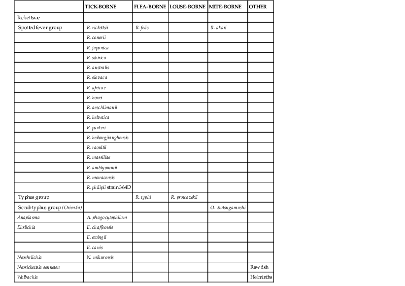
Didier Raoult The definition of the Rickettsiaceae family has been based mainly on nonspecific phenotypic characters. Originally, small gram-negative bacteria, associated (or not) with arthropods and necessitating (or not) eukaryotic cells for growth, were considered Rickettsiaceae. During the past 20 years, gene sequencing and genetic phylogeny have deeply challenged this classification. The controversy has centered on how much difference between strains should constitute a subspecies.1–5 Among the agreed-upon changes, Orientia was created from an independent branch of its phylum. The Ehrlichia group has been reclassified6 into four genera, with Ehrlichia and Anaplasma being associated with ticks, Neorickettsia with helminths, and Wolbachia with both arthropods and helminths. This chapter is limited to the Rickettsiales. All are intracellular alphaproteobacteria associated with eukaryotic hosts (arthropods or helminths). Based on antigenic and genetic data, pathogenic rickettsiae are traditionally divided into three groups—the spotted fever group, the typhus group, and the scrub typhus group (Table 187-1). The spotted fever group accounts for most tick-borne rickettsioses. The typhus group comprises two human pathogens transmitted by insects. Epidemic typhus is caused by Rickettsia prowazekii and is transmitted by the body louse. Murine typhus is caused by Rickettsia typhi and is transmitted by rat and cat fleas. The scrub typhus group comprises Orientia tsutsugamushi only and is transmitted by “chiggers.” New genetic tools and the use of cell culture assays have allowed the description of many new rickettsioses and ehrlichioses during the past 30 years (Table 187-2).7 Three ehrlichioses and 12 rickettsioses have been described since 1980. Three major conditions determined the description and separation of these species. Some were discovered after clinical description in countries where spotted fever had been unknown (Japan and Rickettsia japonica, Flinder’s Island and Rickettsia honei, and Russia and Astrakhan fever). Some were recognized by bacterial identification based on culture and polymerase chain reaction (PCR) in places where the new pathogen was confounded with another known rickettsial pathogen (Rickettsia africae with Rickettsia conorii, Rickettsia heilongjianghensis with Rickettsia sibirica, R. sibirica mongolitimonae and Rickettsia aeschlimannii with R. conorii, Rickettsia felis with Rickettsia typhi, and Anaplasma phagocytophilum and Ehrlichia ewingii with Ehrlichia chaffeensis). Some were identified through association by physicians and microbiologists when an atypical unknown disease (E. chaffeensis, Rickettsia slovaca, Rickettsia raoultii, and Rickettsia helvetica) was being explored.7 TABLE 187-2 Historical Data on Diseases Caused by Rickettsia Species (First and Senior Authors) Data from Raoult D, Roux V. Rickettsioses as paradigms of new or emerging infectious diseases. Clin Microbiol Rev. 1997;10(4):694-719; and Shapiro MR, Fritz CL, Tait K, et al. Rickettsia 364D: a newly recognized cause of eschar-associated illness in California. Clin Infect Dis. 2010;50:541-548.
Introduction to Rickettsioses, Ehrlichioses, and Anaplasmosis
Bacteriology
History and Emerging Diseases
YEAR
DISCOVERY
AUTHORS
1760
Description of exanthematic typhus
Boissier de Sauvage
1879
First report of scrub typhus
Nagayo
1899
Description of Rocky Mountain spotted fever
Maxcy
1906
Isolation of R. rickettsii
Ricketts
1909
Role of body lice in typhus
Nicolle (Nobel Prize)
1909
Description of Mediterranean spotted fever
Conor et al
1910
Serology test based on Proteus
Wilson
1911
Isolation of R. prowazekii
Nicolle
1914
Tick role in Mediterranean spotted fever
Wilson
1916
Weil-Felix test
Weil and Felix
1921
Identification of R. typhi
Mooser
1925
Description of the tâche noire in Mediterranean spotted fever
Pieri
1930
First isolation of Orientia tsutsugamushi (R. orientalis)
Nagayo
1930
Role of chiggers in scrub typhus
Kawarimura
1930
Role of fleas in murine typhus
Dyer
1932
Isolation of R. conorii
Brumpt
1935
Description of Siberian tick typhus
Shmatikov et al
1938
Isolation of R. sibirica
Krontovuka et al
1940
R. phagocytophila
Gordon
1946
Description of rickettsialpox
Huebner
1946
Isolation of R. akari
Huebner
1946
Isolation of R. australis
Plotz and Smadel
1946
Queensland tick typhus
Plotz and Smadel
1956
Ehrlichia sennetsu
Kobayashi
1968
Isolation of R. slovaca
Brezina et al
1974
Culture of R. conorii
Goldwasser
1979
Isolation of R. helvetica
Burgdorfer and Peter
1981
Ehrlichia chaffeensis
Anderson
1984
Japanese spotted fever
Mahara
1985
Culture of R. heilongjianghensis
Udida and Walker
1987
First case of human erhlichiosis in United States
Maeda and McDade
1989
Culture of R. japonica
Lov
1990
First human cases of granulocytic erhlichiosis
Bakken
1990
Isolation of R. africae
Kelly
1991
Flinder’s Island spotted fever
Stewart
1992
Molecular identification of Ehrlichia ewingii
Anderson
1992
First case of infection by R. africae
Kelly and Raoult
1992
Culture and identification of R. conorii
Tarasevitch and Raoult
1992
Culture of R. honei
Baird et al
1993
Culture and identification of R. sibirica mongolitimonae
Yu and Raoult
1994
First case of flea-borne spotted fever
Schriefer and Azad
1996
Infection by R. sibirica mongolitimonae
Raoult et al
1997
First infection by R. slovaca
Raoult et al
1997
Culture of R. aeschlimanii
Beati and Raoult
1999
Astrakhan fever
Tarasevitch and Raoult
1999
First human cases of infection with E. ewingii
Buller
2000
Role of Wolbachia in filariasis
Taylor
2000
First case of acute infection by R. helvetica
Fournier and Raoult
2000
Culture of R. felis
Raoult et al
2002
First case of infection by R. aeschlimanii
Raoult et al
2004
First case of infection by R. parkeri
Paddock et al
2006
Description of infection by R. heilongjianghensis (Far Eastern spotted fever)
Mediannikov et al, 200628
2008
Formal description of R. raoultii
Mediannikov et al, 200829
2008
First case of R. massiliae
Raoult
2008
First case of infection with R. raoultii
Parola et al30
2010
First case of Rickettsia philipii strain 364D
Shapiro et al31
![]()
Stay updated, free articles. Join our Telegram channel

Full access? Get Clinical Tree


Introduction to Rickettsioses, Ehrlichioses, and Anaplasmosis
187


