Intravenous Access and Catheter Management


Intravenous Access and Catheter Management


Rachel P. Rosovsky

David J. Kuter


Central venous catheters (CVCs) have become an integral part of treating patients both in and out of the hospital. They allow for easy administration of medications and uncomplicated withdrawal of blood samples. In cancer patients, who often require long-term chemotherapy, these devices have become the standard of care, and their use is increasing steadily every year. Unfortunately, these instruments are also associated with adverse events, most commonly, mechanical, infectious, and thrombotic complications. Recent research has focused on identifying the risk factors for and incidence, prevention, and treatment of these complications. Several studies have shown, for example, that antimicrobial-impregnated catheters can lower the risk of infection and decrease medical costs.1, 2, 3, 4 Although older studies supported the use of anticoagulants for the prevention of CVC-related thrombosis, newer randomized controlled studies failed to show a benefit in cancer patients.5, 6, 7, 8, 9, 10, 11, 12, 13, 14, 15, 16, 17, 18

This chapter reviews CVCs. We will briefly mention the types of instruments available as well as the major indications for their usage. A major part of the review will focus on the complications of CVC, most notably infectious and thrombotic. We will discuss the risk factors, strategies for prevention, the current options for treatment, and new developments.


Historical Perspective

The history of the CVC begins in 1656 when Christopher Wren (1632 to 1723) administered wine and ale to living dogs via an intravenous cannula. He described in his writings how the animals became somnolent or vomited after being injected with alcoholic substances, opiates, or purgatives.19, 20, 21, 22, 23, 24 Wren’s colleagues performed subsequent experiments with venous catheters involving the transfusion of blood products between animals and humans.25 Unfortunately, some of these studies resulted in the death of either the animal or human. This outcome halted interest in and further investigations of venous cannulae for a number of centuries.

It was not until 1952, when Aubaniac described cannulating the subclavian vein (SC) of a wounded soldier for the purpose of resuscitation that interest in the clinical uses of CVC was renewed.26, 27, 28 Fortunately, this attention led to the development of better technology, and over the past 35 years, a number of devices for the semipermanent cannulation of the central venous system have been introduced. In 1973, Broviac developed the first long-term CVC for parenteral nutrition.29 This was followed by the Hickman catheter in 1979, which was the first permanent venous-access device used for cancer chemotherapy.30 Totally implantable venous access devices (Fig. 41-1) that are placed subcutaneously and contain their own ports attached to a centrally placed catheter, became available in the early 1980s.31 The most recent advancement has been the peripherally implanted central catheters (PICC).32


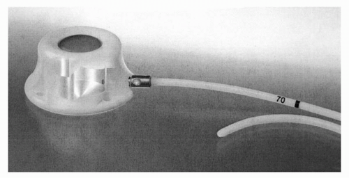
FIGURE 41-1 Cutaway photo of PORT-A-CATH implantable port showing the injection site, reservoir, and attached catheter. (Courtesy of Smith Medical ASD, Inc., St. Paul, Minnesota.)


Current Devices

Various CVCs are currently available (Table 41-1). Short-term devices, including percutaneous lines and PICC, are usually employed for less than 1 month’s duration. Alternatively, the long-term catheters can remain in place for months to years; these include the surgically tunneled catheters described above (the Broviac and the Hickman, as well as, more recently, the Groshong
and the Quinton) and the totally implanted venous access devices that contain their own port (the Mediport, the Infuse-a-Port, and the Port-a-Cath). Several standard techniques presently are practiced for the insertion of a CVC.33 The choice of catheter and the insertion strategy depend on a number of variables, most importantly, the indication for its usage. However, patient characteristics, including any history of failed attempts, previous surgeries, comorbidities, skeletal deformities or scarring, and location of tumor, also need to be accessed.34


TABLE 41.1 Types of CVCs


Short-term devices (1-14 d), nontunneled


Percutaneous internal jugular (IJ), subclavian, femoral lines (singlelumen, double-lumen, or triple-lumen)


Peripherally inserted central catheters (PICC)


Long-term devices (months to years)


Surgically tunneled catheters (Hickman, Broviac, Groshong, Quinton)


Totally implanted venous access devices (Mediport, Infuse-a-Port, Port-a-Cath)


Current Uses

CVCs have become essential for the management of cancer patients. In the United States, more than 5 million CVCs are inserted every year.35 They not only allow for intravenous administration of drugs, antibiotics, blood products, fluids, and nutrition, but also facilitate drawing of blood samples to monitor for potential complications of treatments and disease. Eliminating frequent venipunctures clearly increases a patient’s level of comfort. Whether the use of CVCs translates into extending or improving a patient’s quality of life is currently being investigated.36


Complications

CVC are associated with a number of early and late complications (Table 41-2), which can be both harmful to the patient and costly to treat. Most of the mechanical complications occur during the insertion process; the infectious and thrombotic complications usually occur after the catheter has remained in place for some time. The incidence of catheter-related complications varies greatly among different studies, largely due to the diversity of study designs and diagnostic protocols.


TABLE 41.2 Incidence of complications of CVCs


Early


Arrhythmia: 13%


Arterial puncture: 2.8%-4.7%


Malposition of reservoir: 2%


Pneumothorax: 1%-3%


Wound dehiscence: 1.5%


Hemorrhage: 1.1%-4.4%


Failure of insertion: 1.2%-22%


Late


Infection: 4%-38%


Catheter fracture and embolization: 3%


Migration of catheter tip: 7.4%


Thrombosis: ˜41% (range 12%-74%)


Types


Asymptomatic: ˜29% (range: 5%-62%)


Symptomatic: ˜12% (range: 5%-41%)


Sequelae of CVC-related thrombosis


Postphlebitic syndrome: 15%-35%


Pulmonary embolization: ˜11% (range: 7%-31%)


Symptomatic: ˜6% (range: 3%-14%)


Asymptomatic: ˜5% (range: 3%-15%)


Early Complications


Types of Early Complications

Early complications are primarily related to catheter insertion. Arrhythmias occur in up to 13% of patients and are one of the most common complications that occur during or immediately after the insertion process; however, they are usually self-limiting and rarely cause hemodynamic instability. Most of the other early adverse events are mechanical in nature with an incidence range of 1% to 33%.25,35,37 The most commonly reported ones are venous tear, arterial puncture or cannulation, which can then cause a hematoma or dissection, or a lung or pleural laceration which can result in a pneumothorax (Fig. 41-2) or hemothorax.26,34,38 Injury to adjacent nerves or anatomic structures can also occur during the insertion process. Catheter malposition or breakage can occur either early or late, as can a rare but potentially lethal complication, an air embolus.26,33,35 The incidence of failed attempts approaches 22%37 and strongly predicts for future problems. Lastly, patients rarely experience the serious and occasionally fatal complication of cardiac tamponade.39 Postinsertion care must include a chest x-ray to confirm the correct position of the catheter and the absence of injury to the vessels, lung, and pericardium.


Risk Factors for Early Complications

The risk factors for the early mechanical complications are directly related to the site of CVC insertion as well as patient and catheter-related factors.26,38 For the percutaneous devices, the frequency of mechanical complications is higher for a femoral approach than for using a subclavian or internal jugular (IJ) approach.26,38 However, if one only looks at the serious complications, the femoral and subclavian rates are similar.35,38


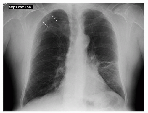
FIGURE 41-2 Pneumothorax in the right upper lobe as outlined by the two white arrows. (Courtesy of Rachel Rosovsky, MD, MPH.)


The type of material used for catheters and the time of their insertion also confer certain risks. Stiff CVC are easier to insert but have a higher rate of mechanical complications.26 When the insertion procedure for short-term percutaneous catheters is performed after-hours or by a physician who has performed less than 50 or if the insertion requires more than two attempts, the complication rate is higher.37,38,40,41 Lastly, thrombocytopenia, altered local anatomy, prior catheterization, recent myocardial infarction, severe obesity, and atherosclerosis are patient-related factors that increase the risk of complications.26


Prevention of Early Complications

A number of strategies can reduce the risks of early complications. Perhaps the most important one is to individualize the approach for each patient based on anatomy, co-morbidities, and other characteristics. Prior surgeries, trauma, or radiotherapy in the clavicular region, for example, may cause an alteration in the anatomy or surface landmarks used to locate a vein. Therefore, the contralateral side should be the preferred location in these patients. Choosing the IJ vein rather than the SC may result in fewer long-term complications.42,43 A recent prospective study evaluating 1,201 patients found that immediate complications were more frequent in the SC than in the IJ approach (respectively, 5.0% versus 1.5%; P < 0.001).42

Another preventive approach uses ultrasound (US) to localize the vessel to be cannulated. US decreases the risks associated with an IJ vein catherization, but does not clearly benefit the SC approach.44 In a recent study of 130 patients in a tertiary emergency department, cannulation of the IJ vein was successful in 61 of 65 patients (93.9%) using US compared to 51 of 65 patients (78.5%) using the landmark technique. A significant difference was also seen in the complication rate: 4.6% in the ultrasonographic group versus 16.9% in the group using anatomic landmarks.45 Obviously, the more experienced the physician, the less risk of a complication. Eisen et al. analyzed 385 CVC attempts and found a complication rate of 39.5% when interns performed the procedure compared to 18.4% with fellows or attendings.37 Another study showed that after three attempts, the incidence of mechanical complications increased to six times the rate after one attempt.34,41

As important as these preprocedure strategies is the ability to recognize and treat the complications. For example, an air embolus can usually be prevented by placing the patient in Trendelenburg’s position during the insertion process and occluding the catheter hub at all times. However, if an air embolus is suspected or occurs, immediately placing the patient in this position and administering 100% oxygen can facilitate the resorption of air and prevent a potentially fatal complication.35


Late Complications: General


Types, Risks, and Prevention of the General Late Complications

Infection and thrombosis are the most common late complications associated with CVCs, and they will be discussed separately. The other late complications occur at a rate of 3% to 8%.25 CVC breakage can develop if defective material is used or if excessive manipulation is applied during the insertion process. CVCs also break due to a phenomenon associated with subclavian catheters termed the “pinch off syndrome.” Because the subclavian catheter is situated between the clavicle and the first rib, over time repeated compression can cause the catheter to fracture, resulting in extravasation of fluids, catheter breakage, and catheter embolization.26,33 Utilizing a more lateral approach or an alternative insertion site is recommended, especially in patients with known narrow thoracic inlet syndrome.33 Extravasation of fluid also develops with CVC dislocation, malposition, or damage. Several other late complications also sometimes occur, though infrequently; these include arteriovenous fistulas, cerebrovascular accidents, cardiac aneurysms, and intracardiac abscesses.26 Lastly, there are additional difficulties associated with CVC removal, but these are beyond the scope of this review.


Late Complications: Thrombosis


Types of Thrombosis


Fibrin Sheath Formation

Several types of thrombi can occur with CVCs. Fibrin sheaths, or sleeve thrombi, form on the outside of catheters. They are ubiquitous, according to both autopsy and imaging studies, but rarely cause any difficulties unless they occlude the tip of the catheter.46, 47, 48, 49

The sheath develops within 24 hours of catheter insertion,48 and is always colonized by cocci.50, 51, 52

The presence of these sheaths, however, does not predict subsequent deep venous thrombosis (DVT) of the vessel in which the catheter is placed. In one study, only 1 of 16 patients with a fibrin sheath developed thrombosis over a median of 12.5 months.49 Furthermore, embolization of the fibrin sheath is uncommon and rarely symptomatic because of the small volume of the embolus.53


Intraluminal Thrombosis

A very common and underreported event is the development of clotting within the lumen of the catheter.54, 55, 56 This event is often uncovered when blood cannot be withdrawn or infusion through a port fails. The frequency of catheter clotting varies widely among different studies. Anderson et al. reported that 40 of 43 (93%) patients had this complication. In a large study by Schwarz et al., 122 out of 923 (13.2%) patients had this problem, for a frequency of 0.81 events per 1,000 catheter days, comparable with the 0.6/1,000 catheter days reported by Ray.54, 55, 56 These intraluminal thrombi can be lysed in most situations (80% to 95%) with local infusion of fibrinolytic agents such as urokinase, streptokinase, and tissue plasminogen activator.57,58

The inability to withdraw blood (“ball valve effect”) does not, however, correlate with the presence of intraluminal thrombosis. In a study by Gould et al., 57% of thrombosed CVC versus 27% of nonthrombosed CVC failed to allow withdrawal of blood.59 When the CVCs that had problems with blood withdrawal were analyzed by venography, 58% were thrombosed but 42% were not,60 leading to the conclusion that nonthrombotic mechanical problems commonly prevented blood flow.


Central Venous Catheter-Related Blood Vessel Thrombosis (Deep Venous Thrombosis)

Catheter-related deep venous thrombosis (DVT) is the most challenging thrombotic complication associated with long-term CVCs.
These mural thrombi may partially or completely block the blood vessel, and their reported incidence ranges from 4% to 75%.17,25,61,62 The wide variability is due, in part, to variation in the catheter type, position, duration of insertion, and the underlying diseases. In addition, uniform standards in defining, identifying, and reporting this information are lacking.


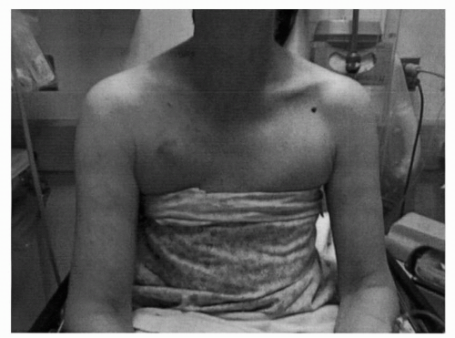
FIGURE 41-3 Venous distention and swelling of the arm due to CVC in the right SC. (Courtesy of Rachel Rosovsky, MD, MPH.)

When diagnostic tests are used to evaluate the patient who presents with symptoms, such as erythema or numbness of the extremity, swelling or pain in the arm (Fig. 41-3), neck or head, phlegmasia, or venous distension, the reported incidence of CVC-related DVT varies from 4% to 41%.17,25,61,62 When surveillance venography or US are used to evaluate patient, irrespective of symptoms, the rate of CVC-related DVT ranges from 12% to 75%.17,25

The time of onset of these CVC-related DVT appears to be within days of insertion. In the most extensive study by De Cicco et al., serial venography was done, on average 8, 30, and 105 days after insertion of CVC.53 Of the DVTs that ultimately developed, 64% occurred by 8 days and 98% by 30 days. In another study of 450 cancer patients where venography was performed on day 8 and 30 after CVC insertion, 96% of the CVC-related DVTs were found on day 8.63 Further analysis of the time course of thrombus formation may be helpful to guide future studies addressing strategies to prevent CVC-related DVT.


Risks for Central Venous Catheter-Related Thrombosis


Patient-Related Risk Factors

Several observational and prospective studies have tried to elucidate the potential risk factors that may be important in the development of DVTs in CVCs. Both patient- and catheter-related risks exist. The presence of malignancy is perhaps the most important patient-related risk factor, and there is some suggestion that some types of malignancy (adenocarcinoma of the lung) have higher rates of catheter-related DVTs than others (head and neck cancer).56 Ovarian cancer is also identified as a significant risk factor for the development of CVC-related thrombosis with an odd ratio of 4.8 in a recent prospective study.62 This finding may be related to the activation of the coagulation system in these different malignancies, tumor-related changes in blood flow in the upper torso, or levels of tissue factor or tissue factor pathway inhibitor. It is probably related to the general increased risk of thrombosis that occurs in oncology patients, as has been investigated extensively.64, 65, 66, 67, 68, 69, 70, 71

The type of chemotherapy also appears to influence the rate of CVC-related thrombosis. Clotting occurred in 6 of 11 (55%) catheters through which sclerosing chemotherapy was infused, but in only 9 of 29 (31%) infused with nonsclerosing chemotherapy.5

May 27, 2016 | Posted by in ONCOLOGY | Comments Off on Intravenous Access and Catheter Management

Full access? Get Clinical Tree

Get Clinical Tree app for offline access