Inherited Disorders of Platelet Function


Inherited Disorders of Platelet Function


Joel S. Bennett

A. Koneti Rao


Prolongation of the bleeding time in a patient with a normal platelet count suggests the presence of either von Willebrand disease (vWD) or impaired platelet function. vWD is discussed in Chapter 52, and acquired disorders of platelet function are discussed in Chapter 66. Inherited disorders of platelet function will be considered in this chapter. For the purposes of this discussion, these disorders will be classified into disorders of platelet adhesion, aggregation, secretion, or procoagulant activity according to the predominant aspect of platelet function affected.


DISORDERS OF PLATELET ADHESION


Bernard-Soulier Syndrome (Glycoprotein Ib-IX-V Deficiency)

The Bernard-Soulier syndrome (BSS), a rare hemostatic disorder characterized by a prolonged bleeding time, very large platelets, and thrombocytopenia, was described by Bernard and Soulier1 in 1948. Patients with BSS bleed because their platelets cannot adhere to von Willebrand factor (vWF), a consequence of deficiency or dysfunction of the platelet membrane glycoprotein (GP) Ib-IX-V (GPIb-IX-V) complex.2,3,4

Following disruption of the vascular endothelium, circulating platelets can adhere to exposed subendothelial connective tissue components such as collagen, fibronectin, and laminins. However, at the higher shear rates present in arterioles and the microcirculation, platelet adhesion requires the additional presence of vWF.5,6 vWF is an elongated multimeric GP, each monomer of which contains two motifs that interact with receptors on platelets7 (see Chapter 52). One motif, located in the A1 domain of vWF, mediates transient vWF binding to the GPIb-IX-V complex on unactivated platelets. The second motif, located in the C1 domain, mediates the stable interaction of vWF with the integrin αIIbβ3 (GPIIb-IIIa) on activated platelets.8 Although vWF in plasma cannot bind spontaneously to GPIb-IX-V,9 exposing soluble vWF to the antibiotic ristocetin or the snake venom protein botrocetin in vitro enables it to bind to GPIb-IX-V.10,11 The physiologic correlates for ristocetin and botrocetin are unclear. It is possible that the A1 domain of subendothelial vWF, but not soluble vWF, has a conformation allowing it to bind to GPIb-IX-V.12 It is also that shear stress changes the conformation of the A1 domain, GPIb-IX-V, or both, enabling vWF binding to GPIb-IX-V.13

GPIb-IX-V is a noncovalent transmembrane heterotrimer consisting of the heavily glycosylated 165,000 mol. wt. protein GPIb,14 the 20,000 mol. wt. protein GPIX,15 and the 82,000 mol. wt. protein GPV.16 The structure of GPIb-IX-V is discussed in detail in Chapter 26A. There are approximately 25,000 copies of GPIb and GPIX on the platelet surface but only half as many copies of GPV, suggesting that individual GPIb-IX-V complexes contain two copies of GPIb, two copies of GPIX, and one copy of GPV. GPIb itself is a heterotrimer containing one copy of a 143,000 mol. wt. α subunit (GPIbα) and two disulfide linked copies of a 22,000 mol. wt. β subunit (GPIbβ).17 GPIbα can be cleaved by trypsin or a calcium-dependent platelet protease into a soluble 135,000 mol. wt. heavily glycosylated amino (N)-terminal fragment glycocalicin.18 The N-terminal portion of glycocalicin contains binding sites for vWF and thrombin, while its C-terminal portion contains a variable number of tandem 13 residue mucin-like repeats to which multiple sialic acid-rich O-linked hexasaccharides are appended.19,20 The cytosolic portion of GPIbα binds to the C-terminus of filamin A (actin-binding protein),21,22 linking GPIbα to the platelet cytoskeleton.23 It binds to the adaptor protein 14-3-3ζ as well, linking GPIbα to platelet signaling pathways.24,25,26 The cytosolic portion of GPIbβ can be phosphorylated at Ser166 by cyclic AMP-dependent protein kinase A,27 a reaction that inhibits collagen-induced platelet actin polymerization.28 A role for GPV in GPIb-IX-V function is unclear. Although GPV can be cleaved by thrombin,29 this is not required for vWF binding to GPIbα, nor does it activate platelets.30

The N-terminal portion of GPIbα, as well as GPIbβ, GPIX, and GPV, contain leucine-rich repeats (LRRs) with the consensus sequence L-L-N-L-LPPGLL-G—L-.16,31 GPIbα contains eight LRR,9 whereas GPIbβ and GPIX contain one each and GPV contains 15. The amino acids flanking the LRR are also conserved.31 A crystal structure for the vWF A1 domain bound to residues 1 to 290 of GPIbα revealed that LRRs 1 and 5 to 8 of GPIbα bind tightly to the A1 domain.9 Conformational changes in a flexible loop located near the C-terminus of the GPIbα fragment, termed the “β-switch”9 or “R-loop,”32 may be needed for vWF binding to GPIbα. This portion of GPIbα is the site of several gain-of-function mutations responsible for platelet-type vWD.9 The function of the LRR in GPIbβ, GPIX, and GPV is unknown, but they do not participate in intermolecular interactions as “leucine-zippers.”

Biosynthetic studies using recombinant GPIb-IX-V revealed that although small amounts of GPIbα can be detected on the surface of cells expressing GPIbα and GPIbβ, GPIbα expression is much more efficient when the cells coexpress GPIbα, GPIbβ, and GPIX.33 Thus, mutations that result in the deficient expression of either GPIbα, GPIbβ, or GPIX can produce the BSS. On the other hand, GPV is not required for GPIb-IX-V expression and why it is absent from BSS platelets is not known.34


Clinical Manifestations

BSS presents in infancy or childhood with ecchymoses, epistaxis, and gingival bleeding.16 Later manifestations include menorrhagia and postpartum, gastrointestinal, and posttraumatic
hemorrhage. Hemarthroses and expanding hematomas are unusual. Although the severity of bleeding is variable and may not always correlate with the extent of thrombocytopenia and the degree of GPIb-IX-V deficiency,35 it can be severe enough to require transfusion and suppression of menses. In a review of 59 patients, for example, there were 10 deaths.36 In some patients with BSS, the severity of hemorrhage inexplicably declines over the course of the disease.36


Laboratory Findings

The bleeding time in patients with BSS is markedly prolonged to >20 minutes.16 Most patients are thrombocytopenic to some degree, and platelet counts <20,000/µL have been reported.37 Nonetheless, platelet counts vary within kindreds and even in individual patients. BSS platelets are large when observed on peripheral blood smears16; 30% to 80% have mean diameters >3.5 µm, and occasional platelets are as large as 20 to 30 µm in diameter.38 There is contradictory evidence about the size of BSS platelets in the circulation. It has been reported that circulating BSS platelets are normal in size39 but have increased membrane, perhaps located in the surface-connected open-canalicular system that is extruded when platelets spread during the preparation of a blood smear.40 Others have reported that the volume of circulating BSS platelets is increased41 and that they have a more spherical shape41 with a more deformable membrane.42 The molecular basis for the macrothrombocytopenia of BSS is not known. However, amelioration of the macrothrombocytopenia when an IL4-GPIbα fusion protein was expressed in a murine BSS model suggests that absence of the GPIbα cytoplasmic domain contributes to this abnormality.43


FIGURE 65.1 Platelet aggregation in various platelet disorders. Platelet aggregation can be studied ex vivo by a variety of techniques. Conventional turbidometric platelet aggregometry is performed in an “aggregometer” using stirred suspensions of either unwashed platelets in plasma or washed platelets in buffer. Immediately after the addition of all platelet agonists except epinephrine, there is a transient decrease in light transmittance that has been attributed to platelet shape change. This is followed by an increase in light transmission as platelet aggregates form. Two “waves” of platelet aggregation have been described. The “first wave” represents primary aggregation and is a direct consequence of agonist stimulation. The “second wave” represents secondary aggregation and is due to the formation of large irreversible aggregates when platelet secretion occurs. If the concentration of platelet agonist is sufficiently high, the first and second waves merge into a single continuous tracing. Measurements of platelet aggregation are expressed as either the extent or the rate of increase in light transmittance that occurs as platelet aggregates form.

The turbidometric aggregometer has been modified to simultaneously measure platelet aggregation and platelet dense granule secretion using the luciferin-luciferase reaction to measure secretion of dense granule ATP (lumiaggregometry). Platelet aggregation can also be measured in diluted anticoagulated whole blood based on the increase in electrical impedance (measured in ohms) that occurs as platelet aggregates accumulate on a pair of electrodes inserted into the suspension of aggregating platelets (impedance aggregometry). Impedance aggregometry tracings are similar to those obtained by the turbidometric method, except that shape change and two waves of aggregation are not seen. Lastly, because aggregometry is insensitive to the formation of small platelet aggregates, methods have been devised to measure the disappearance of single platelets immediately after platelet stimulation. Agonists are added to stirred platelet suspensions and after adding a fixative to stabilize the aggregates, their number and size are measured either visually with a hemocytometer or using an electronic particle counter or flow cytometer.

The platelet aggregation tracings shown in the figure were generated in a conventional turbidometric platelet aggregometer after the addition of collagen, ADP, and ristocetin to stirred aliquots of platelet-rich plasma. vWD, von Willebrand disease; BSS, Bernard-Soulier syndrome; SPD, storage pool disease; ASA, aspirin; TSA, Glanzmann thrombasthenia. (Adapted from Weiss HJ. Platelet physiology and abnormalities of platelet function (second of two parts). N Engl J Med 1975;293:580-588, with permission.)

Erythrocytes and white blood cells are normal in BSS. Distinctive abnormalities have not been observed in marrow megakaryocytes by light microscopy,44 although demarcation membrane system abnormalities have been observed by electron microscopy.45 The laboratory tests that distinguish BSS from normal platelets are the failure of BSS platelets to agglutinate in the presence of ristocetin or botrocetin and the failure of vWF to restore agglutination (FIGURE 65.1).16 Aggregation of BSS platelets in response to ADP, collagen, and epinephrine is normal.16 However, the rate of thrombin-stimulated aggregation can be reduced.46
Although the serum prothrombin time has been reported to be decreased in BSS47 and both collagen-induced platelet procoagulant activity and factor XI binding to be absent,48 prothrombin consumption has also been reported to be normal49 and platelet procoagulant49 and prothrombinase activity to be increased.50 An unexplained decrease in thrombin-, trypsin-, and thromboxane-stimulated phospholipase C (PLC) activity has also been reported.51 GPIb-IX-V can be a target for drug-dependent anti-platelet antibodies52; such antibodies fail to bind to BSS platelets.53


Genetics

BSS is an autosomal recessive disorder.16 In most instances, GPIb-IX-V is not present on the surface of BSS platelets, but in a few cases, 7% to 47% residual GPIb or GPIb-IX has been detected.54 Many, but not all, obligate BSS heterozygotes have platelets that are larger than normal and a content of GPIb intermediate between normal and affected individuals.38,55 Consistent with its rarity, consanguinity has often been present in affected families.

The gene for GPIbα is located on chromosome 17p12-ter, the GPIbβ gene on chromosome 22q11.2, the GPIX gene on chromosome 3q21, and the GPV gene on chromosome 3q29.16 Each gene has a compact “intron-depleted” structure; the genes for GPIbα, GPIbβ, and GPV each contain two exons and the gene for GPV contains three.56 The open reading frame and 3′ untranslated region of each gene is encoded by a single exon except for GPIbβ where its single intron is inserted into the codon for the fourth residue of its signal peptide.57 Like other genes expressed by megakaryocytes, the 5′ flanking region of each gene contains potential binding sites for the transcription factors GATA, Ets, and Sp-1.58,59,60 In addition, polymorphic variation in the region surrounding the GPIbα translation initiation site appears to be a determinant of the level of GPIb-IX-V expression.61


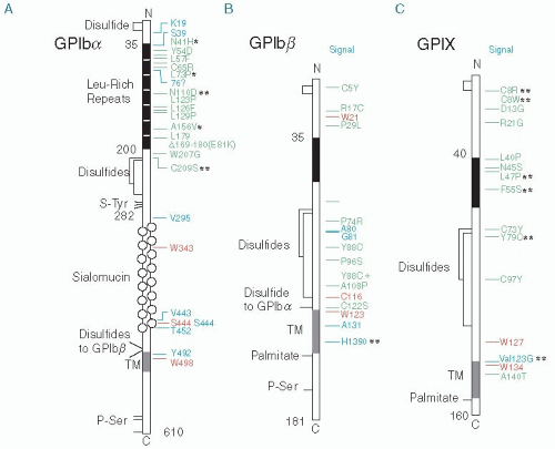
FIGURE 65.2 Mutations in the genes for GPIbα, GPIbβ, and GPIX causing the BSS mapped onto the structures of the mature proteins. Green, missense mutations and short deletions; red, nonsense mutations leading to premature stop; and blue, frameshift mutations leading to stop. *, autosomal dominant inheritance; **, mutations discussed in Savoia A, Pastore A, de Rocco D, et al. Clinical and genetic aspects of Bernard-Soulier syndrome: searching for genotype/phenotype correlations. Haematologica 2011;96:417-423. (Adapted from Berndt MC, Andrews RK. Bernard-Soulier syndrome. Haematologica 2011;96:355-359, with permission.)

As shown in FIGURE 65.2, at least 56 different mutations responsible for BSS have been identified in the genes for GPIbα, GPIbβ, and GPIX.62 Most are missense mutations or nucleotide deletions and insertions resulting in frameshifts and premature stop codons. However, several unique mutations have been identified: deletion of the entire GPIbβ locus on chromosome 22q11.2, occasionally in the setting of the velocardiofacial syndrome63,64; a more circumscribed homozygous deletion of the GPIBB locus that included SEPT5 gene resulting in the BSS, cortical dysplasia, developmental delay, and a platelet secretion defect65; a C to G transversion in the GATA binding site of the GPIbβ promoter resulting in decreased GPIbβ gene transcription66; and an Ala140Thr in the transmembrane domain of GPIX.67,68 The latter is noteworthy because it is consistent with observations that transmembrane domain interactions
are involved in efficient GPIb-IX-V expression69 and with the impairment of GPIb-IX expression observed when the GPIbβ transmembrane domain was mutated in vitro.70 The monoallelic GPIbα mutation Ala156Val, known as the “Bolzano mutation,” has been reported to be the most common cause of inherited thrombocytopenia in Italy. It has been associated with a mild bleeding diathesis, a variable degree of thrombocytopenia, and a modest but consistent increase in platelet diameter and volume.71 It is also noteworthy that many BSS mutations have been reported multiple times, suggesting that there may be “hot spots” for mutation in the GPIbα, GPIbβ, and GPIX genes.


Table 65.1 Differential diagnosis of inherited macrothrombocytopenia


Disorder


Ristocetin Response


Features


Bleeding Diathesis


BSS


No


GPIbα, GPIbβ, GPIX mutations


Yes


MHY9-related disorders


Yes


Leukocyte inclusions, deafness, nephritis, deafness, cataracts


Uncommon


Gray platelet syndrome (GPS)


Yes


Platelet α-granule deficiency


Yes


Montreal platelet syndrome


Yes


Heterozygous for V1316M mutation in exon 28 of vWF producing platelet type vWD


Yes


Mediterranean macrothrombocytopenia


Yes


Heterozygosity for BSS?


No


Mediterranean stomatocytosis/macrothrombocytopenia


Yes


Phytosterolemia due to ABCG 5 and 8 genes mutations


Yes


GATA-1 mutations


Decreased


X-linked inheritance, can be associated with dyserythropoiesis ± anemia


Yes


Sialyl-Lewis-S antigen deficiency


Progressive thrombocytopenia, hypogranular platelets, decreased GPIb, neutropenia


Yes


Paris-Trousseau syndrome


Yes


Part of Jacobson syndrome; caused by loss of the transcription factor FLI1 due to deletion of the long arm of chromosome 11


Yes


Filamin A mutations


Yes


X-linked disorder; usually associated with periventricular nodular heterotopia and the otopalatodigital syndrome


Yes


Platelet-type vWD


Yes


Hypersensitivity to ristocetin; GPIbα mutations causing spontaneous vWF binding to platelets


Yes


Type 2B vWD


Yes


Hypersensitivity to ristocetin; vWF mutations causing spontaneous vWF binding to platelets


Yes


Differential Diagnosis

BSS must be differentiated from other conditions that are associated with large platelets and thrombocytopenia (macrothrombocytopenia). Inherited disorders associated with macrothrombocytopenia are listed in Table 65.1 and can be readily differentiated from BSS because BSS platelets fail to agglutinate in the presence of ristocetin (FIGURE 65.1).72 Many of these conditions are discussed in detail in Chapter 64.

Macrothrombocytopenia is a characteristic feature of mutations in the MYH9 gene located on chromosome 22q12.3-q13.2 encoding the nonmuscle myosin heavy chain IIA (NMMHC-IIA).73,74 Previously, these MYH9 disorders were segregated into four separate autosomal dominant inherited syndromes—the May-Hegglin anomaly and the Sebastian, Fechtner, and Epstein syndromes—based on the presence or absence of Döhle body-like leukocyte inclusions, hereditary nephritis, cataracts, and sensorineural deafness.75 However, because each results from mutations in the MYH9 gene, it now seems appropriate to consider them as a single entity whose variability results from the variable expression of MHY9 mutations and whose common feature is macrothrombocytopenia. In general, the function of platelets affected by MHY9 mutations is normal,76,77,78 and if hemorrhage occurs, it is usually due to thrombocytopenia.79

Macrothrombocytopenia also occurs in the gray platelet syndrome (GPS), a disorder resulting from platelet α-granule deficiency80 and due, at least in some cases, to mutations in the gene NBEAL2 (see below).81,82,83 In contrast to BSS platelets, GPS platelets are pale gray on Wright-stained blood smears and agglutinate normally in the presence of ristocetin.84 In a reevaluation of the Montreal platelet syndrome, large platelets, thrombocytopenia, and spontaneous platelet aggregation were found to be due
to type 2B vWD caused by a heterozygous V1316M mutation in exon 28 of the vWF gene.85,86 Mediterranean macrothrombocytopenia refers to putative differences in platelet count and size between Europeans of Northern and Mediterranean origin87; in Italy, this may be due to heterozygosity for BSS, in particular heterozygosity for the “Bolzano mutation” Ala156Val.88 Mediterranean stomatocytosis/macrothrombocytopenia, the rare combination of stomatocytic hemolysis and macrothrombocytopenia, results from excess plant sterols in the blood and is caused by mutations in the ABCG5 and ABCG8 genes linked to phytosterolemia.89 Several reported patients had significant bleeding histories. Sex-linked macrothrombocytopenia occurs in patients with GATA-1 transcription factor mutations,90,91,92 and macrothrombocytopenia with dystrophic megakaryocytes and neutrophils lacking sialyl-Lewis-S antigen was described in an infant who died at 37 months due to hemorrhage.93 In the Paris-Trousseau syndrome,94 a part of the Jacobsen syndrome,95 a deletion of the long arm of chromosome 11 at q23.3 containing the gene for the transcription factor FLI1 results in mild thrombocytopenia, subpopulations of large platelets and platelets containing giant α-granules, and the expansion of immature megakaryocyte progenitors.96 Mutations in the filamin A gene located on chromosome Xq28 are responsible for periventricular nodular heterotopia and the otopalatodigital syndrome; macrothrombocytopenia and bleeding, misinterpreted as ITP, has been present in some patients.97 In platelet-type vWD, mutations within the vWF binding domain of GPIbα induce spontaneous vWF binding to GPIbα and the clinical picture of type IIB vWD which itself can be associated with macrothrombocytopenia.9,98,99,100,101,102 There are also anecdotal reports of thrombocytopenia and giant platelets, often associated with a mild bleeding diathesis but lacking the features of either BSS or the MHY9 disorders.103,104 In two other reports, macrothrombocytopenia was associated with the presence of large platelet granules whose origin was uncertain.105,106

Acquired BSS-like disorders can be differentiated from the congenital BSS by history. GPIb antibodies occur in some patients with idiopathic thrombocytopenic purpura107; whether the antibodies themselves are responsible for bleeding is not clear because of concomitant thrombocytopenia. Two patients with myelodysplasia and a BSS-like syndrome have been reported,108,109 as has a patient with a lymphoproliferative disorder and a circulating IgG antibody that inhibited ristocetininduced platelet agglutination, but was directed against an unidentified 210,000 mol. wt. platelet protein.110


Therapy

Treatment of hemorrhage in patients with the BSS usually requires platelet transfusion.111 Hormonal control of menses has been effective in managing menorrhagia.112 Splenectomy has been attempted to increase the platelet count113 but has resulted in only transient increases and has not ameliorated the platelet function defect. Corticosteroids are not beneficial in this disorder. Desmopressin acetate (DDAVP) has been reported to shorten the bleeding time in some, but not all, affected individuals in whom it has been administered,114,115,116,117 but its efficacy in patients with hemorrhage is unclear. Antifibrinolytics such as epsilon aminocaproic acid and tranexamic acid can be useful adjuncts to other therapy.118 A number of anecdotal reports suggest that recombinant VIIa may be effective in the treatment of hemorrhage in BSS patients, although the degree of effectiveness has varied.119,120,121,122,123 Lastly, bone marrow transplantation from HLA-identical donors has been performed successfully in patients with BSS.124,125


DISORDERS OF PLATELET AGGREGATION


Glanzmann Thrombasthenia (Integrin αIIbβ3 [Glycoprotein IIb-IIIa] Deficiency)

Thrombasthenia, described by Glanzmann126 in 1918, is a rare bleeding disorder characterized by a prolonged bleeding time, a normal platelet count, and absent macroscopic platelet aggregation (see FIGURE 65.1).127 Platelets from affected individuals are unable to aggregate because of a deficiency or dysfunction of the major platelet integrin αIIbβ3, also as known as GPIIb-IIIa.128

Platelet aggregation is essential for the formation of a hemostatic platelet plug.129 In contrast to platelet adhesion, platelet aggregation is an active metabolic process, requiring platelet stimulation by agonists such as thrombin, collagen, and ADP and exposure of a binding site for fibrinogen or vWF on the integrin αIIbβ3.130,131,132 αIIbβ3-bound fibrinogen or vWF then crosslinks adjacent platelets into an occlusive plug. αIIbβ3, like other members of the integrin family, resides on the cell surface in an equilibrium between inactive (low affinity) and active (high affinity) conformations.133,134 Platelet stimulation shifts αIIbβ3 to its active conformation, enabling it to bind ligands and mediate platelet aggregation.134 Platelet stimulation also enables interaction of the αIIbβ3 cytoplasmic tails with submembranous actin filaments, providing a link between the force of cytoskeletal contraction and a fibrin clot, resulting in clot retraction.135,136

αIIbβ3 is a calcium-dependent heterodimer and dissociates into αIIb and β3 monomers in the presence of calcium chelators.137 In rotary-shadowed electron micrographs, αIIbβ3 appears as a 12 × 8 nm globular head with two 18-nm stalks extending from one side.138 When incorporated into phospholipid vesicles, the globular head extends ≈20 nm above the vesicle surface with the tips of the tails inserted into the phospholipid.139

αIIb itself has an unreduced molecular weight of 136,000 and dissociates into a 125,000 mol. wt. heavy chain (αIIbα) and 23,000 mol. wt. light chain (αIIbβ) following disulfide bond reduction.137 αIIb is synthesized as single chain precursor (Pro-GPIIb) in the rough endoplasmic reticulum (ER)140 where it associates with β3. The resulting heterodimer is transported to the Golgi complex where Pro-αIIb is cleaved into heavy and light chains.141 In contrast to αIIb, β3 is a single chain protein containing 56 cysteine residues and 28 disulfide bonds142; its apparent molecular weight of 90,000 on unreduced sodium dodecyl sulfate gels increases to 110,000 following disulfide bond reduction.137 The disulfide bonds in β3 are concentrated in three regions of the extracellular portion of the molecule: a cysteine-rich, proteaseresistant amino terminus (residues 1 to 62), a protease-sensitive central region (residues 101 to 422), and a disulfide-rich, protease-resistant core (residues 423 to 622).143

In the absence of heterodimer formation, αIIb and β3 monomers are retained in the ER and eventually degraded.144 The factors responsible for the retention of uncomplexed monomers are unknown. However, it is clear from studies of various thrombasthenic mutants that formation of αIIbβ3 heterodimers alone is not sufficient to guarantee egress of αIIbβ3 from the ER.144,145

There are approximately 80,000 copies of αIIbβ3 on the surface of unstimulated platelets.146 Glanzmann thrombasthenia
(GT) results when platelets lack sufficient numbers of functional αIIbβ3 to support platelet aggregation.128 However, the platelets of obligate thrombasthenic heterozygotes aggregate normally, indicating that 50% of the normal amount of αIIbβ3 is at least sufficient to support platelet aggregation.147

Platelets express a second β3-containing integrin, αvβ3, the vitronectin receptor.148 There are 50- to 500-fold fewer copies of αvβ3 than αIIbβ3 on platelets.149,150 While αIIbβ3 expression is restricted to cells of the megakaryocytic lineage,151 αvβ3 is expressed by a variety of cells including endothelial cells, smooth muscle cells, and osteoclasts.152


Clinical Manifestations

The clinical manifestations of GT in a large cohort of patients have been described in detail.127 GT typically presents with mucocutaneous bleeding during the neonatal period or infancy, occasionally with bleeding following circumcision. Spontaneous petechiae are uncommon and bleeding generally results from conditions that would also cause bleeding in an otherwise normal individual. Thus, epistaxis is a frequent type of bleeding, especially during childhood, as are gingival bleeding and menorrhagia. Bleeding at menarche may be severe and require transfusion. Similarly, parturition represents a severe hemorrhagic risk. Other hemorrhagic manifestations include gastrointestinal bleeding and hematuria, but hemarthroses and deep hematomas are unusual. Serious bleeding may follow trauma or surgery. However, the severity of hemorrhage in thrombasthenia is not predictable, even within single kindreds, and does not correlate with the degree of αIIbβ3 deficiency. The apparent decline in the clinical severity of GT with age is likely due to a decrease in the incidence of conditions such as epistaxis with aging.


Laboratory Findings

Platelet counts and platelet morphology on peripheral blood smears are normal. The bleeding time of affected individuals is markedly prolonged.153 A diagnosis of GT is usually suspected when platelet aggregometry reveals absent agonist-stimulated platelet aggregation (see FIGURE 65.1). Platelet secretion induced by strong agonists such as thrombin is normal, but secretion in response to weak agonists such ADP and epinephrine which requires platelet aggregation does not occur. Coagulation tests, such as the prothrombin time and the partial thromboplastin time, are normal in GT, whereas clot retraction in the presence of GT platelets is absent or reduced.

In addition to their inability to aggregate, GT platelets do not spread normally on the subendothelial matrix because of an impaired ability to interact with fibronectin and vWF in the matrix.154 The amount of fibrinogen in the α-granules of GT platelets is decreased to absent.127 Human megakaryocytes do not synthesize fibrinogen, but rather, it is derived from plasma by an αIIbβ3-mediated endocytic process.155,156,157,158,159 A number of biochemical reactions in platelets that require the presence of αIIbβ3 are impaired in GT platelets. For example, the tyrosine phosphorylation of multiple intracellular signaling proteins depends on ligand binding to αIIbβ3 and does not occur in GT platelets.160 Calpain, a calcium-dependent thiol protease, is activated when stirred normal platelets are stimulated by thrombin, but not when GT platelets are treated in a similar manner.161 The kinetics of calcium exchange in unstimulated GT platelets is inexplicably decreased162 since it is not clear what role, if any, αIIbβ3 plays in calcium transport.163,164 Clot retraction in blood containing GT platelets is either absent or reduced.127,135

Aspects of platelet function that do not depend on αIIbβ3 are normal in GT. GT platelets interact normally with collagen153 and undergo secretion when stimulated by “strong agonists” such as thrombin.165 Incubation of GT platelets with ristocetin induces vWF binding to GPIb-IX-V10 and GT platelets adhere to vWF in the subendothelium.154 GT platelets also express normal amounts of procoagulant activity following lysis.50 However, agonist-stimulated prothrombinase activity varies and is influenced by the nature of platelet agonist.50,166


Genetics

The genes for αIIb and β3 are located on the long arm of chromosome 17 at q21→23167 with the αIIb gene at minimum distance of 365 kb downstream from the β3 gene.168 The αIIb gene (ITGA2B) spans ≈18 kb and consists of 30 exons ranging in size from 46 to 220 bp.169 Like the genes for other proteins expressed in megakaryocytes, its 5′ flanking region lacks TATA or CAAT boxes. Instead, it contains a linear array of regulatory elements that includes two motifs recognized by the GATA-1 transcription factor and its cofactor FOG170,171; sequences flanking each GATA-1 binding site are recognized by Fli-1 and perhaps other members of the Ets family of transcription factors172,173,174,175; and an Sp1-binding silencer element is located between the GATA-1 sites.176 The gene for β3 (ITGB3) spans 63 kb and contains 15 exons.151,177 Like the αIIb gene, the 5′ flanking region of the β3 gene lacks TATA or CAAT boxes.178

GT is an autosomal recessive disorder with disease clusters in populations where consanguinity is common. It has been subclassified into three types based on the amount of αIIbβ3 present per platelet and the presence or absence of α-granule fibrinogen and clot retraction.127 In type I, platelets contain <5% of the normal amount of αIIbβ3 and clot retraction and α-granule fibrinogen are absent. In type II, platelets contain 10% to 20% of the normal amount of αIIbβ3, clot retraction is decreased, and α-granule fibrinogen is present. In “variant” thrombasthenia, the platelet content of αIIbβ3 is ≥50% of normal, indicating that the αIIbβ3 abnormality is qualitative, rather than quantitative.

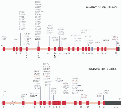
Because the expression of αIIbβ3 on the platelet surface requires the formation of correctly folded heterodimers,179 mutations in the genes for either αIIb or β3 can produce GT. Approximately 200 different mutations responsible for GT have been identified180 (FIGURE 65.3). The vast majority of these mutations consist of a mix of missense and nonsense mutations, nucleotide deletions and insertions, and alternative splicing, resulting in type I and type II GT. However, the most informative mutations have been qualitative αIIbβ3 abnormalities (variant GT) that primarily perturb αIIbβ3 function rather than the level of αIIbβ3 expression.

Mutations located in the extracellular portion of αIIbβ3 cause GT by either impairing fibrinogen or vWF binding to the active αIIbβ3 conformation or by paradoxically inducing constitutive αIIbβ3 activation but causing GT by concurrently decreasing the amount of αIIbβ3 expressed on the platelet surface. Six missense mutations (the αIIb mutation Tyr143His181 and the β3 mutations Asp119Tyr,182 Val193Met,183 Arg214Trp,184,185 Arg214Gln,186 and Aspr217Val187) are located in regions implicated in ligand binding to αIIbβ3 and cause GT by preventing ligand binding.188,189
Similarly, an insertion of two amino acids (Arg-Thr) into the Cys146-Cys167 loop of αIIb produced an inactive αIIbβ3 heterodimer.190 An eighth mutation, β3 Ser123Pro, did not impair αIIbβ3 expression or agonist-induced ligand binding but was associated with absent ADP- and collagen-induced platelet aggregation.191 The β3 mutation, Leu196Pro, was identified independently in two French patients and severely restricted αIIbβ3 expression.192,193 However, the residual αIIbβ3 that made it to the platelet surface was unable to bind ligands, presumably because the mutation prevents αIIbβ3 from assuming its active conformation. By contract, the β3 mutation Cys560Arg locked αIIbβ3 in a high-affinity conformation, but like Leu196Pro, it likely produced a GT-phenotype because the small amount of constitutively active αIIbβ3 on the platelet surface was insufficient to support platelet aggregation.194


FIGURE 65.3 Missense mutations in the ITGA2B and ITGB3 genes resulting in GT. The exons in the ITGA2B and ITGB3 genes are shown schematically as red bars. Mutations shown in black are lack of function mutations, those shown in blue primarily impair αIIbβ3 expression, those in green are characteristic of particular ethnic groups, those in orange cause constitutive αIIbβ3 activation, and those in rose are associated with macrothrombocytopenia. Asterisk indicate the number of times the mutation has been reported. (Adapted from Nurden AT, Fiore M, Nurden P, et al. Glanzmann thrombasthenia: a review of ITGA2B and ITGB3 defects with emphasis on variants, phenotypic variability, and mouse models. Blood 2011;118:5996-6005, with permission.)

The extracellular stalks of αIIbβ3 consist of the thigh, calf-1, and calf-2 domains of αIIb and the epidermal growth factor (EGF)-like domains 1 to 4 and the β TD domain of β3.195 The distal portions of the stalks form an interface containing interacting energetic “hot spots” that are critical for maintaining αIIbβ3 in a stable, inactive conformation.196,197 Thus, it is not surprising that four mutations involving cysteine residues located at the third and fourth EGF domains of β3 (Cys49Arg,198 Cys560Phe,199 Cys560Arg,194 and Cys598Tyr199,199a) as well as a Gly579Ser mutation, not only resulted in type II thrombasthenia but also induced constitutive ligand-binding activity in the residual αIIbβ3. Similarly, a Ser527Phe mutation detected in the third β3 EGF domain of a patient who presented with a mild bleeding diathesis was found to cause constitutive activation of recombinant αIIbβ3 expressed in Chinese hamster ovary cells.200


Naturally occurring mutations involving the β3 cytoplasmic domain have confirmed the singular importance of this structure in regulating αIIbβ3 function. The first mutation identified, a missense mutation that results in the substitution of Pro for Ser at residue 752, prevents αIIbβ3 activation by cellular agonists,201,202,203 likely because it abrogates binding of the cytoplasmic protein kindlin-3 to β3 (see below).204 A second missense mutation, identified separately in two unrelated patients,205,206 converts the Arg724 codon into a stop codon, resulting in the synthesis of a truncated β3 molecule lacking 39 C-terminal amino acids. As a consequence, αIIbβ3 is unable to transduce both “inside-out” and “outside-in signals,” likely because β3 is now unable to interact with both kindlin-3 and talin.204 A third mutation, identified in an Palestinian Arab with GT, produces aberrant mRNA splicing at the intron 14/exon 15 junction and replacement of the β3 sequence distal to residue 741 with 40 unrelated amino acids.207 This results in the absence of the distal talin binding site in the β3 cytoplasmic domain, as well the binding site for kindlin-3, and an αIIbβ3 extracellular domain that appears to be “locked” in its inactive conformation.208

Lastly, several heterozygous mutations located in the highly conserved membrane-proximal regions of the αIIb and β3 cytoplasmic domains have been associated with GT-like phenotypes and macrothrombocytopenia.209 Thus, replacement of αIIb Arg995 with either Gln209,210 or Trp211 and β3 Asp723 with His212,213 results in decreased, but not absent, platelet aggregation, an approximate 50% decrease in the total amount of platelet αIIbβ3 predominantly affecting the surface—rather than the granule—membrane pool, a moderate degree of macrothrombocytopenia and platelet “anisocytosis,” and partial αIIbβ3 activation when αIIbβ3 is expressed in tissue culture cells. A β3 Leu718Pro mutation produced a similar phenotype,214 as did a two residue deletion in the β TD domain of β3.215 How these mutations in αIIbβ3 produce this spectrum of abnormalities is not clear since “classic” GT is not associated with alterations in platelet number or size, but the association of macrothrombocytopenia with BSS and the MYH9 disorders suggests that perturbed αIIbβ3-cytoskeleton interactions could be involved.


Differential Diagnosis

A history of lifelong bleeding, a prolonged bleeding time, and absent platelet aggregation are diagnostic of GT and differentiate it from other disorders of platelet adhesion and secretion. Rare instances of acquired GT have been reported and can be differentiated from congenital GT by history. Although autoantibodies against αIIbβ3 are frequently detected in patients with idiopathic thrombocytopenic purpura,216,217 they rarely induce a GT-like state.218,219,220,221 There are anecdotal reports of autoantibodies producing acquired GT in patients with Hodgkin and non-Hodgkin lymphoma, hairy cell leukemia, and after an allergic reaction to diclophenac.222,223,224 A patient with multiple myeloma has also been reported whose IgG1κ paraprotein was directed against β3 and inhibited αIIbβ3 function.225 Congenital afibrinogenemia may be associated with a prolonged bleeding time and decreased in vitro platelet aggregation due to the absence of sufficient fibrinogen to support normal platelet aggregation.226

In the LAD-III/LAD-1/variant syndrome, patients manifest mucocutaneous bleeding and absent platelet aggregation, despite normal or nearly normal αIIbβ3 expression.227,228,229 Concurrently, they suffer from recurrent and severe bacterial infections due to defective leukocyte integrin function, implying that the syndrome results from a general defect in integrin activation. Although initially attributed to defective expression of the Rap-1 activator CalDAG-GEFI,229,230,231 in several kindreds,232,233 mutations have been identified in the KINDLIN3 gene encoding the protein kindlin-3 that binds to β1, β2, and β3 integrin cytoplasmic tails and is required for agonist-stimulated integrin activation.204,234,235


Therapy

The mainstay of treatment of bleeding in GT remains the transfusion of normal platelets.111 Because bleeding is a lifelong problem, use of HLA-matched platelets should be considered to lessen the chance of refractoriness to transfusion due to platelet alloimmunization. Patients with GT, especially patients with null mutations, not infrequently develop alloantibodies against normal αIIbβ3 after transfusion, potentially limiting the effectiveness of transfused platelets.236,237,238,239 Protein A-Sepharose immunoadsorption has been reported to restore the efficacy of transfused platelets in this situation.240 Oral contraceptives have been useful in controlling menorrhagia. Regular dental care is essential in minimizing gingival bleeding, and fibrinolytic inhibitors, in addition to platelet transfusions, may be useful in controlling bleeding after dental extractions. Corticosteroids are not efficacious in managing bleeding in thrombasthenic patients,153 and there is little evidence that desmopressin (DDAVP) is useful.114,241 Recombinant factor VIIa has been found to be efficacious for the treatment of bleeding episodes and for obtaining surgical hemostasis in patients with GT, particularly in patients who have developed alloantibodies from prior platelet transfusions.111,242,243 The GT phenotype of several severely affected individuals has been corrected by bone marrow,244,245,246,247 stem cell,248,249 and unrelated donor cord blood bone marrow transplantation.250 Lastly, platelet gene therapy has improved hemostatic function in αIIbβ3-deficient dogs,251 suggesting that at some point in the future, gene therapy could be used in humans.


DISORDERS OF PLATELET SECRETION

Platelets contain four types of granules: dense granules containing ADP, ATP, calcium, serotonin, and pyrophosphate; α-granules containing a variety of proteins, some derived from the plasma, others synthesized by the megakaryocyte; lysosomes containing acid hydrolases; and microperoxisomes containing a peroxidase activity. Following platelet activation, the contents of these granules are extruded in an exocytic process known as platelet secretion or the platelet release reaction.252 Disorder of platelet secretion are heterogeneous and are generally manifest as decreased platelet aggregation and absence of the second wave of aggregation when platelets are stimulated with weak agonists like ADP and epinephrine. They can be due to a decrease in the number of granules, a decrease in granule contents, or perturbation of the mechanism of granule secretion.


The Gray Platelet Syndrome (α-Granule Deficiency, α-Storage Pool Disease)

The gray platelet syndrome (GPS), described by Raccuglia80 in 1971, is a rare disorder that results from the specific absence of morphologically recognizable α-granules in the platelets of affected individuals.


Biology and Molecular Aspects of the Disorder

Normal platelets contain ≈50 spherical or elongated structures termed α-granules that contain a variety of proteins, some of which are specific or relatively specific for platelets and others that are also found in plasma.253 The former include platelet factor 4 (PF4), β-thromboglobulin (βTG), platelet-derived growth factor, and thrombospondin (TSP), and the latter include fibrinogen, vWF, albumin, coagulation factor V (FV), IgG, fibronectin, and a number of protease inhibitors.254 The platelet-specific proteins and several of the plasma proteins such as vWF255 are synthesized by megakaryocytes, whereas others, such as albumin and IgG, reach the α-granules via endocytosis of circulating protein.256 The α-granule membrane contains many of the same proteins present in the platelet plasma membrane (αIIbβ3257 and GPIb-IX-V258) and others specific for α-granules (P-selectin259,260 and osteonectin261). Each can be translocated to the platelet surface following platelet activation.

Only gold members can continue reading. Log In or Register to continue

Stay updated, free articles. Join our Telegram channel

Jun 21, 2016 | Posted by in HEMATOLOGY | Comments Off on Inherited Disorders of Platelet Function

Full access? Get Clinical Tree

Get Clinical Tree app for offline access