Infectious Complications in Pediatric Cancer Patients


Infectious Complications in Pediatric Cancer Patients


Monica I. Ardura

Andrew Y. Koh


Infectious diseases are major causes of morbidity and mortality in pediatric patients with cancer. The advances in supportive care during the past several decades have permitted patients to successfully recover from the impact of cytotoxic cancer chemotherapy, hematopoietic stem cell transplantation (HSCT), radiation therapy, surgical intervention, and profound immunosuppression. The advances of pediatric infectious diseases supportive care have contributed substantially to the improved survival and outcome from infectious complications.

This chapter reviews the epidemiology, clinical manifestations, and strategies for managing infectious diseases in pediatric oncology patients. Formidable challenges, however, continue to threaten and undermine these successes, including the inexorable rise of multidrug-resistant bacteria, emergence of invasive fungal infections, and development of refractory viral infections. Moreover, for a number of these infections (especially the invasive mycoses), there continue to be limited therapies. Further compounding these challenges are the increasing uncertainties of the impact of new immunosuppressive therapies on host defenses (Fig. 40.1).


HOST DEFENSES: INNATE AND ADAPTIVE IMMUNITIY

Host defenses against bacterial, fungal, viral, and parasitic pathogens are often classified as innate or adaptive. Innate host defenses include mucocutaneous barriers, phagocytic cells, cytokine regulatory networks, the toll-like receptor (TLR) system, natural killer cells, and nonclonal T and B cells. Adaptive immunity encompasses the production of pathogen-specific antibodies and T-lymphocyte cell-mediated immunity (CMI).


Mucocutaneous Barriers: The First Line of Innate Host Defense

The skin and mucosal surfaces constitute the primary innate host defense against invasion by endogenous and exogenous microorganisms. In addition to providing a physical barrier, mucocutaneous cells express unique biochemical, mechanical, and immunologic defenses against microbial invasion. For example, ciliated and mucus-producing cells of the pseudostratified columnar epithelium of the lower respiratory tract provide a mucociliary “escalator” that propels organisms out of the lungs. Pulmonary surfactant molecules belong to the collectin class of molecules and serve to opsonize bacterial and fungal pathogens.1,2 Pentraxin, a secreted pattern-recognition receptor that has a nonredundant role in resistance to selected microbial agents, in particular to the opportunistic fungal pathogen Aspergillus fumigatus, is another key molecule that has been found to be critical to pulmonary host defenses.3 Other classes of antimicrobial peptides also play key roles in mucosal host defenses.4,5 For example, defensins are cysteine-rich antimicrobial peptides that contribute to host defense against bacterial, fungal, and viral infections. The α-defensins are present in neutrophils (polymorphonuclear leukocytes [PMNs]) and Paneth cells of the small intestine, and β-defensins protect the skin and the mucosal surfaces of the respiratory, gastrointestinal (GI), and urinary tracts. Among the specialized cells of the GI tract, parietal cells elaborate gastric acid that serves as a potent barrier against potential enteric pathogens; Paneth cells of the intestinal tract synthesize and release antimicrobial molecules that protect the luminal surface of the epithelial cells.


Figure 40.1 Effect of patient and pathogen factors on the host defense system that result in the immunocompromised host.

Pediatric oncology patients sustain disruptions of the mucocutaneous integrity because of their underlying cancer and its treatment, resulting in increased susceptibility to infection. For example, mucocutaneous integrity may be disrupted by local tumor invasion or as a result of surgery, irradiation, or cytotoxic chemotherapy. The disruption of mucocutaneous barriers provides a key portal of entry for bacterial and fungal pathogens.

The use of vascular catheters provides a striking example of the impact of cutaneous disruptions. Vascular catheters, whether temporary or chronically indwelling, disrupt cutaneous barriers and provide a direct access of microorganisms through the catheter lumen and into the bloodstream in oncology patients.6 One study reported an estimated fourfold increase in the incidence in bacteremia of neutropenic cancer patients who had catheters compared to those who did not. Foreign devices other than vascular catheters have been implicated in the risk of infectious complications in cancer patients (e.g., Omaya intraventricular reservoirs and prosthetic bone-joint hardware).7 Increasing recognition of the role of biofilms underscores mechanisms by which these organisms may perpetuate seeding of the bloodstream and promote emergence of resistance.8,9

Several antineoplastic compounds administered in patients with hematologic malignancies may induce severe mucosal disruption. These include high-dose methotrexate, cytosine arabinoside (Ara-C), anthracyclines (daunarubicin, doxorubicin), and etoposide. Radiation therapy to the abdomen or thorax may result in similar disruption of the GI epithelium in the esophagus or in the small and large intestines, respectively. GI barriers may also be disrupted by mucosal disruption due to herpes simplex virus (HSV) or cytomegalovirus (CMV). Graft-versus-host disease
(GVHD) may also compromise GI mucosal integrity and permit bacteria and/or fungi to translocate from the mesenteric capillary bed and portal venous system.

Other disruptions of mucocutaneous barriers in pediatric oncology patients include ventricular drains, ventriculostomies, nasogastric tubes, endotracheal tubes, chest tubes, surgical drains, dialysis catheters, nephrostomy tubes, urinary catheters, finger sticks, venipunctures, and bone marrow aspirations. These disruptions alter the epidermal integument and provide a potential nidus for colonization, local infection, and hematogenous dissemination of bacterial and fungal pathogens.


Microbial Colonization: Normal Microflora Serving as an Extension of Mucocutaneous Barriers

Most bacterial and fungal infectious episodes in immunocompromised patients are preceded by colonization with the infecting organism. Colonization by normal bacterial flora provides a competitive microbiologic barrier to colonization by extrinsically acquired bacterial and fungal organisms. This normal bacterial flora may be abrogated by antibiotics or by the onset of illness. The mechanisms by which this bacterial barrier is maintained are not well understood. In healthy persons, integumentary and mucosal attachment sites are populated with a quantitative predominance of aerobic Gram-positive bacteria and a variety of anaerobic bacteria with relatively low virulence. However, within 24 hours of hospitalization, seriously ill patients undergo a change in their indigenous microflora toward one of aerobic Gram-negative organisms. The mechanisms of this microbiologic shift are unclear; however, underlying disease and exposure to broad-spectrum antibiotics likely contribute to these changes in bacterial adherence and colonization.

Approximately one half of the responsible pathogens causing infections are acquired by oncology patients after initial admission to the hospital. More than 80% of the microbiologically documented infections that occur in adult patients with acute myelogenous leukemia (AML) are caused by organisms that were a component of the endogenous mucosal organisms, usually at sites at or near the source of infection.


Phagocytic Cells: The Second Line of Innate Host Defense

The innate host defense system includes the repertoire of phagocytic cells consisting of PMNs, circulating monocytes (MNCs), and tissue macrophages. These cells assert their antimicrobial effects of phagocytosis, oxidative and nonoxidative mediators, and the release of cytokines. PMNs possess the greatest degree of oxidative capacity and response to a microbial stimulus followed by MNCs and then by macrophages. Pulmonary alveolar macrophages, splenic macrophages, and Küpfer cells constitute an important residual phagocytic barrier during neutropenia, when PMNs and MNCs may be depleted. Both MNCs and macrophages serve as important immunoregulatory cells by expressing cytokines and chemokines to activate and regulate host response.

Phagocytic cells may be quantitatively or qualitatively impaired. Patients with quantitative or qualitative defects of their PMNs are subject primarily to infections due to bacteria (Escherichia coli, Klebsiella pneumoniae, Pseudomonas aeruginosa, and more recently an expanding range of resistant Gram-negative organisms) and fungi (particularly Candida spp., Aspergillus spp., Fusarium spp., and Zygomycetes), and does not, per se, appear to increase the incidence or severity of viral and parasitic infections.

The most important determinants of risk of infection associated with neutropenia are (1) the absolute neutrophil count (ANC), (2) the rate of decline of the ANC, and (3) duration of neutropenia. The ANC is a critical determinant of susceptibility to most bacterial and fungal pathogens in patients receiving cytotoxic chemotherapy. Cytotoxic chemotherapy, total body irradiation, aplastic anemia, and bone marrow infiltration by leukemic cells may all cause neutropenia. The seminal work of Bodey and colleagues10 demonstrated that a decline of an ANC to less than 500 cells per mL significantly increased the risk of infection and that profound neutropenia (less than 100 cells per mL) precipitously increased the risk of bacterial and fungal infections. The rate of decline of circulating PMNs is also critical. For example, patients with rapidly declining ANCs following therapy for acute leukemia appear to be at greater risk for infectious complications than patients with chronic neutropenia (e.g., aplastic anemia). Finally, neutropenia that persists for more than 7 to 10 days is associated with increasing risk for severe, recurrent, or new bacterial and fungal infections. On the other hand, those in whom no infection has been documented, who defervesce rapidly on empirical antimicrobial therapy, and recover from neutropenia within 1 week of the febrile episode are more likely to pursue an uncomplicated course. As discussed later, duration of neutropenia is an important variable in assessment of patients for oral administration of empirical antibacterial therapy and for preventive strategies for management of invasive fungal infections.

In addition to quantitative declines of PMNs and MNCs, functional changes may occur in pediatric oncology patients. Qualitative abnormalities of PMN function may occur as a consequence of the underlying malignancy (especially acute leukemia) or secondary to antineoplastic therapy. For example, the PMNs from patients with leukemia or lymphoma exhibit suboptimal chemoattractant responsiveness, bactericidal activity, and superoxide production. Antineoplastic chemotherapy and radiotherapy also cause qualitative abnormalities of PMN function. Deficiencies of PMN function iatrogenically induced by the administration of various medications (e.g., opiates, corticosteroids, antibiotics) may have a detrimental effect. For example, corticosteroids suppress oxidative metabolism in superoxide production, impair phagocytosis, and reduce microbicidal activity of PMNs against bacteria and fungi.11

The advent of corticosteroid-sparing immunosuppressive agents may also indirectly impair PMN and MNC function because of their suppressive effects on activating cytokines. For example, infliximab, which is used in the management of corticosteroid-refractory GVHD through inhibition of tumor necrosis factor alpha (TNF-α), may result in an increased risk for reactivation of tuberculosis and for development of invasive aspergillosis.


Cytokines and TLRs

The innate host defense mechanisms include an elaborate network of immunoregulatory cytokines and chemokines.12 TNF-α, interleukin-1 (IL-1), interferon-gamma (IFN-α), interleukin 2, interleukin 6 (IL-6), and interleukin 12 (IL-12) are proinflammatory cytokines that upregulate phagocytic response against bacteria, mycobacteria, fungi, and viruses. IL-1, TNF-α, interferon-γ, and IL-12 promote increased microbicidal activity against most bacteria, mycobacteria, and fungi mediated by a T-helper 1 type response (Th1). By comparison the molecules interleukin 4 and interleukin 10 promote a downregulation of host response against these pathogens; these molecules are characteristic of a T-helper 2 type response (Th2).

TLRs are key components of innate host response that are responsible for recognition of pathogens and cytokine response to surface molecules.13,14 The family of TLRs recognizes pathogen-associated molecular patterns (PAMPs). Different pathogens interact with certain TLRs through distinct patterns of molecules on their cell surface in the form of carbohydrates, proteins, and glycoproteins. Binding of PAMPs to TLRs activates signal transduction pathways that lead to cytokines, interferons, and chemokines that in turn may activate host response to the infecting pathogen. As this process occurs independent of adaptive
immunity, the TLR system may be especially relevant to immunocompromised patients when adaptive immunity is significantly suppressed. For instance, one study suggests an association between a donor TLR4 haplotype and the risk of invasive aspergillosis among recipients of hematopoeitic stem cell transplants from unrelated donors.15 Similarly, other TLR4 single-nucleotide polymorphisms have been associated with susceptibility to infections caused by Gram-negative bacteria,16 C. albicans,17 and respiratory syncytial virus (RSV).18


Adaptive Immunity

Adaptive immunity is composed of B- and T-cell populations that mediate humoral immunity and CMI, respectively. The B cell-plasma cell axis of humoral immunity provides a host with antibody and response to bacterial, viral, and some fungal pathogens. During the course of chemotherapy or during hypogammaglobulinemia, there may be substantial qualitative and quantitative defects in antibody response. Therefore, children with malignancies receiving chemotherapy or undergoing HSCT may have suboptimal antibody responses to vaccines. Defective immunoglobulin synthesis or hypogammaglobulinemia also leads to an increase in the susceptibility to encapsulated bacteria, particularly Streptococcus pneumoniae, Haemophilus influenzae type B (HIB), and Neisseria meningitidis. Children with cancer can safely receive inactivated vaccines, and these should be administered during less intense phases of chemotherapy and after HSCT to allow augmentation of the immunologic response to vaccination. Vaccines administered during chemotherapy do not count toward completion of the routine childhood vaccination schedule; routine childhood vaccination schedule should be reinitiated 3 months after completion of chemotherapy, when humoral and cellular immunity have recovered. Live vaccines are contraindicated during chemotherapy, but can be safely administered 3 to 6 months after chemotherapy completion. Most pediatric studies have evaluated the humoral immune response to vaccination after completion of chemotherapy and found that vaccination can elicit protective antibody responses.19

Certain seasonal infections, like influenza, require yearly vaccination. A review of available pediatric clinical trials concluded that immunogenicity to influenza vaccination was suboptimal in children with malignancy receiving chemotherapy when compared with healthy controls, but all studies demonstrated some degree of immunologic response to influenza vaccination.19 More recent data from the 2009 influenza H1N1 pandemic demonstrated modest immunogenicity to inactivated influenza vaccine in children with malignancies receiving chemotherapy.20 Nonetheless, given an excellent safety profile, potential for efficacy, and the higher incidence, morbidity, and mortality of influenza disease in pediatric oncology patients, annual inactivated influenza vaccine is strongly recommended in pediatric oncology patients aged ≥6 months. Exceptions are those patients who are receiving intensive chemotherapy (e.g., induction or consolidation for acute leukemia) and those receiving anti-B cell antibodies in the preceding 6 months. In all patients, immunization of household contacts and hospital staff should be strongly encouraged. Further studies are necessary evaluating potential predictors of immunogenic response to permit optimization of vaccination during chemotherapy.

Patients with Hodgkin’s disease or non-Hodgkin’s lymphoma have an impaired CMI that can persist even when the cancer is in remission. Patients with deficiencies of cellular immunity are prone to fungal, viral, and intracellularly replicating bacterial pathogens (e.g., Mycobacterium tuberculosis, Listeria monocytogenes, Salmonella species). Corticosteroids and radiotherapy can also contribute to lymphocyte dysfunction. Allogeneic (and to a much lesser degree autologous) bone marrow transplantation is associated with a high risk for herpes virus infections, because both humoral and CMI are impaired. Patients receiving T-cell—depleted bone marrow transplants are particularly susceptible to viral pathogens, especially CMV. Furthermore, pathogens such as CMV that infect patients with altered cellular immunity can further suppress host defenses.

Cytotoxic chemotherapy may further deplete helper T cells (CD4+). Although PMN, MNC, and platelet numbers consistently recover to greater than 50% of pretreatment values after sequential cycles of therapy, lymphocyte numbers do not recover promptly, and lymphopenia may persist for many months after the completion of a chemotherapy regimen. The capacity for CD4+ T-cell regeneration after chemotherapy seems to diminish with age, such that younger children have significantly greater recovery of T cells 6 months after chemotherapy than young adults, who have persistent, severe T-cell depletion. Prolonged T-cell depletion probably contributes to the development of opportunistic infections such as herpes zoster or Pneumocystis carinii pneumonia during the months after chemotherapy.


Spleen and Reticuloendothelial System

The spleen is a key organ of both innate and adaptive immunity. The spleen acts as a mechanical filter and as an immune effector organ. At the same time, the spleen also is the principal organ involved in the production of antibodies to polysaccharide antigens, filtering damaged cells, and opsonized organisms from the circulation. Splenectomized patients are deficient in antibody production when challenged with particulate antigens and have decreased levels of immunoglobulin M (IgM) and properdin (a component of the alternate complement pathway). Asplenic patients are at increased risk for fulminant and rapidly fatal septicemia caused by encapsulated bacterial pathogens, especially S. pneumoniae, H. influenzae, and N. meningitides.21 S. pneumoniae, which is the most common cause of sepsis in the asplenic patient, may be penicillin-resistant, particularly in patients on chronic penicillin prophylaxis. Several other pathogens are also associated with a fulminant course in asplenic patients: Capnocytophaga canimorsus (DF-2), Salmonella species, Babesia microti, and Plasmodium falciparum. Fulminant sepsis due to C. canimorsus is typically associated with dog bites. B. microti, which is transmitted by the bite of Ixodes tick, occurs in the same regions as Lyme disease. Patients with severe combined immunodeficiency (SCID) are predisposed to developing bacteremia, or even splenic abscesses, secondary to functional asplenia.


Neurologic, Mechanical, and Nutritional Factors Contributing to Immune Impairment

Alterations in central nervous system (CNS) function or decreased levels of awareness due to tumor or opioids increase the risk of aspiration pneumonia. Aspirated pharyngeal organisms, most commonly aerobic Gram-negative bacilli, can colonize, invade, and disseminate from a pulmonary source. The risk of an aspiration pneumonia and subsequent disseminated infection is heightened by decreased mucosal clearance mechanisms and damage mediated by antineoplastic therapy.

Mechanical obstruction of hollow viscus by a primary or metastatic tumor mass can promote an infection by the organisms colonizing the site of obstruction. For example, obstruction of the bronchus by osteogenic sarcoma may lead to postobstructive pneumonia, obstruction of the GI tract by a pelvic rhabdomyosarcoma may cause perforation and peritonitis, and obstruction of the ureter may lead to hydronephrosis and pyelonephritis.

The compromising effect of a malnourished state on immune function is well documented. Nutritional deficiencies affect B and T lymphocytes, PMNs, mononuclear phagocytes, complement system function, and cytokine immunoregulation. Increasing data indicate that proteosome activation may severely deplete total-body protein22 with profound implications for mechanical and cellular host defenses.


CARE OF FEBRILE CANCER PATIENTS


General Principles

Fever in the neutropenic patient is a common manifestation of infection in pediatric oncology. Left untreated, febrile, neutropenic patients may sustain devastating complications of bacterial sepsis. Hence, fever in a neutropenic patient should be managed as a potential medical emergency. The localizing signs initially may be muted owing to the paucity of inflammatory cells. A careful history and physical examination directed toward identifying possible foci of infection is important as a guide to selection of antimicrobial therapy and any adjunctive supportive measures, such as surgical intervention. Less commonly but importantly, neutropenic patients can have serious and life-threatening infection in the absence of fever (e.g., afebrile, neutropenic patients with Clostridium septicum can present with severe abdominal pain and can ultimately develop septic shock and rapidly progressive necrotizing fasciitis with myonecrosis). Accordingly, if a neutropenic patient develops signs or symptoms suggestive of a localizing infection, they should be managed according to the same principles as the neutropenic patient who presents with fever. Supportive culture data and diagnostic imaging procedures will further define the microbiologic etiology and location of the infection. Ultimately, continued reevaluation of these patients is critical to their successful outcome.


Mechanisms of Fever

Fever (defined as a single oral temperature greater than 38.3°C or 38°C sustained for over a 1-hour period) is a common clinical manifestation among children with cancer. The production of fever in humans is mediated by the actions of several proinflammatory cytokines, primarily IL-1, TNF-α, and IL-6. Although MNCs and macrophages are the major cellular source for these cytokines, multiple other cell types produce these proinflammatory cytokines. These cytokines share a number of proinflammatory properties that inhibit bacterial replication, including the induction of fever; hepatic synthesis of acute-phase reactants (e.g., C-reactive protein and fibrinogen); activation of T and B cells; and metabolic changes such as mobilization of amino acids, decreases in serum iron and zinc, and an increase in serum copper. By inducing the synthesis of other cytokines and chemokines, IL-1 and TNF-α also stimulate PMN, lymphocyte, and MNC migration and activate mature PMN functions such as chemotaxis, phagocytosis, and killing of bacteria and fungi. IL-1 and TNF-α also appear to mediate the development of septic shock in humans. The elaboration of these cytokines results in a coordinated proinflammatory and immunoregulatory host response against an infectious pathogen.


Initial Manifestations of Infection in Neutropenic Patients

Although fever in cancer patients may frequently be caused by infection, noninfectious causes must also be considered in the differential diagnosis. The initial manifestations of acute lymphoblastic leukemia (ALL) commonly include fever due to the primary neoplastic process; by comparison, fever in patients with AML is seldom due to the leukemic process and is more likely to be due to an infection. In addition to the malignant process, pyrogenic medications (e.g., bleomycin and Ara-C), blood product transfusions, and allergic reactions also are potential sources of a febrile response. Nonetheless, in a neutropenic patient, fever may be the first and only sign of infection. Other clinical signs and symptoms frequently indicative of an infectious process (i.e., pain, erythema, swelling) may be blunted or lacking. Alternatively, localized pain and signs of inflammation may occur in the absence of fever in a neutropenic patient. For example, neutropenic patients with intra-abdominal sepsis may complain only of localizing pain despite a perforated bowel. Thus, afebrile, neutropenic patient with localizing pain, hemodynamic instability, altered mental status, or other new signs or symptoms suggestive of infection should be evaluated and treated as high-risk patients, with prompt initiation of empirical antibacterial therapy.

Patients with an ANC, including PMNs plus band forms, of 500 cells per mL or less are considered to be neutropenic and at increased risk for infection. However, patients who have recently received chemotherapy may present with an ANC greater than 500 but less than 1,000 cells per mL. Because these patients likely have rapidly declining counts, they are managed in a manner similar to those whose counts are already declining.


Pediatric Versus Adult Patients

Pediatric cancer patients are different from their adult counterparts in numerous ways. These include the spectrum of oncologic diagnoses, the intensity of chemotherapeutic regimens (with a larger percentage of children with cancer who are treated very intensely with a curative goal), and the incidence and severity of comorbid medical conditions preceding the diagnosis of cancer. In addition, the use of prophylactic antimicrobials, the percentage of patients with indwelling central venous catheters, the community exposures to infectious pathogens, and maturation of the immune system may be different based on age.

These differences between adult and pediatric patients affect the frequency and nature of episodes of fever and neutropenia. A review of results from four European Organization for Research and Treatment of Cancer (EORTC) studies reported that the sites of infection and spectrum of infecting organisms are different in children and adults. Children more often do not have a clinically apparent site of infection and consequently have a higher rate of fever without a source. When a defined site is present, children were more likely than their adult counterparts to have upper respiratory tract findings. The overall incidence of bacteremia is similar; however, the rate of death during fever and neutropenia was 1% in children compared with 4% in adults.


Evaluation of Patients with Fever and Neutropenia

A careful history and meticulous physical examination are essential to the initial assessment of patients with fever and neutropenia. Neutropenic hosts have a decreased ability to manifest an inflammatory response, and thus even subtle signs and symptoms should be considered significant. The history and physical examination should focus on areas at special risk in patients receiving cytotoxic therapy, including the oropharynx, respiratory tract, perianal area, central venous line sites, any site of recent invasive procedures, and the skin and soft tissues.

Blood cultures should be obtained from all lumens of central venous lines, when present. Volume of blood cultures is the most important factor for detection of circulating bacteria and fungi. To maximize the sensitivity and specificity of blood cultures in the diagnosis of bacteremia, the 2010 Infectious Diseases Society of America (IDSA) guidelines recommend that two sets of blood cultures be collected from each lumen of an existing central venous site and concomitantly from a peripheral vein site before initiating antibiotics in patients with fever and neutropenia.23 Practice guidelines put forth by the National Comprehensive Cancer Network and the 2012 International Pediatric Fever and Neutropenia Guideline Panel allow for consideration of obtaining peripheral blood cultures in addition to a set of blood cultures already obtained from all lumens of a central venous catheter.24,25 Some centers do not perform peripheral blood cultures given the additional discomfort to the patient and risk of possible contamination. Data regarding the true utility of peripheral blood cultures in the diagnosis of bacteremia are conflicting, with some pediatric data confirming their utility26,27,28 and others showing little benefit to two-site
culturing in patients with cancer with Vascular Access Devices (VADs). Obtaining blood cultures from a peripheral site concomitantly with blood cultures from the central venous catheter may be helpful in differentiating central line-associated bloodstream infections (CLA-BSI) from non-CLA-BSI.29 This distinction may be important in optimizing management and identifying patients who could safely undergo catheter salvage from those who would benefit from catheter removal or adjunctive therapies.

Urine cultures should be obtained routinely, preferably before commencing antibiotics, but should not delay treatment. Concomitant urinalysis is not useful, as pyuria may be lacking in neutropenic children.30 Other cultures (e.g., stool, cerebrospinal fluid [CSF], etc.) or diagnostic testing (viral polymerase chain reaction [PCR] or non-culture-based diagnostics) should be obtained based on clinical suspicion.

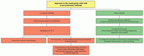
A chest radiograph should be performed for all symptomatic patients; the yield of routine chest radiographs in asymptomatic patients with neutropenia is less than 5%.31 The presence of a pulmonary infiltrate should prompt consideration for subsequent evaluation by computed tomography (CT) or even bronchoscopy for a more definitive microbiologic diagnosis.

Following the completion of the history, physical examination, and cultures, broad-spectrum antibiotics should be started promptly in all febrile, neutropenic patients. Should the chest radiograph prove to be positive, additional coverage for community-acquired pneumonia or invasive fungal infections should also be considered.


Evaluation of Afebrile, Neutropenic Patients with Localizing Signs

Fever may be absent in some cases of subsequently documented infection in neutropenic patients, particularly those with profound neutropenia and those receiving corticosteroids. The presence of infection in this setting may be detected only by attention to seemingly minor complaints from the patient or by subtle physical findings. It is critical that the physician acknowledge these complaints or findings seriously and pursue them vigorously. Abdominal pain, for example, may signify an evolving intra-abdominal infection (e.g., typhlitis), whereas erythema and tenderness along a subcutaneous catheter tunnel track usually indicates the presence of a deep soft tissue infection, even if the patient is afebrile. In these situations, it is most prudent to obtain cultures of blood and any other pertinent sites, and then to immediately begin antibiotics directed against probable pathogens. Although colonization with microorganisms often precedes development of significant infection, routine surveillance cultures are rarely helpful in a neutropenic patient. Any delay in antibiotic therapy while awaiting the results of cultures may permit the unchecked progression of infection in the neutropenic host.


Risk Assessment in Cancer Patients with Fever and Neutropenia

Traditionally, empirical antibacterial therapy for oncology patients with fever and neutropenia has involved admission to the hospital and administration of broad-spectrum intravenous (IV) antibiotics. Given the heterogeneity of this population, it has become clear that not all patients with fever and neutropenia are at equal risk for significant morbidity or mortality from infection.32 Patients with fever and neutropenia can generally be divided into high- and low-risk categories for poor outcomes based on age, underlying malignancy and type of chemotherapy, medical comorbidities, severity and anticipated duration of neutropenia, and presenting clinical signs. The identification of a low-risk subset poses the question of whether this group should be treated differently—with the ultimate goal of reduced therapy-related toxicity, an improved quality of life, and decreased cost of treatment.

A retrospective study performed by Talcott et al.32 evaluated risk factors for serious medical complications and death during episodes of fever and neutropenia in adult oncology patients. A “high-risk” group was defined as those patients who were inpatients at the time of diagnosis with fever and neutropenia and those presenting as outpatients with either concurrent comorbidity or uncontrolled cancer. The “low-risk” group was, by exclusion, those patients presenting with fever and neutropenia as outpatients without comorbidity or progressive cancer. Importantly, the information required to stratify a patient as either high or low risk was available to the clinician at the time of the patient’s presentation with fever and neutropenia. The medical course in the two groups was found to be significantly different. The rates of serious complications ranged from 31% to 55% in the high-risk group, compared to 2% in the low-risk group. Similarly, rates of death ranged from 14% to 23% in the high-risk group compared to no deaths in the low-risk group.

Although six distinct low-risk stratification schemas have been formulated for children with cancer,33,34,35,36 there is no single, uniformly validated risk stratification strategy that has been universally adopted into routine clinical practice in pediatrics. Nonetheless, several key factors appear to increase the risk of infectious complications in pediatric cancer patients: anticipated duration of neutropenia; significant medical comorbidity32; cancer status and cancer type; documented infection on presentation (i.e., pneumonia, IV catheter-site infection); evidence of bone marrow recovery (e.g., absolute monocyte count)37; and magnitude of fever.

Numerous studies have investigated the use of oral antibiotic therapy (pefloxacin/amoxicillin-clavulanic acid, ofloxacin, ciprofloxacin,38 cefixime,39,40 and moxifloxacin41) for empiric therapy in low-risk febrile cancer patients with neutropenia. Although the results of these studies are promising, many of these trials were conducted primarily in adults, statistically underpowered, and limited by methodologic issues. In contrast, two large, prospective, randomized clinical trials of low-risk febrile and neutropenic cancer patients being treated with oral ciprofloxacin plus oral amoxicillin-clavulanate compared to parenteral antibiotic therapies administered in the inpatient setting showed that the efficacy of the oral and IV regimens were comparable. But the authors of both of these trials were quick to warn that their findings should not be used to justify the widespread implementation of the use of empirical oral antibiotic therapy in treating low-risk febrile and neutropenic patients.

In pediatric cancer patients, two prospective randomized trials compared early hospital discharge (at 24 to 72 hours) versus continued hospitalization in low-risk children with fever and neutropenia,42,43 and both studies concluded that ambulatory management was safe, effective, and cost-efficient. In regard to the optimal route of antibiotic administration, two meta-analyses of randomized controlled trials comparing oral with parenteral antibiotics for management of fever and neutropenia in the inpatient and outpatient settings44,45, demonstrated no significant difference in treatment failure, overall mortality, or adverse events from antibiotic therapy, yet oral outpatient management was associated with a higher rate of hospital readmission compared with parenteral outpatient therapy.44,46

Therefore, before adapting an institutional policy for managing febrile, neutropenic children in an outpatient setting with oral therapy, careful consideration of infrastructural support is needed. The first challenge in administering oral antibacterial empirical therapy is to identify and validate criteria for the truly low-risk patients within one’s own institution. Practitioners must have a validated system to accurately prognosticate a pediatric cancer patient’s risk of serious complications or death from infection. The length of time of inpatient observation, if any, and design of outpatient follow-up need to be determined to ensure the efficacy and safety of such regimens. In addition, the potential burden on patients and families, satisfaction with care in the inpatient versus outpatient settings, and cost, including level of reimbursement for services and out-of-pocket expenses for patients and their families, need to be assessed. Other factors that need to be addressed are the availability of reliable telecommunications, proximity to a hospital for close follow-up and emergency transfer, a reliable caregiver to
ensure compliance with oral therapy, and the availability of transportation to a medical facility.


Figure 40.2 Algorithm for the management of the child with neutropenia and a pulmonary infiltrate.


Evaluation of Febrile, Non-neutropenic Patients

Evaluation of a febrile, non-neutropenic cancer patient begins with a careful history and physical examination. Blood cultures are generally obtained on febrile, non-neutropenic pediatric oncology patients—especially those with indwelling catheters. Patients with localized symptoms or signs should undergo the appropriate diagnostic procedures (e.g., stool cultures in patients with diarrhea, lumbar puncture for patients with meningeal irritation). Patients with focal findings should receive appropriate therapy based on the site involved and likely pathogen. If blood cultures are negative in such patients, then the empirical antibacterial therapy may be discontinued. Patients who are non-neutropenic, clinically well, without any identifiable focus of infection and without an indwelling central venous catheter, may be observed without empirical antibacterial therapy. By comparison, patients who are febrile and non-neutropenic, clinically well, without any identifiable focus of infection but who have an indwelling central venous catheter should receive empirical antibacterial therapy (e.g., ceftriaxone), pending results of blood cultures.

Patients with an indwelling venous access catheter (i.e., Hickman, Broviac) who become febrile present a special problem. The frequency of infectious complications in patients with intravascular devices can be high with central line-associated bacterial bloodstream infections being of greatest concern. Blood cultures should be obtained from each port of a multilumen catheter. The catheter exit site should be examined carefully for signs of erythema, tenderness, or discharge, and any discharge material should be cultured. If signs of infection or clinical instability are observed, an antibiotic regimen designed to cover the most commonly encountered line-related pathogens (i.e., Staphylococcus aureus, Staphylococcus epidermidis, and Gram-negative aerobes) should be initiated. Vancomycin should be added to the empirical antibiotic therapy of patients with evidence of skin/soft tissue infections around the catheter exit site or who are clinically ill, have a prior history of methicillin-resistant Staphylococcus aureus (MRSA) infection, and in health care settings with very high prevalence of MRSA when a Gram-positive infection is suspected. Daptomycin or linezolid may be considered as alternative agents, particularly in cases of infection due to vancomycin-resistant enterococcus (VRE) or MRSA with vancomycin minimum inhibitory concentrations (MICs) >2 µg/mL. A broad-spectrum third-generation cephalosporin (e.g., ceftriaxone) offers adequate initial coverage in the absence of an obvious tunnel infection. Antibiotics should be continued for a 48- to 72-hour trial. If the preantibiotic blood and catheter culture results are negative and no site of infection is determined, the antibiotics may be withdrawn, whether or not fever persists. This allows for a thorough evaluation of the cause of fever, without the confounding influence of antibiotics. If the cultures are positive, a full therapeutic course is warranted (Fig. 40.2). For further details, please refer to the 2009 guidelines of the IDSA for the diagnosis and management of intravascular catheter-related infection.27


EMPIRICAL ANTIBACTERIAL TREATMENT OF FEBRILE, NEUTROPENIC PATIENTS


General Considerations

Perhaps the single most important advance in infectious diseases oncology supportive care leading to improved survival has been the prompt initiation of empirical antibacterial antibiotics when the neutropenic cancer patient becomes febrile. Before this approach was instituted in the early 1970s, the mortality rate from Gram-negative infections, especially that of P. aeruginosa, E. coli, and K. pneumoniae, approached 80%. With the widespread use of effective empirical antibiotics, the overall mortality rate has declined to approximately 10% to 40% for infections caused by Gram-negative bacteria.

An ideal empirical antibacterial regimen should provide a broad spectrum of activity against a wide variety of pathogenic organisms, including but not limited to Pseudomonas, be bactericidal in the absence of neutrophils, and have low potential for adverse effects. The choice of a specific agent or combination should also be predicated on the specific patterns of bacterial infection in one’s own institution, as well as antibiotic susceptibility profiles, cost, toxicity, and standards used at one’s center. The patient’s prior history of infection or colonization with resistant bacteria and prophylactic antimicrobial regimen should also be considered.

Approximately 85% to 90% of pathogens that are documented to be associated with new fevers in neutropenia patients are Gram-positive and Gram-negative bacteria (Table 40.1). Hence, an empirical antibiotic regimen must cover a broad spectrum of bacteria, provide high serum bactericidal drug levels, be stable against the emergence of resistant bacteria, and be as nontoxic and as simple to administer as possible. These conditions have traditionally required the combination of two or more antibiotics.
Several regimens, usually consisting of a cephalosporin, an aminoglycoside, and extended-spectrum penicillin, have been employed.


TABLE 40.1 Predominant Pathogens in Pediatric Cancer Patients


Gram-positive bacteria


Staphylococcus spp. (e.g., S. epidermidis and S. aureus)


Streptococcus spp. (α-hemolytic; e.g., S. mitis)


Enterococcus spp. (e.g., E. faecium, E. faecalis)


Corynebacterium spp. (e.g., C. jeikeium)


Listeria monocytogenes


Bacillus spp. (B. cereus, B. circulans, B. licheniformis)


Clostridium spp. (C. difficile, C. septicum, C. tertium)


Gram-negative bacteria


Enterobacteriaceae (Escherichia coli, Klebsiella spp., Enterobacter spp., Serratia spp.)


Pseudomonas aeruginosa (similar oxidase-positive multiresistant Gram-negative organisms), Stenotrophomonas maltophilia


Anaerobes (e.g., Bacteroides spp., Clostridium spp., and Prevotella spp.)


Fungi


Candida spp. (e.g., C. albicans, C. glabrata, C. tropicalis, C. krusei, C. parapsilosis)


Aspergillus spp. (e.g., A. fumigatus, A. flavus, and A. terreus)


Zygomycetes (e.g., Rhizopus oryzae, Mucor spp.)


Fusarium spp. (e.g., F. solani and F. oxysporum)


Scedosporium spp. (S. apiospermum and S. inflatum)


Cryptococcus neoformans


Pneumocystis jiroveci (formerly, P. carinii)


Viruses


HSV


Varicella zoster virus


CMV


EBV


RSV


Adenovirus


Influenza virus


Parainfluenza viruses


Human herpes virus 6


Polyoma viruses (e.g., BKV, JCV)


Protozoa and helminths


Toxoplasma gondii


Cryptosporidium spp.


Strongyloides stercoralis


Combination Therapy

Combination antibacterial therapy was especially important in earlier days of antibiotic development. The objective was to provide expanded antibacterial spectrum, enhance potential synergistic interaction, and prevent the emergence of resistance. Antimicrobial synergy was particularly relevant to Gram-negative bacteria. A synergistic combination would enhance the efficacy and lower the effective MIC of each compound when used in combination. However, with the advent of third- and fourth-generation cephalosporins and carbapenems, which have significantly lower MICs than earlier β-lactams, combining two agents to achieve enhanced synergistic activity was no longer important. Moreover, third- and fourth-generation cephalosporins and carbapenems possess the broader spectrum of the combination of conventional β-lactam/aminoglycoside combinations. Finally, third-and fourth-generation cephalosporins and carbapenems are more resistant to the β-lactamases that emerged in organisms resistant to the earlier carboxypenicillins and acylureidopenicillins.


Antibiotic Monotherapy

Fourth-generation cephalosporins (e.g., cefepime) and carbapenems (e.g., imipenem and meropenem) provide alternatives to the more traditional aminoglycoside-containing combination regimens (Table 40.2). Several of these compounds are able to provide breadth of antimicrobial spectrum and high bactericidal levels when used as single agents. The use of antibiotic monotherapy for the empirical management of the neutropenic patient with fever is attractive because of the ease of administration, lower cost, and lesser toxicity of a single drug, particularly without the use of an aminoglycoside. Additionally, empiric use of monotherapy with antipseudomonal β-lactam antibiotics was found to be as effective as combination antimicrobial therapy in adult patients with fever and neutropenia who were hemodynamically stable and had no evidence for sepsis, skin/soft tissue infection, or catheter-related infections.35

As oncology centers have different patterns of microbial isolates and antibiotic resistance, the clinical decisions about appropriate antibacterial empirical regimens must ultimately be individualized for each institution. The 2010 guidelines of the IDSA for the use of antimicrobial agents in neutropenic patients with cancer underscore the need to adjust empirical therapy to local patterns of infection, institutional antimicrobial susceptibilities, and to individual patient characteristics.23 This document identified the following antipseudomonal β-lactam antibiotics to be appropriate as empirical monotherapy in neutropenic patients with fever based on large randomized controlled trials: cefepime, piperacillin-tazobactam, and carbapenems (imipenem-cilastatin and meropenem).


Cefepime

Cefepime is a potent broad-spectrum fourth-generation cephalosporin with enhanced activity against Gram-positive and Gram-negative aerobes, including some pathogens resistant to other cephalosporins. Its in vitro spectrum includes viridans streptococci (e.g., Streptococcus mitis and Gram-negative bacilli expressing Amp-C type 1 β-lactamases). Of note, cefepime does not provide activity against anaerobic bacteria and may not be active against Gram-negative bacteria that produce extended-spectrum β-lactamase (ESBL) enzymes as can be seen with Enterobacteriaceae spp. like Klebsiella and E. coli. The toxicity profile of cefepime is similar to that of ceftazidime. Cefepime has been compared to ceftazidime, imipenem, and piperacillin/tazobactam in a series of clinical trials evaluating monotherapy for fever and neutropenia. In each of these studies, the regimens were similar in terms of efficacy or toxicity.47 Cefepime is the only cephalosporin that is Food and Drug Administration (FDA) approved for empirical antibacterial therapy in febrile, neutropenic patients. A recent meta-analysis investigating the efficacy and safety of cefepime came to the conclusion that all-cause mortality was higher with cefepime than with other β-lactams.48 In November 2007, the FDA posted an alert regarding the use of cefepime and the announcement of an investigation to review the safety of administering cefepime. In June 2009, the FDA released a meta-analyses based on additional data beyond those included in the Yahav et al. publication.48 In the FDA analyses, no statistically significant increase in mortality was seen in cefepime-treated patients compared with comparator-treated patients.49


Piperacillin/Tazobactam

Tazobactam is an irreversible inhibitor of TEM-1 (Bush-Medeiros type 2) β-lactamases that has been paired with the extended-spectrum antipseudomonal piperacillin to yield a broad-spectrum agent that is relatively stable and active in the presence of many of the clinically important Gram-positive and Gram-negative aerobes and anaerobes. Piperacillin/tazobactam penetrates most tissues, but penetrates poorly into the CSF and should not be used in cases of suspected meningitis.


TABLE 40.2 Commonly Used Antimicrobial Agents for Pediatric Cancer Patients


Class


Agent


Spectruma


Daily Dose (Maximum)


Comments


Antibacterial agents


Third-generation cephalosporin


Ceftazidime


Enteric bacteria, some Gram-positive aerobes, no anaerobic coverage


100 mg/kg divided every 8 h (max 6 g/d)


Only third-generation cephalosporin with activity against


Pseudomonas aeruginosa


Ceftazidime may no longer be reliable for empirical monotherapy because of decreasing activity against Gram-negative bacteria and poor coverage of Gram-positive bacteria (refer to local, institutional epidemiology and antimicrobial susceptibility patterns)


Fourth-generation cephalosporin


Cefepime


Enteric bacteria, Gram-positive aerobes


100 mg/kg divided every 8 h (6 g/d)


Active against some P. aeruginosa, Enterobacter spp., and Serratia spp. resistant to ceftazidime; broader Gram-positive spectrum


Carbapenems


Imipenem Most


Gram-negative and Gram-positive aerobes, including P. aeruginosa, enterococci; excellent anaerobic coverage


50 mg/kg divided every 6 h (4 g/d)


Stenotrophomonas maltophilia and Burkholderia cepacia not covered


Meropenem


Similar to imipenem


60-120 mg/kg divided every 8 h (3 g/d)


Less likely than imipenem to cause seizures


Extended-spectrum penicillins


Piperacillin, azlocillin, mezlocillin


Enteric aerobes, including some P. aeruginosa, Enterobacter spp., Serratia spp.; anaerobes


300 mg/kg divided every 4 h (21 g/d)


Must be paired with an aminoglycoside for coverage of P. aeruginosa


Piperacillin-tazobactam


Similar to piperacillin, increased activity versus some β-lactamase-producing Gram-positive cocci, Gram-negative bacilli, and anaerobes


300 mg/kg divided every 4 h (12 g/d)


Monobactams


Aztreonam


Exclusively aerobic Gram-negative aerobes including P. aeruginosa


100-150 mg/kg divided every 6 h (4 g/d)


Limited spectrum requires pairing with Gram-positive agent, not cross-reactive with β-lactams, so can be used in penicillin or cephalosporin allergic patients


Glycopeptide


Vancomycin


Exclusively Gram-positive


25-40 mg/kg divided every 6-12 h (3 g/d) IV


No need to add vancomycin routinely for empirical coverage for fever and neutropenia


Lipopeptide


Daptomycin


Exclusively Gram-positive, including MRSA and susceptible strains of VRE


4 mg/kg/d IV


Data in pediatrics are limited. Should not be u sed for pneumonia


Oxazolidinone


Linezolid


Exclusively Gram-positive, including MRSA, susceptible strains of VRE, and penicillin and cephalosporin-resistant S. pneumoniae


10 mg/kg q12h


Excellent oral bioavailability


Streptogramin


Quinupristin/dalfopristin


Exclusively Gram-positive, similar to linezolid but spectrum does not include E. faecalis


7.5 mg/kg q8h


Venous irritation, should be given via central venous catheter


Antifungal agents


Amphotericin B


Deoxycholate amphote-ricin B


Very broad antifungal activity including Candida spp., Aspergillus spp., Zygomycetes, Cryptococcus neoformans, Histoplasma capsulatum


0.5 mg/kg once daily for empirical therapy, higher doses (1.0-1.5 mg/kg) are necessary for documented infections due to Aspergillus spp. and other filamentous fungi


Significant nephrotoxicity may be reduced by saline hydration before daily infusion


Lipid formulations (ABLC, ABCD, and liposomal amphote-ricin B)


Same spectrum as deoxycholate formulation


3 mg/kg/d for empirical therapy, 5 mg/kg/d (or greater) for documented infections due to Aspergillus spp. and other filamentous fungi


Significantly less nephrotoxicity with efficacy at least equal to that of D-AmB


Triazole


Fluconazole


Candida spp. (not C. krusei and not some strains of C. glabrata); C. neoformans, Trichosporon spp., and Coccidioides immitis


3-12 mg/kg/d. Dosage of 12 mg/kg/d is required for life-threatening infections to achieve comparable plasma drug exposure attained in adults with 400 mg/d (see text)


Excellent bioavailability, independent of gastric acidity


Itraconazole


Aspergillus spp., Candida spp., H. capsulatum, Blastomyces dermatitidis, and C. immitis


3-5 mg/kg/d POb


Absorption erratic but increased with taking drug with meals or by using cyclodextrin liquid formulation


Voriconazole


Candida spp., Aspergillus spp., Trichosporon spp. and some strains of Scedosporium spp., and Fusarium spp.


NO activity against Zygomycetes


9 mg/kg q12h for two doses (loading dose), then 8 mg/kg q12h IV for documented infections; higher dosages in pediatric patients are necessary in order to achieve comparable adult drug exposures (see text)


Pediatric suspension is available; bioavailability is enhanced when taken on an empty stomach


Posaconazole


Candida spp., Aspergillus spp., Zygomycetes


For patients ≥13 y old, prophylactic dosing of suspension: 200 mg PO 3 times a day


Data in pediatrics are limited at this time


Echinocandin


Caspofungin


Candida spp. and Aspergillus spp.


70 mg/m2 IV on day 1, followed by 50 mg/m2/d IV to achieve comparable plasma concentrations to adults; dosing for candidemia of 100 mg/d IV, 150 mg IV/d for filamentous fungi


Micafungin


Candida spp. and Aspergillus spp.


Dosing by age: 3-10 mg/kg/dose (max 100 mg/d)


Antiviral agents


Antiherpetic


Acyclovir


HSV, VZV


HSV: 750 mg/m2 divided q8h or 5 mg/kg q8h


IV dose for VZV is twice that for HSV. Hydration should be ensured when administering high doses


VZV: 1500 mg/m2 divided q8h or 10 mg/kg q8h


Ganciclovir


CMV, HSV, VZV, HHV-6


For CMV: 5 mg/kg q12h for 14 days induction, then 5 mg/kg/d for maintenance


Granulocytopenia is the major dose-limiting toxicity; not routinely used for HSV, VZV but dose used for CMV is effective for the other herpesviruses


Foscarnetw


HSV, VZV, CMV (including most acyclovir- and ganciclovir-resistant strains)


CMV: 60 mg/kg/d q8h for 14 d then 90-120 mg/kg/d for maintenance


Nephrotoxicity is dose-limiting effect, renal function and electrolytes require close monitoring


VZV, HSV: 40 mg/kg q8h


Anti-PCP agents


Trimethoprim-sulfameth-oxazole


Pneumocystis jiroveci (formerly, P. carinii) is also active against many Gram-positive and Gram-negative bacteria, including S. maltophilia and B. cepacia


20 mg/kg/d IV in two divided doses for PCP treatment


May cause bone marrow suppression in high doses


Pentamidine


P. jiroveci


4 mg/kg/d IV for treatment


Adverse effects include pancreatitis, hypoglycemia, hypocalcemia, infusional hypotension


Dapsone


P. jiroveci


2 mg/kg/d (for prophylaxis)


High incidence of hemolytic reactions, can also cause methemoglobinemia


Atovaquone


P. jiroveci


30 mg/kg/d, max 1500 mg/d


Suspension formulation has better bioavailability


a Spectrum depicted here is for summary purposes and is not a complete list; bIV formulation for itraconazole is available but dosage and pharmacokinetics have not been defined in pediatric patients.


VRE, vancomycin-resistant enterococci; HSV, herpes simplex virus; HHV-6, human herpes virus 6.


Piperacillin/tazobactam in combination with aminoglycosides and as monotherapy has been evaluated in a series of small trials.50 Piperacillin/tazobactam was compared with cefepime in an open-label, randomized study of empirical therapy in febrile, neutropenic patients with hematologic malignancies.51 The analyses demonstrated noninferiority for piperacillin-tazobactam at all time points. Adverse events were similar. This large clinical trial supports the use of piperacillin/tazobactam as an acceptable agent of monotherapy for empirical antibacterial therapy in febrile, neutropenic patients.


Carbapenems: Imipenem and Meropenem

Imipenem and meropenem are members of the class of β-lactam antibiotics called carbapenems. Although their mechanism of bactericidal activity involves interference with bacterial cell wall synthesis, imipenem and meropenem uniquely possess the broadest antimicrobial spectrum of any currently available β-lactam antibiotic. In addition to activity against Gram-negative aerobes, including P. aeruginosa and the Enterobacteriaceae, imipenem and meropenem act against many Gram-positive organisms (e.g., methicillin-susceptible S. aureus, some Enterococcus spp., some coagulase-negative staphylococci) and most anaerobic organisms.

Imipenem is impervious to destruction by Amp-C chromosomally mediated β-lactamases that are commonly produced by Enterobacter, Citrobacter, and Serratia species. Treatment of infections caused by these pathogens by ceftazidime (or another β-lactam agent) may induce β-lactamase production and, accordingly, lead to therapeutic failures. Because imipenem offers the advantage of β-lactamase stability as well as efficacy against most of these organisms, it is an appropriate treatment for serious infections caused by Enterobacter spp. and related species or as empirical therapy in ill patients previously treated with multiple antibiotics, in whom the likelihood of resistant organisms is increased.

Several adverse effects of imipenem therapy have been identified that may limit its use. Nausea and vomiting are well-documented adverse effects of imipenem. There is also an increased frequency of Clostridium difficile colitis observed among imipenem recipients, presumably related to alterations in normal anaerobic bowel flora. Imipenem is also known to decrease the seizure threshold in seriously ill patients and in those with CNS pathology, and it should be avoided in these patients. Imipenem-associated seizures also occur in the setting of renal impairment and increased plasma concentrations.

Meropenem is another carbapenem that shares similar in vitro antimicrobial properties of imipenem. A large trial, evaluating more than 1,000 episodes of fever and neutropenia, that compared monotherapy with meropenem versus combination therapy with ceftazidime and amikacin showed the two regimens to be similarly effective and both well tolerated. Meropenem has the potential advantage over imipenem of less GI toxicity and does not appear to alter the seizure threshold.

As Stenotrophomonas maltophilia is usually resistant to imipenem and meropenem, this organism should be considered among the most likely pathogens emerging as a cause of infection in immunocompromised patients receiving one of these carbapenems. Hence, an oxidase-negative Gram-negative bacillus causing bacteremia in a febrile, neutropenic patient who is already receiving imipenem is likely to be S. maltophilia. Trimethoprim-sulfamethoxazole (TMP-SMX) is the preferred antibiotic against S. maltophilia. Some strains of S. maltophilia initially may appear susceptible to other antibiotics, such as fluoroquinolones; however, emergence of resistance may occur during the course of therapy.52 Multidrug-resistant P. aeruginosa may also emerge during the course of carbapenem therapy.

Ertapenem is a new carbapenem with the advantage of once-daily IV administration in comparison to the 3 times daily IV administration of imipenem and meropenem.53 Although similar to those of meropenem and imipenem, the in vitro antimicrobial spectrum of ertapenem does not include P. aeruginosa, which ultimately may exclude its use in febrile, neutropenic cancer patients.


Modifications to the empiric antibiotic regimen should be guided by clinical and microbiologic data and may include addition of other antibiotics, which are summarized in the following section. Persistent fever alone in a patient who is clinically stable with no new findings on physical examination and negative initial blood cultures and other diagnostics does not require modification in empiric antibiotics. On the other hand, patients who remain or become hemodynamically unstable after initial empiric antibiotics are dispensed should have their antimicrobial regimen broadened to include coverage for resistant Gram-negative, Gram-positive, and anaerobic bacteria and fungi.


Vancomycin

Vancomycin is a glycopeptide antibiotic with microbicidal activity against a broad range of Gram-positive bacteria. Its widespread use has been associated in the last decade with the emergence of VRE species (Enterococcus faecium and Enterococcus faecalis), as well as vancomycin-intermediate S. aureus and vancomycin-resistant S. aureus. Other Gram-positive bacteria that are resistant to vancomycin include Enterococcus gallinarum, Enterococcus casseliflavus, Leuconostoc mesenteroides, Pediococcus spp., Lactobacillus spp., and Erysipelothrix rhusiopathiae.

Empirical administration of vancomycin is not recommended for routine use in patients with fever and neutropenia.23 Randomized trials comparing empirical antibiotic regimens with and without vancomycin as part of the initial therapy do not demonstrate significant differences in duration of fever or outcomes, including overall mortality. The 2010 Clinical Practice Guidelines for the Use of Antimicrobial Agents in Neutropenic Patients with Cancer from IDSA recommend that vancomycin be considered for empiric use in the following clinical scenarios: patients with hemodynamic instability, radiographic evidence of pneumonia, and skin and soft tissue infection or clinically suspected serious catheter-related infection (e.g., pocket and tunnel infections or cellulitis of exit site). When initial blood cultures demonstrate Gram-positive bacteria, repeat blood cultures should be obtained, and vancomycin initiated until final identification and susceptibility results are available to guide further therapy. Additional indications for vancomycin may include severe mucositis in a clinically ill patient, particularly if the patient has had a previous infection with penicillin-resistant streptococci. If vancomycin is initiated empirically and a Gram-positive infection is not subsequently microbiologically documented, it may be discontinued. If a Gram-positive organism is identified that is oxacillin-susceptible, then a penicillinase-resistant β-lactam or alternative agent is recommended. A patient with a documented history of an immediate-type hypersensitivity reaction (e.g., hives, wheezing) in whom β-lactam and carbapenem therapy would be contraindicated is a candidate for vancomycin as initial empirical therapy, in combination with aztreonam.


Daptomycin, Linezolid, and Quinupristin-Dalfopristin

As the challenges of infections caused by VRE, MRSA, and other resistant Gram-positive cocci continue to mount worldwide, daptomycin, linezolid, and quinupristin-dalfopristin have become increasingly important resources in management of immunocompromised patients.

Daptomycin is a lipopeptide antimicrobial agent with bactericidal activity against MRSA and Enterococcus species, including VRE.54 Administered parenterally once daily, daptomycin may be particularly useful in modification of initial therapy in neutropenic patients after VRE is documented as a cause of serious infection. Although combination therapy is necessary to achieve microbicidal activity for treatment of enterococcal sepsis in neutropenic patients (e.g., ampicillin plus aminoglycoside), daptomycin achieves bactericidal activity as a single agent against Enterococcus spp. As multidrug-resistant VRE may preclude such combination therapy, daptomycin may be particularly effective in neutropenic hosts as a single agent for treatment of this emerging pathogen. The use of daptomycin for pulmonary infections (e.g., MRSA pneumonia), however, is not recommended since pulmonary surfactant inactivates daptomycin.55 The pharmacokinetics and optimal dosage of daptomycin in children remain to be studied.

Linezolid is the first of a new class of synthetic antimicrobial agents, the oxazolidinones, which exert a unique mechanism of action through inhibition of protein synthesis via interference of formation of the initiation complex.54 Because of its unique mode of action, linezolid does not exhibit cross-resistance with other antimicrobial agents. It is active against Gram-positive bacteria, including MRSA, VRE (including E. faecium and E. faecalis), as well as β-lactam-resistant strains of S. pneumonia.56 Linezolid is administered parenterally and orally with excellent oral bioavailability. In a randomized clinical trial of patients with MRSA infections, linezolid demonstrated efficacy similar to that of vancomycin, as well as in treatment of patients with nosocomial pneumonia in which linezolid and vancomycin were each paired with aztreonam.57,58 In an open-label salvage therapy study for treatment of resistant Gram-positive infections, linezolid was effective and well tolerated in treating resistant Gram-positive infections in neutropenic patients with neoplastic diseases.59 The toxicity profile of linezolid is favorable; however, reversible thrombocytopenia, anemia, and neutropenia may occur in patients receiving prolonged therapy, especially exceeding 2 weeks. Peripheral neuropathy and optic neuropathy also may develop during prolonged administration. As linezolid also is a weakly active but reversible monoamine oxidase inhibitor, it may interact with adrenergic or serotonergic agents.

Quinupristin-dalfopristin is a 30:70 mixture of quinupristin and dalfopristin, which are semisynthetic streptogramin antibiotics.54 The antibacterial spectrum of quinupristin-dalfopristin is similar to that of linezolid; however, quinupristin-dalfopristin is active against isolates of E. faecium but not of E. faecalis. Quinupristin-dalfopristin is effective in treatment of serious infections caused by vancomycin-resistant E. faecium, nosocomial pneumonia, as well as in complicated soft tissue infections due to resistant Gram-positive cocci.60,61 The toxicity profile of quinupristin-dalfopristin includes myalgias and arthralgias in approximately 10% of patients, as well as conjugated hyperbilirubinemia and cholestatic jaundice. Because quinupristin-dalfopristin causes local pain, inflammation, and thrombophlebitis during peripheral venous infusion, it should be administered through a central venous catheter. Because of its inhibition of metabolism of agents cleared through cytochrome P-450 3A4, adverse drug interactions may occur.


Fluoroquinolones

The fluoroquinolones constitute a group of synthetic antibiotics that possess a broad spectrum of microbicidal activity against most medically important aerobic Gram-positive and Gram-negative bacteria, but are virtually devoid of activity against the clinically important anaerobic bacteria. Ciprofloxacin, norfloxacin, and ofloxacin comprise the first generation of fluoroquinolones that were developed. The second generation of fluoroquinolones includes levofloxacin, gatifloxacin, and moxifloxacin. This newer generation of fluoroquinolones has reliable activity against most isolates of penicillin-resistant pneumococci; however, quinolone-resistant pneumococcal isolates have become more frequent in association with increased use of these agents. Levofloxacin and moxifloxacin provide excellent bioavailability, once-daily dosing, tolerability, and improved Gram-positive spectrum, including most strains of penicillin-resistant S. pneumoniae.

Fluoroquinolones exert bactericidal activity through a unique mechanism of action via inhibition of the DNA gyrase responsible for supercoiling and packaging of bacterial DNA. Because they are structurally unrelated to the β-lactams or to any other antibiotic class, cross-resistance through a common mechanism between
the fluoroquinolones and other antibiotics is uncommon. Fluoroquinolones are usually active against multiresistant organisms; ciprofloxacin, for example, exhibits activity against many of the resistant Gram-negative rods, including P. aeruginosa, Serratia, Enterobacter, and Klebsiella species that are responsible for serious infections in neutropenic and otherwise immunocompromised patients. Ciprofloxacin activity against streptococcal species, particularly α-hemolytic streptococci (e.g., Streptococcus mitis, Streptococcus sanguis), is often inadequate.

As the result of the paucity of reliable activity against streptococcal and staphylococcal pathogens, ciprofloxacin is not useful as a single agent for empirical therapy in febrile, neutropenic patients. However, the addition of vancomycin or a penicillinase-protected penicillin to IV ciprofloxacin yields a regimen that compares favorably with more traditional combinations of a β-lactam plus an aminoglycoside and is a suitable alternative for patients who have hypersensitivity reactions to β-lactam antibiotics.

Neither aminoglycosides nor fluoroquinolones are recommended for initial empirical therapy for febrile, neutropenic patients; they may be appropriate in the settings of hemodynamic instability or in a critically ill patient with prior history of severe infection with multiresistant Gram-negative bacteria. The combination of an antipseudomonal β-lactam with a quinolone empirical therapy for febrile, neutropenic patients provides broad-spectrum activity against Gram-negative bacilli and avoids aminoglycoside-related nephrotoxicity. In a large randomized study consisting of 471 evaluable febrile episodes, ciprofloxacin plus piperacillin was as effective as tobramycin plus piperacillin as empirical therapy for febrile, neutropenic patients.62 However, the combination of ciprofloxacin plus piperacillin-tazobactam confers substantially greater Gram-positive activity to this fluoroquinolone-based regimen.

Laboratory findings of chondrotoxicity in beagle puppies treated with fluoroquinolones have historically limited use of this class of antibiotics in pediatric patients. Nonetheless, ciprofloxacin is being given with increasing frequency to children with cystic fibrosis who are colonized with P. aeruginosa and to children with other refractory Gram-negative infections. A report on more than 1,700 young patients who received ciprofloxacin therapy had only a few reports of transient arthralgias that resolved promptly after discontinuation of the drug. These encouraging findings, however, do not preclude a low frequency of clinically delayed development of arthropathy in pediatric patients treated with fluoroquinolone. Thus, for children with cancer who are at risk for severe infections and in whom a very potent oral antibiotic would facilitate the possibility of outpatient therapy, the benefits of fluoroquinolone therapy in selected cases may be found to outweigh the risk of chondrotoxicity. Such patients should also be monitored for Achilles tendon rupture, which may also ensue in patients of any age and could be potentiated in patients receiving glucocorticoid therapy.30


Aztreonam

Aztreonam is a monobactam antimicrobial agent with activity that is exclusively against aerobic Gram-negative bacteria. The in vitro antimicrobial spectrum of aztreonam includes the Enterobacteriaceae (e.g., most isolates of E. coli, Klebsiella, Serratia, Enterobacter) and P. aeruginosa. Because there is no Gram-positive or anaerobic coverage, aztreonam is not used as a single agent for empirical antibacterial therapy in febrile, neutropenic patients. Aztreonam has been used successfully in combination with vancomycin for empirical treatment of febrile, neutropenic patients with cancer.

The absence of the thiazolidine component in the monobactam molecule substantially reduces the antigenic cross-reactivity between aztreonam and β-lactam antibiotics. Thus, aztreonam is most useful for patients with significant allergy to penicillin or other β-lactams in whom an antipseudomonal agent is desirable or required. The Gram-negative spectrum and absence of renal toxicity allow the use of aztreonam as an alternative to aminoglycosides in certain instances.


ANTIFUNGAL AGENTS

During the past 30 years, the armamentarium of systemically administered antifungal compounds has expanded from amphotericin B as the only available compound for treatment of life-threatening invasive fungal infections to the new classes of lipid formulations of amphotericin B (LFABs), antifungal triazoles, and echinocandins. The antifungal and pharmacologic properties of these compounds are summarized in the following section.


Amphotericin B

Amphotericin B is a polyene antifungal agent with a broad spectrum of activity against yeast-like and filamentous fungi. Deoxycholate amphotericin B (D-AmB) has been the cornerstone of antifungal therapy in most critically ill pediatric patients with deeply invasive fungal infections for the past 50 years. However, newer agents, including the LFABs, antifungal triazoles, and echinocandins with an improved therapeutic index, are providing safer alternatives with similar or superior efficacy. The principal mechanism of action of amphotericin B, as of other polyenes, is the binding to ergosterol, which is found in fungal cell membranes but not in mammalian cell membranes. Amphotericin B forms porelike ionic channels, which result in altered membrane permeability and leakage of monovalent and divalent cations from the fungal cell. Amphotericin B also binds to a lesser extent to other sterols, such as cholesterol, which accounts for much of the toxicity associated with its usage. Yet, another mechanism of action of amphotericin B is the induction of lipoperoxidation of the fungal cell membrane. Amphotericin B also modulates host response by cytokine- and oxidation-dependent enhancement of the effector functions of macrophages, MNCs, and PMNs.63,64

The pharmacokinetic profile of amphotericin B in children differs from that in adults. Children older than 3 months of age have a smaller volume of distribution and a faster clearance than what is usually found in adults. There is a strong inverse correlation between patient age and total clearance of amphotericin B, suggesting that higher dosages may be better tolerated in patients between 3 months and 9 years of age.

Acute or infusion-related toxicity of D-AmB is characterized by fever, chills, rigor, nausea, vomiting, and headache. Fever, chills, and rigors may be mediated by TNF and IL-1, cytokines that are released from human peripheral MNCs or tissue macrophages in response to D-AmB. These acute reactions may possibly be blunted by corticosteroids, acetaminophen, aspirin, other nonsteroidal antiinflammatory drugs, or meperidine. Corticosteroids should be used only in relatively low dosages (e.g., 0.5 to 1.0 mg per kg of hydrocortisone). Meperidine in low doses (0.2 to 0.5 mg per kg) interdicts development of rigors. Acetaminophen may decrease fever but appears to have little effect on rigors. Aspirin should be avoided in thrombocytopenic patients. Although diphenhydramine is used in many centers for prevention of acute infusion reactions, the rationale for this anti-H1-receptor inhibitor is not pharmacologically clear, as most acute reactions due to D-AmB are not histamine-mediated. Any perceived benefits of diphenhydramine for D-AmB-associated acute infusion reactions are more likely related to its sedating effects.

Nephrotoxicity (glomerular or tubular) is the most significant dose-limiting adverse effect of D-AmB. The clinical and laboratory manifestations of glomerular toxicity include a decrease in glomerular filtration rate and renal blood flow, as evidenced by azotemia. Tubular toxicity is manifested as the presence of urinary casts, hypokalemia, hypomagnesemia, renal tubular acidosis, and nephrocalcinosis. Hypomagnesemia may be more profound in patients with cancer who develop a divalent cation-losing nephropathy
associated with cisplatin or ifosfamide. Although azotemia in most pediatric patients is usually reversible, renal function may not return to normal levels after cessation of D-AmB in children who have received previous repeated or prolonged courses of this polyene. Moreover, return to pretreatment levels may require several months in some cases.

Foremost among the important drug interactions with amphotericin B is the nephrotoxicity caused by concomitant aminoglycosides, cyclosporine, and foscarnet; where possible, D-AmB should not be used in conjunction with these nephrotoxic agents. Acute pulmonary reactions (hypoxemia, acute dyspnea, and radiographic evidence of pulmonary infiltrates) have been associated with simultaneous transfusion of granulocytes and infusion of amphotericin B. Although some investigators have disputed the causality of amphotericin B in such reactions, a rational approach is to separate the infusions of amphotericin B and granulocytes by the longest possible time period.


Lipid Formulations of Amphotericin B

Three engineered LFABs have been approved in North America and Europe: a small unilamellar vesicle formulation of liposomal amphotericin B (L-AmB or AmBisome), amphotericin B lipid complex (ABLC or Abelcet), and amphotericin B colloidal dispersion (ABCD, Amphotec, or Amphocil). The introduction of LFABs has been an important advance in improving the therapeutic index of amphotericin B. Because toxicity is the major dose-limiting factor of this drug, lipid formulations have been developed to reduce toxicity and permit larger doses to be administered. Although classically considered as liposomal formulations of amphotericin B, the investigational and clinically approved formulations of amphotericin B have a wider diversity of lipid structure. Liposomes (defined as phospholipid bilayers of one or more closed concentric structures) and other lipid formulations have been used as vehicles for amphotericin B with encouraging results. The lipid formulation may provide a selective diffusion gradient toward the fungal cell membrane and away from mammalian cell membrane.

ABLC was the first lipid formulation approved in the United States by the FDA in November 1995 for both children and adults. ABLC, 5 mg per kg per day IV, under an emergency compassionate release protocol, was found to be active in treatment of immunocompromised pediatric patients with refractory mycoses and those with intolerance to conventional amphotericin B. This study found little dose-limiting nephrotoxicity of ABLC. These findings were subsequently confirmed in a large open-label prospective study of pediatric patients receiving ABLC. A phase I-II study of ABLC in children with hepatosplenic (chronic disseminated) candidiasis found that the compound administered at 2.5 mg per kg for 6 weeks was effective, had no dose-limiting nephrotoxicity, and appeared to reach steady-state plasma pharmacokinetics by 7 days.

Two phase I-II studies of the safety, tolerability, and activity of L-AmB in oncology patients demonstrated that dosages of as much as 15 mg per kg per day were not dose limiting and were effective in empirical antifungal therapy as well as in treatment of invasive filamentous fungal infections. Although the optimal dosage for treatment of invasive fungal infections is controversial, we recommend that treatment of invasive candidiasis should begin with 3 mg per kg per day and invasive aspergillosis and other filamentous fungal infections at 5 mg per kg per day.

Although LFABs are associated with less nephrotoxicity, they may confer their own patterns of toxicity. For example, the multilamellar lipid formulation of amphotericin B induced reversible hypoxemia, pulmonary hypertension, and depression of cardiac output during infusion. ABLC is more commonly associated with the infusion-related reactions of the well-known fever, chills, and rigors typical of D-AmB. Although historically associated with less infusion-related toxicity than that of D-AmB and ABLC, L-AmB may cause a syndrome of severe acute infusion-related reactions characterized by substernal chest pain, hypoxia, flank and abdominal pain, and urticarial eruptions.65 Infusion-related toxicity with ABCD was found to be more severe than that of D-AmB.

Despite the relatively greater expense of the LFABs, an expanding body of data underscores the risk of D-AmB-induced irreversible nephrotoxicity and the expense of renal impairment in management of seriously ill patients. A lipid formulation of amphotericin B is appropriate as initial empirical therapy or as definitive therapy for proven mycoses in high-risk patients receiving concomitant nephrotoxic agents ([e.g., cyclosporine, aminoglycosides] in diabetes mellitus, preexisting renal impairment), those with preexisting renal impairment, and those with an anticipated course of protracted neutropenia during which dose-limiting nephrotoxicity may ensue.


Flucytosine

Historically, 5FC has been most frequently used as an adjunct to amphotericin B therapy in the treatment of cryptococcal meningitis. This combination was originally proposed because of the observation that amphotericin B potentiates the uptake of 5FC by increasing fungal cell membrane permeability. Because of rapid emergence of resistant strains, 5FC is used only in combination with another antifungal agent, most commonly amphotericin B. The strongest clinical data support the use of D-AmB plus 5FC in treatment of CNS cryptococcosis. Although experimental data support the use of 5FC in combination with D-AmB in treatment of experimental disseminated candidiasis, there are no controlled clinical trials to test this hypothesis.

Dose-dependent myelosuppression is the most serious toxicity associated with administration of 5FC. GI side effects, such as diarrhea, nausea, and vomiting, are the most common symptomatic side effects and are dose dependent. Abnormal elevation of hepatic transaminases also has been reported in approximately 5% of patients receiving the drug. Other than its use in treatment of cryptococcal meningoencephalitis, 5FC is seldom used owing in part to its narrow therapeutic index.


Antifungal Imidazoles and Triazoles

The antifungal azoles include imidazoles (clotrimazole, miconazole, and ketoconazole) and triazoles (itraconazole, fluconazole, voriconazole, and posaconazole). Clotrimazole and miconazole are available in topical applications. Ketoconazole has been generally supplanted in its use by itraconazole and fluconazole in the pediatric oncology setting. Accordingly, only fluconazole and itraconazole are discussed in this section.

The antifungal azoles are synthetic compounds that demonstrate less toxicity than D-AmB, have flexibility for oral administration, and have comparable or superior efficacy against certain infections. The antifungal azole agents function principally by inhibition of the fungal cytochrome P-450 enzyme lanosterol 14a-demethylase, which is involved in the synthesis of ergosterol.


Fluconazole

Fluconazole, available in both oral and parenteral formulations, has been shown to be effective against infections caused by Candida spp., Cryptococcus neoformans, and other fungi in patients with neoplastic diseases, human immunodeficiency virus (HIV) infection, and other immunocompromised states. Fluconazole is a relatively small molecule with rapid absorption and excellent bioavailability. The concentration-time curves of orally and parenterally administered fluconazole are almost superimposable. Fluconazole exhibits linear plasma kinetics and is only slightly metabolized. In the setting of renal impairment, the dosage of fluconazole is adjusted to reflect glomerular filtration. A 50% reduction of dosage is recommended in patients with a creatinine clearance of 21 to 50 mL per minute and a 75% reduction in those with a clearance of less than 21 mL per minute. Oral absorption of fluconazole does not depend on a low intragastric pH, feeding, fasting, or mucosal integrity.


The plasma half-life of fluconazole in children aged 5 to 15 years was substantially reduced in comparison with the half-life in adults (17 hours in children vs. reports of 27 and 37 hours in adults). In light of this more rapid clearance of fluconazole, life-threatening fungal infections in children are treated with 12 mg per kg per day in two divided doses (assuming normal renal function) to approximate the dosage equivalency in adults of 400 mg once daily (˜ 6 mg per kg in a 70-kg adult).

Fluconazole penetrates well into CSF. It has been well tolerated with few dose-limiting side effects in different pediatric populations. Nausea, other GI symptoms, and elevated hepatic transaminases occur infrequently and are usually reversible.

The drug interactions of fluconazole are noteworthy and are similar to those of other azoles. For example, fluconazole has been reported to precipitate phenytoin toxicity because of inhibition of metabolism, thus warranting monitoring of phenytoin concentrations during coadministration of fluconazole. Concentrations of cyclosporin may be increased, and the effects of warfarin may be potentiated.


Itraconazole

Itraconazole, although structurally similar to ketoconazole, has a broader spectrum of antifungal activity, less toxicity, a longer plasma half-life, and the capacity to penetrate into brain tissue. The spectrum of itraconazole includes Candida spp., C. neoformans, Trichosporon spp., Aspergillus spp., dematiaceous molds, and the endemic dimorphic fungi, including Histoplasma capsulatum, Blastomyces dermatitidis, Coccidioides immitis, and Paracoccidioides brasiliensis. Despite this extended spectrum and greater safety profile, itraconazole has limited bioavailability. Itraconazole is soluble only at low pH, as in the normal gastric milieu.

Itraconazole is available in capsule and oral solution formulations; however, these are not bioequivalent and thus not interchangeable. Plasma levels of itraconazole are substantially decreased by antacids and by histamine H2-receptor blocking agents (e.g., ranitidine) or proton-pump inhibitors because of elevated gastric pH, which impairs absorption of the drug. Thus, itraconazole capsules should be taken in the setting of high gastric acidity to maximize absorption. Alternatively, itraconazole capsules should be given on an empty stomach and generally result in higher drug concentrations than the capsules. This erratic bioavailability compromises the role of itraconazole in neutropenic patients, particularly those with chemotherapy- or radiotherapy-induced mucosal disruption.

Attainment of adequate plasma concentrations is critical for optimal antifungal effect of itraconazole. Because itraconazole is highly protein bound with only 0.2% available as free drug, concentrations in body fluids equivalent to body water, such as saliva and CSF, are negligible. However, tissue concentrations, including those of the CNS, are 2 to 5 times higher than those in plasma, and they persist for longer, explaining the efficacy of the drug despite low plasma concentrations. Because the primary route of excretion is the biliary tract, no adjustment of dosage is necessary in patients with renal impairment.

Itraconazole has properties of drug interaction with cyclosporin, rifampin, phenytoin, phenobarbital, antihistamines, coumadin, and oral hypoglycemic agents. Important drug interactions between itraconazole and other agents can prolong the plasma half-life of cyclosporin, which may lead to cyclosporin-induced nephrotoxicity or neurotoxicity. Consequently, serum cyclosporin levels should be closely monitored, and dosages of cyclosporin should be adjusted in patients receiving itraconazole. Itraconazole’s inhibition of the metabolism of some concomitant antihistamines may lead to widening QT intervals and cardiac ventricular arrhythmias, including torsades de pointes: pimozide, dofetilide, quinidine, and cisapride. The serum concentrations of itraconazole may be markedly decreased with concomitant administration of drugs that induce hepatic microsomal enzymes, such as rifampin, phenobarbital, and phenytoin. Caution should also be exerted in the coadministration of itraconazole with vinca alkaloids, coumadin, and oral hypoglycemic agents, because the increased concentrations of these drugs may cause increased neuropathy, bleeding, and hypoglycemia, respectively.

Itraconazole is well tolerated with long-term use and has a relatively low incidence of hepatic toxicity. Most of the reported adverse reactions are transient: GI disturbances, dizziness, and headache, and no adverse effect on steroidogenesis.

A parenteral formulation of itraconazole has become available, but because of the potential nephrotoxicity of the vehicle, use of the compound is not approved beyond 2 weeks. Although studies of the safety, pharmacokinetics, and tolerability of the oral formulations of itraconazole have been completed in immunocompromised pediatric patients,66 the parenteral formulation of itraconazole has not been studied in children.


Voriconazole

Voriconazole was developed from fluconazole by substituting a fluoropyrimidine ring for one of the azole groups to enhance the spectrum, and adding an α-methyl group to provide fungicidal activity against Aspergillus and other filamentous fungi. Voriconazole has broad in vitro, in vivo, and clinical antifungal activity against most medically important yeasts (Candida spp., C. neoformans, Trichosporon spp., and endemic dimorphic fungi) as well as Aspergillus spp., Fusarium spp., Scedosporium apiospermum, and other filamentous fungi. Voriconazole, however, has no activity against Zygomycetes (the agents of zygomycosis or mucormycosis).

Voriconazole is administered parenterally and orally, in tablet and liquid formulations. Oral voriconazole is rapidly absorbed in the fasted state. High-fat meals result in reduced plasma concentrations. Voriconazole penetrates well into multiple tissues, including those of the CNS. Metabolism and elimination of voriconazole is primarily via hepatic cytochrome P-450 enzymes. CYP 2C19 plays a major role in voriconazole metabolism and demonstrates allelic polymorphism, with individuals of Asian descent having a greater likelihood of being poor metabolizers and requiring higher levels. As voriconazole is a substrate for CYP 2C19, 2C9, and 3A4, drug interactions are also probable.

In adults, voriconazole pharmacokinetics are nonlinear. The dosing regimen most widely studied for IV voriconazole is a loading dose of 6 mg per kg IV every 12 hours for two doses followed by 4 mg per kg IV every 12 hours. In children, however, voriconazole pharmacokinetics are linear, so that a dosage of 4 mg per kg in a pediatric patient results in lower drug exposure.67 Higher doses are required in children 2 to 12 years of age: a loading dose of 9 mg per kg IV every 12 hours for two doses followed by a maintenance dose of 8 mg per kg IV every 12 hours are necessary to achieve comparable adult drug exposure.68 Further voriconazole dosage adjustments may be required in young children69,70,71 and obese patients.72,73 Oral bioavailability of voriconazole is also lower in children compared with adults and requires dosage adjustment.68,74

It is recommended that patients receiving voriconazole for the treatment of proven or probable invasive fungal infections undergo therapeutic drug monitoring.75 Increased morbidity and mortality have been demonstrated to occur in patients with voriconazole trough concentrations under 1 meg per mL, and breakthrough fungal infections have been documented in patients with voriconazole trough concentrations under 2 meg per mL; conversely, high trough concentrations above 5.5 mcg per mL have been correlated with toxicity.75,76

Based on a large international, randomized, open-label trial against D-AmB that resulted in a significant 22% survival advantage, voriconazole is considered the drug of choice for primary treatment of invasive aspergillosis.77 Further supporting the role of voriconazole in primary treatment of invasive aspergillosis is the observation that some species, such as A. terreus, are resistant to amphotericin B but susceptible to antifungal triazoles.78,79

Although voriconazole is generally well tolerated, there are four specific concerns of safety with its use: hepatotoxicity, visual adverse
events, cutaneous reactions, and visual hallucinations. Hepatic enzyme abnormalities (elevated aspartate transaminase and alanine transaminase) are dose-limiting adverse events for voriconazole. Transient altered perception of light, photopsia, or photophobia may occur following oral or IV dosing; these visual effects tend to appear and resolve early in the course of therapy over several days. Erythematous macular and desquamative rashes, most of which are mild, may occur in as many as 15% to 20% of patients receiving voriconazole. However, severe cutaneous reactions, including Stevens-Johnson, toxic epidermal necrolysis, and intense photosensitivity reactions may occur.80 Visual hallucinations occur significantly more often in patients receiving voriconazole versus L-AmB81; these hallucinations are distinct from the infusion-related visual side effects and may be dose related.


Posaconazole

Posaconazole is structurally similar to itraconazole and has broad in vitro, in vivo, and clinical efficacy against most yeasts and filamentous fungi. This triazole is distinct in having activity against Zygomycetes. Posaconazole was found to be efficacious when used for prophylaxis against invasive mold infections in clinical trials of patients with neutropenia and HSCT recipients with severe GVHD.82,83 In 2006, the FDA approved the use of posaconazole for prophylaxis against the development of invasive Aspergillus and Candida infections in immunocompromised patients 13 years of age and older. Though posaconazole is not indicated in the primary treatment of zygomycosis, it has been successfully used in salvage treatment of zygomycosis and in patients with invasive aspergillosis who were intolerant to conventional antifungal therapy.84,85,86 Posaconazole’s efficacy as an empiric antifungal agent in febrile and neutropenic patients has not been investigated. In children, dosages of 12 to 24 mg per kg per day divided in three to four doses have been proposed;87,88,89 however, clinical experience with posaconazole in young children is limited by lack of pediatric pharmacokinetic and pharmacodynamic data. Posaconazole is now available in the United States in parenteral and oral formulations; a delayed-release tablet may be used in those patients who are unable to take the suspension with a fatty meal to increase its absorption.


Echinocandins

Echinocandins are semisynthetic cyclic hexapeptide antifungal compounds that interrupt cell biosynthesis by noncompetitive inhibition of 1,3 β-d-glucan synthase. The polymer, 1,3 β-d-glucan, is a key component of the fungal cell wall of Candida spp. and Aspergillus spp. Echinocandins have N-acyl aliphatic or aryl side chains that expand the antifungal spectrum to include Candida species, Aspergillus species, and P. carinii, but not C. neoformans, Trichosporon spp, and Rhodotorula spp. Three echinocandins have been studied in clinical trials: caspofungin, micafungin, and anidulafungin. These three echinocandins, which are currently available only in parenteral formulations, have been generally well tolerated and associated with few drug interactions.


Caspofungin

Caspofungin has documented in vitro, in vivo, and clinical activity against Candida spp. (including azole-resistant non-albicans Candida spp.) and Aspergillus spp.90,91 Caspofungin was found to be effective with an overall success rate of 45% (similar to success rates using LFABs in salvage treatment) in the treatment of 90 patients with invasive aspergillosis refractory to or intolerant of standard therapy.90

An international randomized, double-blind study demonstrated that caspofungin was similar in efficacy to D-AmB for treatment of invasive candidiasis in adults.91 Approximately 50% of patients in this trial also had non-albicans Candida spp.; most had a successful outcome. However, caution is warranted in the treatment of candidemia due to C. parapsilosis. Approximately 20% in each group had C. parapsilosis isolated at baseline, and five caspofungin versus no D-AmB patients had persistently positive blood cultures for C. parapsilosis. In another large, international randomized, double-blind trial in patients with persistent fever and neutropenia, caspofungin was also found to be as effective, but better tolerated than L-AmB when given as empirical antifungal therapy.92

There are an increasing number of studies of caspofungin for treatment of fungal infections in pediatric patients. A prospective, multicenter study by the International Pediatric Fungal Network evaluated the epidemiology and management of pediatric and neonatal patients with candidemia and found that outcomes were similar using echinocandins and polyenes.93 In a prospective, randomized multicenter trial comparing caspofungin versus L-AmB for empiric antifungal therapy in children 2 to 17 years of age with persistent fever and neutropenia, caspofungin was comparable in tolerability, safety, and efficacy to the comparator.94 Adverse effects attributable to caspofungin in febrile, neutropenic children have included rashes and hepatotoxicity occurring during concomitant cyclosporine therapy.95

Caspofungin undergoes hepatic metabolism and is not removed with hemodialysis. No dosage adjustments are recommended for renal insufficiency or during hemodialysis. The plasma pharmacokinetics of caspofungin in children is different from those of adults. To achieve comparable drug exposure, an initial dose of 70 mg per m2 IV is given on day 1, followed by 50 mg per m2 IV once daily, which is recommended for pediatric patients who are 3 months to 17 years of age.96


Micafungin and Anidulafungin

These two echinocandins also demonstrate in vitro, in vivo, and clinical activity against Candida spp. and Aspergillus spp. Both compounds have been found to be effective in treatment of esophageal candidiasis. Micafungin was found to be effective in prevention of invasive fungal infections in neutropenic HSCT recipients in a randomized controlled trial against fluconazole.97 Anidulafungin was highly active for treatment of candidemia, achieving an approximately 80% response rate, in an open-label study,98 and shown to be synergistic with voriconazole (at a dose of 5 mg/kg/d) and antagonistic at higher doses (10 mg/kg/d) in an experimental invasive pulmonary aspergillosis model in rabbits.99 Current studies indicate that both echinocandins are well tolerated in pediatric oncology patients.100,101


Combination Antifungal Therapy

Not all antifungal combinations are beneficial; indeed, some combinations may be antagonistic and potentially deleterious to improved patient outcome. With the exception of D-AmB plus 5FC for cryptococcal meningoencephalitis, combination antifungal therapy is clinically unproved and expensive; as such, it should be considered as an investigational modality. Different organisms respond differently to antifungal combinations. For example, the combination of fluconazole plus 5FC appears experimentally to be synergistic against cryptococcal meningitis, whereas the same combination has no benefit above single agent against disseminated candidiasis. The combination of amphotericin B and antifungal triazoles should be considered potentially antagonistic against Aspergillus spp., and is thus not recommended for directed therapy. The combination of an echinocandin with a triazole or with a formulation of amphotericin may be additive or synergistic in vitro and in vivo against experimental invasive aspergillosis102; however, these combinations have not been shown or studied in prospective randomized clinical trials to be superior to standard monotherapy.


ANTIVIRAL AGENTS

Immunocompromised pediatric oncology patients are at risk for a wide range of viral infections: those due to the herpesviruses group
(HSV, CMV, varicella zoster virus [VZV], human herpesvirus 6 [HHV-6], and Epstein-Barr virus [EBV]), community-acquired respiratory viruses (influenza, parainfluenza, and RSV), adenoviruses, and polyoma viruses (JC virus [JCV] and BK virus [BKV]).

Members of the herpesviruses group cause acute infections and are subsequently maintained in a state of latency, specifically within dorsal root ganglia in the case of HSV and VZV and probably within MNCs in the case of CMV. Latent herpesviruses are apparently held in abeyance and prevented from reactivation by the presence of effective cellular immune function. Immunosuppression, as a consequence of either cancer chemotherapy or the underlying malignancy itself, has a permissive effect in inducing reactivation of herpesvirus from latency. Reactivation of viral replication may be detected as asymptomatic shedding of virus, circulating antigenemia, or elevated PCR signal without disease (e.g., CMV), or clinically overt end-organ disease. The common disease manifestations of the herpesvirus group in immunocompromised pediatric oncology patients are HSV stomatitis and esophagitis, localized or disseminated zoster, CMV-related interstitial pneumonitis and GI hemorrhage, HHV-6-associated encephalitis, and EBV-related lymphoproliferative disorders.

Influenza, parainfluenza, and RSV can cause lethal pneumonic processes. In addition to causing respiratory tract infections, adenoviruses may also cause diarrhea and hepatitis in immunocompromised children. JCV and BKV are etiologic agents, respectively, in progressive multifocal leukoencephalopathy and hemorrhagic cystitis, particularly in HSCT recipients. Antiviral agents have been developed for some but certainly not all of these viral infections. These agents are discussed in the following sections.


Acyclovir

Acyclovir was the first widely available antiviral agent effective against HSV and VZV, and has become an essential element in the supportive care of children and adults with cancer. Acyclovir is a guanine nucleoside analog that, when triphosphorylated, is selectively recognized by viral DNA polymerase as a nucleotide. Acyclovir triphosphate acts as an inhibitor of herpesvirus DNA polymerase and stops viral DNA synthesis. The selective antiviral action of acyclovir and other similar compounds is caused by preferential phosphorylation (i.e., activation of the drug) by the virus-encoded thymidine kinase (TK) enzyme. This virus-specific TK-dependent activation of acyclovir also carries important implications for resistance, as a common mechanism of HSV resistance to acyclovir is low expression of TK activity.

Acyclovir is effective for prophylaxis and treatment of both primary infections and reactivations of HSV types 1 and 2 in immunocompromised patients. It is used prophylactically in seropositive persons who are undergoing intensive therapy or bone marrow transplantation. Although acyclovir itself has no therapeutic efficacy against established CMV disease, it also may prevent reactivations of CMV in HSCT recipients who receive it prophylactically.103 When valacyclovir (the valine esterified analog of acyclovir with high oral bioavailability) was compared with acyclovir as prophylaxis in a randomized study of allogeneic HSCT recipients, it was more effective than acyclovir in preventing CMV reactivation (28% vs. 40%, respectively).103 However, as neither acyclovir nor valacyclovir are completely adequate agents for CMV prophylaxis, prospective surveillance and preemptive therapy with ganciclovir or foscarnet are still indicated.

IV acyclovir treatment of localized zoster in immunocompromised patients has been shown to prevent dissemination and reducemortality from visceral VZV infection. In immunocompetent children with chickenpox, oral acyclovir marginally reduces the duration of symptoms, whereas IV acyclovir remains the standard of care in immunocompromised patients. Because VZV is relatively less susceptible to acyclovir, the IV dosages of acyclovir required to treat VZV disease (500 mg per m2 every 8 hours or 10 mg per kg every 8 hours) are double those used for HSV therapy (250 mg per m2 every 8 hours or 5 mg per kg every 8 hours). Acyclovir is associated with few adverse effects; however, high IV doses should be given with adequate hydration to avoid nephrotoxicity, particularly involving the renal tubules. Seizures and ataxia may ensue if acyclovir dosage is not adjusted for renal impairment.


Ganciclovir

Ganciclovir is a deoxyguanosine nucleoside analog, which is active against HSV, VZV, and CMV but which is also more potent than acyclovir against CMV. The significant myelotoxic effects of ganciclovir preclude its routine use to treat HSV or VZV. It is used almost exclusively for treatment and prevention of disease caused by CMV. For invasive CMV disease (i.e., colitis, pneumonitis, hepatitis, retinitis), ganciclovir induction therapy at 5 mg per kg twice daily for 2 to 4 weeks is often followed by a prolonged maintenance therapy period until resolution of signs, symptoms, and CMV antigenemia or PCR signal. For preemptive therapy, ganciclovir is initiated in allogeneic HSCT recipients based on the presence of CMV antigenemia in a non-neutropenic host or in the presence of a positive PCR signal in a neutropenic host.103 Because of its dose-dependent effects of myelosuppression, the use of ganciclovir for routine prophylaxis in allogeneic HSCT recipients is not recommended. Instead, the use of antigen or PCR-guided preemptive therapy provides a more balanced approach between myelosuppression and prevention of CMV disease. The oral formulation of valganciclovir provides a more effective means of managing patients on maintenance therapy on an ambulatory basis.


Foscarnet

Foscarnet directly inhibits the DNA polymerases of the herpesviruses. Unlike acyclovir and ganciclovir, foscarnet does not require TK and phosphorylation for activation. The unique function of foscarnet has made it particularly useful for the treatment of infections caused by HSV, VZV, and CMV that have become resistant to the standard nucleoside analogs. Typically, these resistant herpesviruses have, by mutation, lost the viral TK activity that normally activates acyclovir and ganciclovir.

Although electrolyte disturbances, hypocalcemia, and azotemia are the major toxicities associated with foscarnet, a more recent study suggests that these toxicities may be manageable in a preemptive setting. Reusser et al.104 report in a randomized clinical trial of foscarnet versus ganciclovir for preemptive CMV therapy in allogeneic HSCT recipients that ganciclovir was more frequently discontinued prematurely for either neutropenia or thrombocytopenia. Notably, renal impairment was only observed in 5% of foscarnet recipients.

For many allogeneic HSCT recipients requiring IV therapy for CMV patients receiving concomitant nephrotoxic agents, including cyclosporine, amphotericin B, and aminoglycosides, there appears to be increased risk of foscarnet-associated nephrotoxicity and they may better tolerate ganciclovir. Alternatively, foscarnet may be preferred in patients with limited marrow reserve and preexisting neutropenia or thrombocytopenia.


Valacyclovir and Famciclovir

Valacyclovir and famciclovir are prodrugs designed to enhance the bioavailability of their parent antiviral compounds. Valacyclovir and famciclovir have pharmacokinetic profiles that permit less frequent oral dosing for treatment of herpes zoster in immunocompetent patients. Twice-daily dosing of famciclovir and thrice-daily dosing of valacyclovir provide clear advantages for the patient when compared with the 5 times daily oral acyclovir dose that is recommended for immunocompetent adults. Both agents appear to be as effective as orally administered acyclovir for treatment of HSV infection in these patients.


The plasma pharmacokinetics of valacyclovir administered as tablets have been studied in immunocompromised pediatric patients.105,106 Eksborg et al.105 found that the bioavailability of acyclovir after oral administration of valacyclovir was 45% in neutropenic children with mucositis. However, it should be noted that a pediatric formulation of valacyclovir is unavailable and that crushed tablets (used for children) have a very unpleasant taste. There are also few data on the pharmacokinetics and use of famciclovir in children.107


Cidofovir

Cidofovir is a nucleotide analog of deoxycytidine monophosphate with demonstrable activity against all of the human herpesviruses and other DNA viruses, including adenoviruses. Cidofovir’s activity does not depend on virus-specific phosphorylation, and hence it is active against TK-deficient and TK-mutated strains of HSV, as well as ganciclovir-resistant CMV due to mutations in the UL-97 gene encoding viral phosphotransferase. It acts as a competitive inhibitor of dCTP and viral DNA polymerase. Cidofovir’s long intracellular half-life of as much as 60 hours (plasma half-life of ˜ 2.5 hours) permits a once-weekly dosing of 5 mg per kg. For induction therapy, the dosage is 5 mg per kg once weekly with maintenance dose of 5 mg per kg once every 2 weeks. Cidofovir is cleared by glomerular filtration and tubular secretion. Cidofovir-induced nephrotoxicity is characterized by azotemia, proteinuria, and proximal tubular toxicity manifested by glycosuria and metabolic acidosis. Hydration and probenecid (which reduces tubular secretion, increases plasma concentrations, and diminishes nephrotoxicity) are used to reduce the nephrotoxic effects of cidofovir. Cidofovir is used in treatment of CMV disease after failure or intolerance to ganciclovir or foscarnet.108 Encouraging data have emerged in the use of cidofovir for treatment of adenovirus infections in pediatric and adult allogeneic HSCT recipients.109,110 An alternative dosage of 1 mg per kg 3 times weekly has been attempted in some patients as a strategy for reduction of nephrotoxicity without an apparent loss of efficacy;109 however, breakthrough CMV and HSV infections have occurred using this dosing regimen.111


Ribavirin

Ribavirin is a synthetic virostatic nucleoside with antiviral properties in vitro against a variety of RNA and DNA viruses. It is a small-particle aerosol usually given in a dose of 20 mg per mL in 300 mL of distilled water nebulized in an oxygen hood, tent, or mask over 12 to 18 hours for every 24-hour period. Shorter-duration therapy with high-dose aerosolized ribavirin appears to be efficacious and is more convenient.112 Ribavirin is associated with few side effects: nausea, headache, and bronchospasm occur at low frequency. Data have suggested that health care workers are not at significant risk for adverse effects with the minimal exposure that occurs during care of a child receiving aerosolized ribavirin, although pregnant women are advised to avoid areas where ribavirin therapy is administered because of concerns about the uncertain teratogenic potential of the drug in humans.113

The use of ribavirin for treatment of RSV pneumonia was originally studied in infants with severe disease. The efficacy of this drug has shown mixed results, with some studies showing improvement in overall severity of illness and others showing no difference in the ribavirin-treated group. The ambiguity of the data prompted the American Academy of Pediatrics to change its recommendation from ribavirin “should be used” to “may be considered” for selected infants and young children at high risk for serious disease. These include children with chronic lung disease, congenital heart disease, prematurity, or those who are immunosuppressed. The mortality rate from RSV pneumonia in adults undergoing therapy for AML and in bone marrow transplant patients is high. Progression to lower respiratory tract infection (LRTI) is associated with increased morbidity and mortality in immunocompromised patients. Factors associated with progression to LRTI and worse outcomes include absolute lymphocyte count less than 100 per mm3, supplemental oxygen requirement, and in HSCT recipients, conditioning regimen and stem cell source.114,115 The data regarding the efficacy of ribavirin therapy in these patients are based primarily on reports of case series compared to historical controls. There is a suggestion from pilot studies that the early initiation of ribavirin therapy may have some beneficial effect.


Palivizumab (Synagis) and RSV Immune Globulin (Respigam)

Palivizumab is a monoclonal antibody directed at the F glycoprotein of RSV, a surface protein highly conserved among RSV isolates, and was licensed by the FDA in 1998 for the prevention of RSV in premature infants and in those with chronic lung disease. It is given monthly during the RSV season at a dose of 15 mg per kg, administered intramuscularly (IM). With this regimen it was shown in a randomized placebo-controlled trial involving 1,502 patients with chronic lung disease who were younger than 24 months or in patients with a gestational age less than 35 weeks and were less than 6 months old that prophylaxis during the RSV season decreased hospitalization, intensive care unit days, and severity of disease. RSV can be a serious pathogen in oncology patients, especially in those with acute leukemia and those undergoing bone marrow transplantation. Although palivizumab appears to be well tolerated in cancer patients,116 there is, however, no data substantiating the utility of this agent for prophylaxis in these patient groups.

RSV immune globulin (RSV-IG) is a blood product prepared from donors selected for high titers of RSV neutralizing antibodies. The range of RSV antibody titer is 1:2,400 to 1:8,000, whereas unselected immune globulin usually has anti-RSV antibody titers of less than 1:1,000. Licensed by the FDA in 1996 also for the prevention of RSV pneumonia in premature infants and for those with chronic lung disease, RSV-IG is given intravenously once a month at a dose of 750 mg per kg. Again, however, there are no data evaluating this agent for prophylaxis in oncology patients.

The American Academy of Pediatrics recommends palivizumab for infants and young children with hemodynamically significant congenital heart disease and further stated that palivizumab is preferred for most high-risk infants and children because of ease of IM administration. Monthly administration of palivizumab during RSV season resulted in a 45% to 55% decrease in the rate of hospitalization attributable to RSV.

There are a number of small case series in adult and pediatric oncology and bone marrow transplant patients using RSV-Ig or palivizumab116,117 in combination with ribavirin for the treatment of severe lower tract disease with a suggestion of improved outcomes over controls or patients treated with ribavirin alone.118 However, as palivizumab and RSV-IG are expensive and difficult to obtain, a more practical approach in immunocompromised pediatric oncology patients may be to detect RSV infection early and promptly initiate aerosolized ribavirin with IVIG (in which anti-RSV titers approach those of RSV-IG). One study found that use of IV immunoglobulin or palivizumab was not associated with improved outcomes in HSCT recipients with LRTI who were receiving ribavirin.114

Only gold members can continue reading. Log In or Register to continue

Stay updated, free articles. Join our Telegram channel

Aug 25, 2016 | Posted by in ONCOLOGY | Comments Off on Infectious Complications in Pediatric Cancer Patients

Full access? Get Clinical Tree

Get Clinical Tree app for offline access