Infection Associated with Neuropathy
Charcot Arthropathy
A wide spectrum of neurologic disorders are associated with the late development of neuropathic joint:
Spinal cord level or higher
Syphilis (tabes dorsalis)
Syringomyelia
Meningomyelocele
Cerebral palsy
Peripheral neuropathy
Diabetes
Leprosy
Alcohol abuse
Any other cause of peripheral neuropathy
Neuropathic joint (Charcot arthropathy) is defined as a progressive disease of bone and joints characterized by painless bone and joint destruction arising in limbs that have lost sensory and autonomic innervation.
Pathophysiology
The fundamental first step leading to joint and periarticular destruction is a profound regional osteopenia arising as a result of a loss of vasomotor control. A sustained regional hypervascular flush (active hyperemia) is associated with marked osteoclast activity. There is a loss of important structural subchondral bone with collapse under physiologic load of the affected joint. The loss of sensation to the region allows little perception of what is, in essence, a stress fracture situation. Normal weight bearing continues with mechanical displacement of shards of shredded and displaced articular cartilage, evoking a foreign body granulomatous response in the periarticular soft tissues. Once destruction has occurred, the biology of fracture healing occurs, modulated by the abnormal biomechanics of the region and ongoing abnormal vasore-gulation. Over months to years the region may develop bony and soft tissue stability or may require surgery or external bracing to provide this.
Diagnosis
Clinical Presentation
Acute
The manifestations begin with synovitis and progressively involve instability, subluxation, dislocation, and complete destruction of the joint.
The first manifestation of Charcot arthropathy can be swelling, erythema, warmth, and pain.
Infection can be considered in the differential diagnosis for this presentation but is less likely the cause because the pain is not severe.
Gout, inflammatory arthritis, and trauma are also considered in the differential.
Chronic
The acute presentation may be missed or not perceived, and the patient may present with an established deformity.
Radiologic Findings
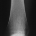
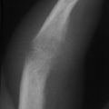
Plain films (Fig. 21-17) can show an alarming degree of regional osteopenia and destruction of joint and periarticular structures. The destructive process can happen over a very short period (weeks).
Once destruction has occurred, the radiographs will demonstrate the evolution over time of fracture healing in this region.
Diagnostic Work-up
Recognition of the underlying neurological condition
Stay updated, free articles. Join our Telegram channel

Full access? Get Clinical Tree


