Fig. 13.1
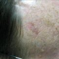
Small 2 mm pigmented lesion on forearm
Dermatoscopy
On examination with a dermatoscope (Heine Delta 20 dermatoscope, manufactured by Heine, Optotechnic GmbH, Herrsching, Germany), the lesion being discussed had no obvious melanin network, but it had asymmetry of color; further the blueness suggested that is was probably both melanocytic and atypical (Fig. 13.2). As we discussed earlier, small melanomas are not only missed by the ABCD rule, but dermatoscopy is notoriously difficult, with most dermatoscopic algorithms not being useful.
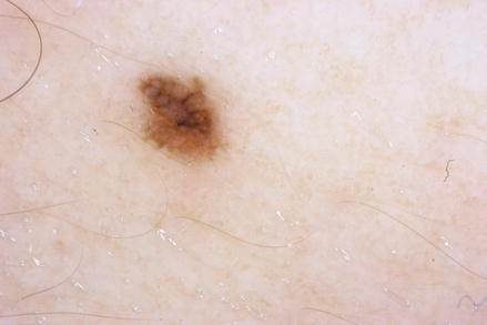
Fig. 13.2
Dermatoscopy
Looking for ‘Chaos and Clues’ in dermatoscopy has been described as an extremely useful method [17]. In this method ‘chaos’ is defined as the presence of asymmetry in structure or color. In the presence of ‘chaos’ one looks for any of the following eight clues:
1.
Thick reticular lines
2.
Grey or blue structures of any kind
3.
Pseudopods or radial lines at the periphery
4.
Black dots in the periphery
5.
Eccentric structure-less area of any color
6.
Polymorphous vascular pattern
7.
White lines
8.
Parallel lines on ridges
In the case being described here, the lesion exhibited ‘chaos’ (asymmetry of color or structure) and also a ‘clue’ (grey or blue structure of any kind). Therefore excision biopsy was done. Interestingly, this lady had very few (<5) nevi and none of the other equally small and pigmented nevi exhibited any asymmetry.
In dermatoscopy, most two-step algorithms commonly recommended were established to differentiate melanocytic from non-melanocytic lesions as a first step. However, using a ‘chaos and clues’ method helps us differentiate malignant from benign lesions first – by looking for chaos over symmetry. In comparing these methods, Kittler and others commented that looking for chaos and clues is preferable over other methods – given that the first step of the traditional dermatoscopic 2-step algorithm, if applied consistently, has a low specificity especially in patients with severely sun-damaged skin, as is often found in Australasia [18].
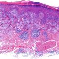
Histopathology
Argenziano and others suggest that small melanomas need more stringent criteria and a ‘consensus approach’ to diagnosis among examining pathologists, as there is no gold standard. In their study they suggest that severe cytologic atypia represents a useful clue in differentiating small melanomas from small dysplastic nevi [19].
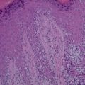
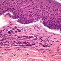
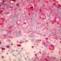
Sections here show superficial sun-damaged skin bearing a small proliferation of atypical melanocytes showing pagetoid scatter to the granular layer along with trans-epidermal elimination of melanin pigment. Superficial dermis shows melanophages and there is no dermal invasion. The appearance is suggestive of a melanoma in-situ because of the combination of cytologic atypia and epidermal invasion (Figs. 13.3, 13.4, and 13.5). Figure 13.3 shows the biopsy specimen; Fig. 13.4 shows atypical hyperchromatic melanocytes singly and in nests and Fig. 13.5 shows transepidermal (pagetoid) invasion.
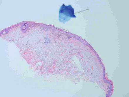
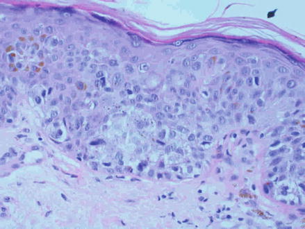
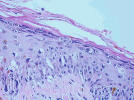
Fig. 13.3
2-mm pigmented skin lesion
Fig. 13.4
Atypical hyperchromatic melanocytes singly and in nests
Fig. 13.5
Transepidermal pagetoid invasion
Discussion
Tiny melanomas (‘micromelanomas’, the term suggested by the author) as discussed earlier, naturally will not fit the ABCD acronym. In a study of pigmented lesions 3–6 mm in diameter, the authors showed that clinical criteria for diagnosing melanoma are not as reliable in the diagnosis of pigmented lesions of less than 6 mm diameter [20]. In this case of a tiny pigmented lesion of 2 mm diameter, the unusual features were the absence of several nevi, which is usually the case in other reported cases. In this situation, this was the only nevus that exhibited any chaos and therefore using the ‘chaos and clues’ algorithm proved decisive. The lesion turned out to be a melanoma-in-situ and was managed by wide surgical excision to ensure 5 mm margins all around.
Related posts:
 Topical Treatment of Skin Cancers and the Risks of ‘Fighting Fire with Fire’
 Amelanotic Malignant Melanoma of the Toe Presenting as an Ulcer: Management and Biopsy Guidelines
 Zosteriform Cutaneous Metastasis
 Metastatic Cutaneous Adenocarcinoma
 Multiple Basal Cell Carcinomas and Superficial Radiotherapy (SRT)
 Balloon Cell Nevi and Balloon Cell Melanomas: What Are They?
Stay updated, free articles. Join our Telegram channel

Full access? Get Clinical Tree


