Hodgkin Lymphoma in Children


Hodgkin Lymphoma in Children


Debra L. Friedman


INTRODUCTION

The first description of Hodgkin lymphoma (HL) was in 1832 when Thomas Hodgkin described seven patients with enlarged lymph glands and spleen. This was followed with histologic descriptions of multinucleated giant cells, by Sternberg in 18981 and Reed in 1902.2 In the 1960s, the clonality of the Reed-Sternberg cell was established.3 More recently, work has focused on the molecular biology of the disease, including the role of immunoglobulin genes, transcription factors, apoptotic pathways, and Epstein-Barr virus (EBV) incorporation.4, 5, 6, 7

Therapeutic advances in HL began in 1902 when Pusey reported on the use of radiotherapy,8 followed by the use of single-agent chemotherapy, mechlorethamine (nitrogen mustard) in 1946,9 then combination chemotherapy with MOPP in 1964,10 and ABVD in the 1970s.11 Donaldson and colleagues at Stanford introduced the concept of combined modality therapy for pediatric patients using the MOPP backbone and low-dose radiation therapy (RT)12 (see Table 94.1 for acronyms of treatment protocols). Multimodality, risk-adapted therapies are now the standard of care for pediatric and adolescent HL. The goals of contemporary clinical trials are to balance short- and long-term toxicity with efficacy, in order to maximize cure and minimize adverse sequelae of treatment simultaneously.


EPIDEMIOLOGY


Incidence

Supported by data from United States Surveillance, Epidemiology End Results [SEER],13 HL makes up 8.8% of all childhood cancer under the age of 20 years, but 17.7% of cancer in children between ages 15 and 17 years. The overall annual incidence rate in the United States is 12.1 per million for children under 20 years and increases to 32 per million when limiting the analysis to adolescents between 15 and 19 years. Overall there is a slight female predominance when considering all children less than 20 years (M:F = 0.9). The Caucasian:African American ratio is 1.3:1.13


TABLE 94.1 COMMON CHEMOTHERAPY PROTOCOLS AND ACRONYMS


Acronym


Chemotherapy Agents


ABV


Doxorubicin, bleomycin, vinblastine


ABVD


Doxorubicin, bleomycin, vinblastine, dacarbazine (DTIC)


BEACOPP


Bleomycin, etoposide, doxorubicin, cyclophosphamide, vincristine, procarbazine, prednisone


CHOP


Cyclophosphamide, doxorubicin, vincristine, prednisone


COP (P)


Cyclophosphamide, vincristine, procarbazine (prednisone)


COPDAC


Cyclophosphamide, vincristine, prednisone, dacarbazine


D(A)BVE (PC)


Doxorubicin, bleomycin, vincristine, etoposide (prednisone, cyclophosphamide)


MOPP


Mechlorethamine (nitrogen mustard), vincristine, procarbazine, prednisone


VAMP


Vincristine, doxorubicin, methotrexate, prednisone


VEPA


Vinblastine, etoposide, prednisone, doxorubicin


OEPA


Vincristine, etoposide, prednisone, doxorubicin


OPPA


Vincristine, procarbazine, prednisone, doxorubicin


ICE


Ifosfamide, carboplatin, etoposide


IV


Ifosfamide, vinorelbine


BEAM


Carmustine, etoposide, cytarabine, melphalan


Adolescent and young adults are most likely to have disease of the nodular sclerosing subtype. Although among children <20 years of age, the nodular sclerosing subtype accounts for 70% of cases, this subtype accounts for 74% of cases in those 15 to 19 years of age. Under the age of 20 years, the mixed cellularity subtype accounts for 16% of cases, but under the age of 10 years, 32% of cases and across the pediatric age group, it is more common in males.13


Risk Factors

There are several factors that are known to increase the risk of HL, which include family history of HL, Epstein-Barr virus (EBV) infections, socioeconomic status, and social contacts. For young adult disease (ages 16 to 44 years), there is a 99-fold increased risk among monozygotic twins and a seven-fold increased risk among other siblings.14, 15, 16 There is a fascinating interaction between EBV and HL epidemiology and biology. EBV-associated HL, with incorporation of the EBV in the genome, is most commonly reported with the mixed cellularity histologic subtype in children from underdeveloped and developing nations and in young adult males. Conversely, in young adult HL, incorporation of EBV in the tumor genome is unusual but a history of infectious mononucleosis and high-titer antibodies to EBV are associated.7, 17, 18, 19, 20 Interestingly, the association between HL and socioeconomic status also differs by age. In children under 10 years, the disease is associated with lower socioeconomic status and large sibship.21, 22 In contrast, risk in young adult patients increases with socioeconomic status and with the related characteristics of a small nuclear family, single-family housing, and fewer siblings or childhood playmates. These findings may be related to an association with infections, where increased infections in early childhood may reduce risk of young adult HL.23, 24 There are inconsistent data regarding clustering of young adult cases.25, 26


Biology

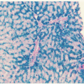
The hallmark of classical HL is Reed-Sternberg (RS) cell which most commonly derives from a neoplastic clone originating from B-lymphocytes in lymph node germinal centers, but is then embedded within a reactive infiltrate of lymphocytes, macrophages, granulocytes, and eosinophils.27 The RS cell is a binucleated or multinucleated giant cell that is often characterized with a bilobed nucleus, with two large nucleoli, described commonly as an owl’s eye appearance.28 Sequence analyses of RS cell clones reveal rearrangements of immunoglobulin variable-region genes resulting in deficient immunoglobulin production. RS cells then evade the apoptotic pathway, leading to the genesis of HL, and perhaps the paraneoplastic immune-mediated phenomena that sometimes accompany the disease.4, 5 The B lymphoid cells from which RS arise have high levels of constitutive nuclear NF-κB, a transcription factor known to mediate gene expression related to inflammatory and immune responses, and deregulation of NF-κB has been postulated as a mechanism by which RS cells evade apoptosis.6, 29 NF-κB dimers are held in an inactive cytoplasmic complex with inhibitory proteins, the IκBs.29 B-cell stimulation by
diverse signals results in rapid activation of the IκB kinase (IKK). The IKK complex phosphorylates two critical serine residues of IκBs,30, 31, 32 thereby targeting them for rapid ubiquitin-mediated proteasomal degradation. Active NF-κB dimers are then released and translocated to the nucleus, where they activate gene transcription. Activation of NF-κB appears to be a final common effect of co-stimulatory interactions, genetic aberrations, or viral proteins that operate in HL.33

RS cell survival is dependent on several downstream pathways. RS cells express CD40 and CD40 ligand (CD40L) is expressed on inflammatory T and dendritic cells that surround them. CD40/CD40L interactions normally provide a second signal from activated helper T-cells to normal B-cells, resulting in activation of NF-κB. NF-κB in turn causes proliferation and induces expression of BCL-xL, which protects B-cells from apoptosis.34 Tumor necrosis factor receptor-associated factor 1 (TRAF 1) is overexpressed in EBV-transformed lymphoid cells and RS cells35 and is associated with activation of NF-κB and protection of lymphoid cells from antigen-induced apoptosis. Activation of NF-κB, in turn, leads to expression of TRAF1, thereby establishing a positive feedback loop that maximizes NF-κB-dependent gene expression.36 EBV latent membrane protein 1 (LMP1) interacts with TRAF1, and tumors with TRAF1-LMP1 aggregates exhibit high NF-κB activity.37,38 LMP1 activates NF-κB by promoting IκBa turnover.39 RS cells express CD30 and CD30 ligation promotes proliferation of HL-derived cells with constitutive activation of NF-κB.40

EBV genome fragments can be found in approximately 30% to 50% of HL specimens, and may play a role in the rescue and repair of RS cells, further aiding in their evasion of apoptosis and enhanced survival.7, 41, 42, 43 Three latent viral antigens are expressed in EBV-positive HL in RS cells: Epstein-Barr nuclear antigen-1 (EBNA1), required for viral episome maintenance, LMP1 with transforming properties, and LMP2, which is nontransforming.44, 45


Histology

HL can broadly be divided into two pathologic classes: classical Hodgkin lymphoma (CHL) and nodular lymphocyte predominant Hodgkin lymphoma (NLPHL).46, 47 In turn, CHL can be further divided into four subtypes: lymphocyte rich (LRCHL), nodular sclerosis (NSHL), mixed cellularity (MCHL), and lymphocyte depleted (LDHL). See Chapter 86 for descriptions of the subtypes. As noted above, different cell types are more common in younger or older children and adults with HL.

In CHL, the RS cells do not express B-cell antigens such as CD45, CD19, and CD 79A, but virtually all express CD30 and approximately 70% express CD15, with only 20% to 30% expressing CD20.48 In comparison, in NLPHL, the tumor cells do express B-cell antigens such as CD19, CD20, CD22, and CD79A, may or may not express CD30 and do not express CD15.49 In addition, the OCT.2 and BOB.1 oncogenes are down-regulated in CHL but not in NLPHL, correlating with immunoglobulin transcription.50


CLINICAL CONSIDERATIONS


Presentation and Staging

The most common presentation of HL, occurring in 80% of patients, is painless adenopathy. Mediastinal involvement is present in approximately 76% of adolescents but only in 33% of children 10 years of age and younger. A large mediastinal mass with a maximum diameter that is greater than one third of the chest diameter and/or a node or nodal aggregate greater than 10 cm occurs in about 20% of patients.51, 52 B symptoms, defined as (1) unexplained loss of >10% of body weight in the 6 months preceding the diagnosis; (2) unexplained fever with temperatures >38°Celsius for more than 3 days; or (3) drenching night sweats, are seen in 20% of patients at the time of initial disease presentation.52, 53, 54 Staging is performed clinically, based on the Ann Arbor staging system55 as revised in 1989.53 Approximately 80% to 85% of children and adolescents with HL have involvement limited to or with direct extension from the lymph nodes and/or the spleen (Stages I to III), whereas 15% to 20% of patients are Stage IV with involvement of the lung, bone marrow, bone, or liver.55 Although staging definitions by nodal region, B symptoms, and definition of extranodal involvement are well defined, bulk disease and substaging have not been consistent across studies (Table 94.2).


Diagnostic Evaluation

A detailed history is required to elucidate B symptoms, which becomes important in risk stratification. A thorough physical evaluation should be performed, documenting the location and size of adenopathy, presence of splenomegaly and any evidence of organ dysfunction. This should be complemented by computed tomography (CT) scans of the neck, thorax, abdomen, and pelvis. FDG-PET imaging is the standard of care in HL and response to disease is now judged by FDG-PET alone or in combination with CT. With the advent of combined CT-PET scans, areas of disease can be evaluated simultaneously with both modalities in an overlapping fashion.56, 57, 58, 59 Technicium-99 bone scintigraphy can be considered in patients with bone pain or elevated alkaline phosphatase, although PET may obviate the need for this. In addition, an upright chest radiograph (CXR) with posteroanterior (PA) and lateral views has traditionally been required for documentation of a large mediastinal mass (bulk mediastinal disease) for clinical trials, defined as tumor diameter >1/3 the thoracic diameter (measured transversely at the level of the dome of the diaphragm on a 6-foot upright PA CXR),60 although the ongoing value of this remains unclear.

Bone marrow biopsy is recommended for all Stage III and IV patients or patients with B symptoms. There are less consistent recommendations for lower-stage patients without B symptoms.61, 62 Laboratory studies include a complete blood count, blood chemistries to evaluate hepatic and renal function and may include acute phase reactants such as ferritin, erythrocyte sedimentation rate, and serum copper, which may be seen as nonspecific markers of tumor activity, but may correlate with prognosis or response.


Prognostic Factors

Adverse prognostic markers established from clinical trials often form the basis for risk stratification and subsequent modification of therapeutic algorithms. In sequential trials, as treatment is then risk-based, these adverse factors are abrogated by the changes in therapy. Pre-treatment factors that have been shown to be associated with adverse outcome include advanced stage, B symptoms, bulk disease, extranodal extension, male sex, and elevated erythrocyte sedimentation rate; and in some studies, hemoglobin <110 g/L or white blood cell count >11.5 × 10⁹/L, age 5 to 10 years, and increased numbers of sites of disease.63, 64, 65, 66, 67, 68, 69 Patients with NLPHL appear to have an overall better prognosis than those with CHL, and among those with CHL, histologic subtype is not consistently associated with prognosis.67, 70 Serum markers that may confer adverse prognostic risk include soluble vascular adhesion molecule-1, tumor necrosis factor, soluble CD-30, beta-2 microglobulin, transferin, serum IL-10, and serum CD 8 antigen.71, 72, 73, 74, 75, 76 High RS cell levels of caspase 3 may be associated with a more favorable outcome.77 Early response to treatment, which may be a correlate for biology, may also be an important prognostic factor, allowing titration of therapy to the individual.68, 78, 79, 80


TABLE 94.2 CLINICAL AND STAGING CRITERIA FOR HODGKIN LYMPHOMA


A. Stage Grouping


Stage I:


Involvement of single lymph node region (I) or localized involvement of a single extralymphatic organ or site (IE).


Stage II:


Involvement of two or more lymph node regions on the same side of the diaphragm (II) or localized contiguous involvement of a single extralymphatic organ or site and its regional lymph node(s) with involvement of one or more lymph node regions on the same side of the diaphragm (IIE).


Stage III:


Involvement of lymph node regions on both sides of the diaphragm (III), which may also be accompanied by localized contiguous involvement of an extralymphatic organ or site (IIIE), by involvement of the spleen (IIIS), or both (IIIE+S).


Stage IV:


Disseminated (multifocal) involvement of one or more extralymphatic organs or tissues, with or without associated lymph node involvement, or isolated extralymphatic organ involvement with distant (nonregional) nodal involvement.


B. Symptoms and Presentations


“A” Symptoms: Lack of “B” symptoms.


“B” Symptoms: At least one of the following:


Unexplained weight loss >10% in the preceding 6 mo


Unexplained recurrent fever >38°C


Drenching night sweats.


X Bulk disease (see C below)


E Involvement of a single extranodal site that is contiguous or proximal to the known nodal site.


C. Bulk disease


One or both of the following presentations are considered “bulk” disease:


Large mediastinal mass: tumor diameter >1/3 the thoracic diameter (measured transversely at the level of the dome of the diaphragm on a 6-foot upright PA CXR) In the presence of hilar nodal disease the maximal mediastinal tumor measurement may be taken at the level of the hilus. This should be measured as the maximum mediastinal width (at a level containing the tumor and any normal mediastinal structures at the level) over the maximum thoracic ratio.


Large extramediastinal nodal aggregate: A continuous aggregate of nodal tissue that measures >6 cma in the longest transverse diameter in any nodal area.


a Some studies use 10 cm for definition of extramediastinal bulk disease.


INITIAL TREATMENT FOR PEDIATRIC HODGKIN LYMPHOMA

To our knowledge, the biology and natural history of HL does not differ between children and adults. As a result, early therapeutic approaches for pediatric HL were similar to or modeled after those developed for adults with HL. This seemed particularly logical and HL is most commonly a disease of adolescents as opposed to adults. However, with high cure rates, contemporary standard approaches to adolescent and young adult HL include multiagent chemotherapy with or without low-dose involved field radiotherapy. The overriding principles of these treatment regimens are to balance efficacy with both acute and, perhaps, more important, long-term toxicities.81, 82, 83 A summary of common protocols for upfront therapy is found in Table 94.3. Figure 94.1 shows the timeline of evolution of therapy for pediatric HL.

Only gold members can continue reading. Log In or Register to continue

Stay updated, free articles. Join our Telegram channel

Oct 21, 2016 | Posted by in HEMATOLOGY | Comments Off on Hodgkin Lymphoma in Children

Full access? Get Clinical Tree

Get Clinical Tree app for offline access