Hodgkin and Non-Hodgkin Lymphoma


Hodgkin and Non-Hodgkin Lymphoma


Lauren C. Pinter-Brown

Dennis A. Casciato


EVALUATION OF SUSPECTED LYMPHOMA


I. SYMPTOMS AND SIGNS

A. History

1. Painless lymphadenopathy, involving any of the superficial lymph nodes, is the most common chief complaint of patients with Hodgkin lymphoma (HL) and non-Hodgkin lymphoma (NHL).

2. Systemic symptoms. Fevers, night sweats, and weight loss are characteristic in advanced presentations of HL and aggressive NHL but may be encountered in all stages and pathologic types of lymphoma. Marked fatigue and general weakness may also be reported, not always correlating with the degree of anemia.

a. Pruritus, often intense, may be the presenting symptom in HL, particularly the nodular sclerosis subtype, and may antedate diagnosis by months or years.

b. Pel-Ebstein fever is periodic and uncommon but characteristic of HL.

3. Pain

a. Alcohol-induced pain in areas of involvement is infrequent but is characteristic of HL.

b. Abdominal pain or discomfort may be due to splenomegaly, bowel dysfunction due to adenopathy or bowel involvement, or hydronephrosis.

c. Bone pain may reflect localized areas of bone destruction or invasion or diffuse marrow infiltration.

d. Neurogenic pain is caused by spinal cord compression, plexopathies, nerve root infiltration, meningeal involvement, and complicating varicella zoster.

e. Back pain suggests massive retroperitoneal nodal involvement, often with psoas muscle invasion.

B. Physical examination. should evaluate for hepatosplenomegaly, the presence of effusions, evidence of neuropathy, and signs of obstruction (e.g., extremity edema, superior vena cava syndrome, spinal cord compression, hollow viscera dysfunction). Lymph node chains must be carefully examined, including the submental, supraclavicular, infraclavicular, epitrochlear, iliac, femoral, and popliteal nodes.

1. The lymph nodes are examined for size, multiplicity, consistency, and tenderness. Lymphomatous involvement typically imparts a rubbery consistency, not the rock-hard quality of carcinoma.

2. The tonsils and oropharynx are thoroughly examined. Waldeyer ring involvement mandates complete evaluation of the nasopharynx, oropharynx, and hypopharynx by endoscopy.


II. DIFFERENTIAL DIAGNOSIS

(Table 21.1) compares clinical features of HL and NHL.

A. Lymphadenopathy

1. Infections. Patients, particularly young children with apparent viral or other infections, may develop striking lymphadenopathy. Such patients should be evaluated for infectious processes and observed for clear-cut resolution. Microorganisms associated with prominent lymphadenopathy include Epstein-Barr virus (EBV; infectious mononucleosis), cytomegalovirus (CMV), human immunodeficiency virus (HIV), hepatitis virus, secondary syphilis, mycobacteria, some fungi, and Toxoplasma, Brucella, and Rochalimaea species infection. In some cases, biopsy is required for diagnosis of specific infectious diseases.

2. Systemic immune disorders, such as rheumatoid arthritis, Sjögren syndrome, and systemic lupus erythematosus, are associated with both benign lymphadenopathy and lymphoma. Progressive or asymmetric lymphadenopathy mandates biopsy.

3. Patients at risk for HIV infection present problems requiring individualization in management. Persistent generalized lymphadenopathy is a part of the acquired immunodeficiency syndrome (AIDS) spectrum, but lymphadenopathy can also be caused by opportunistic infections, Kaposi sarcoma, or lymphoma.

4. Lymph nodes that are usually benign

a. Occipital. Consider scalp infection.

b. Posterior auricular. Usually viral or scalp infection

c. Shotty inguinal nodes. Often present with no obvious cause but may suggest external genital or lower extremity infections

5. Cervical nodes. Patients with isolated enlargement of high or middle cervical lymph nodes often harbor occult primary carcinoma of the head and neck. The special approach required for these patients is discussed in Chapter 7, Section X.

B. Midline masses

1. Retroperitoneal masses (see Chapter 19, Section II)

2. Mediastinal masses may occur in a variety of nonneoplastic and neoplastic (both primary and metastatic) conditions (see Chapter 19, Section I).


Table 21.1 Comparison of Hodgkin and Non-Hodgkin Lymphomas


In Non-Hodgkin Lymphoma


Characteristic


In Hodgkin Lymphoma


Low Grade


Others


Site of origin


Nodal


Extranodal (˜10%)


Extranodal (˜35%)v


Nodal distribution


Centripetal (axial)


Centrifugal


Centrifugal


Nodal spread


Contiguous


Noncontiguous


Noncontiguous


CNS involvement


Rare (<1%)


Rare (<1%)


Uncommon (<10%)


Hepatic involvement


Uncommon


Common (>50%)


Uncommon


Bone marrow involvement


Uncommon (<10%)


Common (>50%)


Uncommon (<20%)


Marrow involvement adversely affects prognosis


Yes


No


Yes


Curable by chemotherapy


Yes


No


Yes


3. Hilar masses. Isolated symmetric bilateral hilar lymphadenopathy (without mediastinal mass) is strongly suggestive of sarcoidosis, and many experts believe that observation alone could suffice in this clinical setting. Unilateral hilar masses are frequently secondary to lung cancer; metastatic disease must also be considered. Coccidioidomycosis and histoplasmosis enter the differential diagnosis in the appropriate clinical and geographic milieu.

C. Splenomegaly. The diagnosis can usually be made with careful history taking and physical examination, laboratory evaluation, CT scans of abdomen, bone marrow biopsy or aspiration with flow cytometric analysis, and, occasionally, liver biopsy. When a diagnosis cannot be established by these means, careful follow-up of the patient is warranted. Splenectomy should be considered for diagnosis in patients with massive or progressive isolated splenomegaly.

1. Normal. A palpable spleen is occasionally seen in otherwise healthy young adults of thin body habitus.

2. Infections include most pathogens listed in Section II.A.1, bacterial endocarditis, malaria, and abscess.

3. Secondary to portal hypertension (congestive splenomegaly). Patients with chronic liver disease or portal or splenic vein thrombosis may have no other findings to direct the diagnostic search. Portal hypertension may be documented by ultrasound of the abdomen with Doppler or by liver-spleen scanning, which reveals redistribution of the radionuclide to the spleen and marrow.

4. Storage diseases, particularly Gaucher disease, may produce prominent splenomegaly; characteristic cells are seen in the bone marrow in most cases.

5. Tumors are predominantly hematologic, including lymphomas and leukemias. Metastases, particularly from melanoma and breast cancer, and primary splenic sarcomas may also occur.

6. Myeloproliferative disorders such as polycythemia vera, agnogenic myeloid metaplasia (myelofibrosis), and chronic myelogenous leukemia may cause marked splenomegaly.

7. Autoimmune disorders. Rheumatoid arthritis (Felty syndrome), systemic lupus erythematosus, and autoimmune hemolytic anemia may produce splenomegaly (not isolated autoimmune thrombocytopenia) and can usually be diagnosed by history and associated laboratory findings.

8. Miscellaneous. Splenic cysts, thyrotoxicosis, sarcoidosis, chronic nonim-mune hemolysis, and amyloidosis are unusual causes of splenomegaly.


III. BIOPSY PROCEDURES

A. Sites and methods of diagnostic biopsy. Tissues or organs that are suspected of involvement are subjected to generous open biopsy for primary diagnosis wherever possible. Fine-needle aspiration cytology is mainly used for staging evaluation or for proving recurrence but may sometimes allow cytologic diagnosis if expertise in interpretation is available.

1. Peripheral node biopsy. One of the largest accessible lymph nodes is excised whenever peripheral lymphadenopathy is present. Small lymph nodes may be more readily removed but may be uninvolved.

2. Inguinal lymph nodes are frequently enlarged because of chronic inflammatory processes in the lower extremities. These nodes should be excised only when other sites are not suspected or when pathologic involvement is clearly anticipated.


3. Bone marrow biopsy combined with aspiration is used for staging and may lead to diagnosis, particularly in the presence of abnormal circulating cells or cytopenias.

4. Mediastinoscopy or limited thoracotomy (e.g., Chamberlain procedure) for definitive diagnosis is required for a substantial proportion of patients with mediastinal masses.

5. Laparotomy is used to diagnose some cases of lymphoma restricted to the abdomen and may include biopsies of the liver and random lymph nodes, as well as the primary area in question.

6. Laparoscopy assesses the liver and peritoneum and allows extensive biopsy, obviating the need for laparotomy in some patients.

7. Endoscopic gastric biopsy with staining for Helicobacter pylori may be helpful in the diagnosis of gastric MALToma. Repeated attempts with deeper biopsies and immunoperoxidase staining for leukocyte common antigen and keratin intermediate filaments may be helpful in the differential diagnosis between lymphoma and carcinoma. Small bowel involvement beyond the duodenum usually requires open biopsy, although capsule biopsies may be suggestive of lymphoma in some cases.

8. Retroperitoneal and mesenteric masses may be evaluated by imagingguided Trucut biopsy or fine-needle aspiration with immunologic analysis of the specimens, obviating the need for laparotomy.

B. Handling the biopsy material. The procured biopsy specimen is submitted to the pathologist directly and not placed in a fixative by the operating surgeon to ensure the best use of the available tissue. Prior communication with the pathologist is advantageous. Maintaining frozen sections is preferable for subsequent analysis. Pathology tissue processing includes the following procedures:

1. Touch preparations (imprints), which provide cytologic detail and material for immunologic phenotyping

2. Immunologic phenotyping with monoclonal antibodies can be crucial to diagnosis. Lymphoid cells are characterized immunologically using flow cytometry. Discriminatory immunophenotypes in lymphoma are shown in Appendix C5. A common NHL panel should include assessment of expression of CD2 or CD3, CD5, CD19 or CD20, and CD23 in blood, bone marrow, or biopsy specimens. Classic Reed-Sternberg (RS) cells are usually CD15-positive and CD30-positive. More surface markers may need to be analyzed if this screening is inconclusive or if rare entities (such as natural killer [NK] cell or hairy cell leukemia) are considered.

3. Special handling of tissues for procedures that may occasionally be used in difficult diagnostic problems or research such as cytogenetics, molecular genetic analysis, and electron microscopy

4. Microbial culture of submitted material when the clinical picture or tissue suggests infection


IV. CLINICAL EVALUATION.

The extent of the staging evaluation is determined by the individual case presentation, the histopathologic diagnosis, and the effect of the stage on treatment planning.

A. Evaluation of blood tests

1. Hematologic manifestations are discussed in Chapter 34.

2. Diagnostically abnormal circulating lymphoid cells or lymphocytosis is seen in some patients with either indolent or aggressive forms of NHL.
Lymphoid cells are characterized immunologically using flow cytometry, and monoclonality may be established by κ:λ ratios (B cell) or gene rearrangement technology (T and B cell); these techniques are capable of detecting minute clones of circulating lymphoma cells not detectable by inspection of the blood smears.

3. Acute-phase reactants, such as the erythrocyte sedimentation rate (ESR), fibrinogen, haptoglobin, and serum copper levels, may parallel disease activ ity, especially in HL.

4. Liver function tests are unreliable in predicting lymphomatous involvement of the liver. Marked elevation of alkaline phosphatase and occasionally frank cholestatic jaundice may complicate HL as a paraneoplastic event without direct liver involvement. Extrahepatic biliary obstruction may also occur with lymphoma caused by enlarged nodes in the porta hepatis.

5. Renal function tests. Elevated creatinine and blood urea nitrogen levels suggest ureteral obstruction and, less commonly, direct renal involvement. Uric acid nephropathy or hypercalcemia may contribute to renal insufficiency. Frank nephrotic syndrome as a paraneoplastic phenomenon may complicate HL and other lymphomas (see Chapter 31).

6. Serum uric acid. Hyperuricemia is a common manifestation of high-turnover-rate (aggressive) NHL and may also be seen with extensive lower grade lymphomas. Treatment of high-grade NHL or treatment of sensitive bulky low-grade lymphoma may provoke brisk tumor lysis, leading to further elevation of uric acid and renal shutdown (see Chapter 27, Section XIII). Hypouricemia may be seen in HL.

7. Hypercalcemia has been noted in some cases of lymphoma and may be secondary to production of parathyroid hormone-related peptide or activation of vitamin D by lymphoma tissue.

8. Serum lactate dehydrogenase (LDH) levels reflect tumor bulk and turnover, particularly in the aggressive NHL, and are considered an independent prognostic factor.

9. Serum immunoglobulins. Polyclonal hypergammaglobulinemia is commonly seen in HL and NHL. Hypogammaglobulinemia is particularly common in the small lymphocytic lymphomas (SLLs) and late in the disease. Monoclonal spikes are seen occasionally in NHL patients.

B. Evaluation of the chest

1. Chest radiographs may demonstrate mediastinal and hilar lymphadenopathy, pleural effusions, and parenchymal lesions. A cavitating lesion is more typical of infection than lymphomas.

2. CT scans tend to replace chest x-rays because they can demonstrate parenchymal and mediastinal abnormalities in more detail.

3. Thoracentesis and pleural biopsy may demonstrate direct lymphomatous involvement of the pleura. Obstruction of mediastinal lymphatic-venous drainage may result in cytologically negative or chylous effusions.

C. Evaluation of the abdomen and retroperitoneum

1. CT scans are useful in delineating abnormal enlargement of nodes in retroperitoneal, mesenteric, portal, and other lymph node sites. The CT scan also detects splenomegaly and, with constant enhancement, may define spaceoccupying lesions in the liver, spleen, and kidneys.

2. Bipedal lymphangiography has been abandoned because of improvements and availability of alternative imaging techniques and because expertise in performance and evaluation is frequently unavailable.


3. Abdominal ultrasonography is too insensitive to be useful in routinely assessing abdominal lymphadenopathy. It is occasionally helpful in distinguishing hepatic or splenic lesions (cystic vs. solid) and in excluding an obstructive basis for renal insufficiency and jaundice.

4. MRI with contrast may be useful in distinguishing benign from malignant hepatic lesions.

D. Evaluation of the gastrointestinal (GI) tract. Direct involvement of the GI tract is uncommon in HL but is common in NHL. Patients with Waldeyer ring lymphoma, suggestive GI symptoms, extensive abdominal nodal involvement, unexplained iron deficiency, or GI bleeding are evaluated with upper GI series and complete small bowel follow-through. Barium enema may be necessary. Endoscopic examination and biopsy of accessible abnormalities are performed. Routine GI tract evaluation for patients with mantle cell lymphoma is performed in some centers.

E. Evaluation of the central nervous system (CNS). Spinal fluid examination is routinely used to exclude occult lymphomatous involvement of the meninges in patients with Burkitt lymphoma (BL) or lymphoblastic lymphoma and is often performed in patients with intermediate-grade or high-grade lymphoma involving the testes or paranasal sinuses (B-cell histology), with extensive bone marrow involvement, or with multiple sites of extranodal involvement and elevated LDH. In these cases, the incidence of CNS disease is in the 5% range. Patients with AIDS-related lymphoma may require CT or MRI scans of the brain and spinal fluid analysis. Symptoms suggestive of intracranial, spinal cord, or peripheral nerve involvement require immediate diagnostic evaluation.

F. Nuclear scans

1. Positron emission tomography (PET) using 18F-fluorodeoxyglucose scans tends to replace gallium scans. It seems to be more sensitive in detecting unsuspected metastasis or in differentiating active versus uninvolved nodes with accuracy approaching 95% depending on nodal histology. Similar to gallium scans, PET is somewhat less reliable in indolent lymphoma. False-positive results can be produced by any inflammation, whereas faint normal uptake of muscles, the bowel, and bone marrow recovering from chemotherapy should be differentiated from involvement. The advent of combined PET/CT scan is thought to increase the accuracy of the procedure and may eventually become the gold standard for staging and following patients.

2. Gallium scans. 67Ga scans are primarily used in assessing residual radiographic mediastinal and, less often, retroperitoneal abnormalities after therapy. Persistent 67Ga uptake in these areas strongly suggests residual tumor instead of fibrosis or necrosis. To be useful in such follow-up, a 67Ga body scan is recommended before therapy. 67Ga scans can be unreliable below the diaphragm because of competing uptake in the GI tract, liver, and spleen.


HODGKIN LYMPHOMA


I. EPIDEMIOLOGY AND ETIOLOGY

A. Incidence. HL accounts for about 1% of new cancer cases annually in the United States, or 7,000 cases per year.

1. Age. HL demonstrates a bimodal age-incidence curve in the United States and some industrialized European nations. The first peak, constituting predominantly the nodular sclerosis subtype, occurs in the 20s and the second
peak occurs after 50 years of age. In Third World countries, the first peak is absent, but there is a significant incidence of mixed cellularity and lymphocyte-depleted HL in men.

2. Sex. About 85% of children with HL are boys. In adults, the nodular sclerosing subtype of HL shows a slight female predominance, whereas the other histologic subtypes are more common in men.

B. Risk factors. In Western countries, the first peak of HL is associated with a higher social class, advanced education, and small family size; a delayed exposure to a common infectious or other environmental agent has been suggested. HL may be associated with EBV infection, but the significance of this association is unclear. A slightly increased incidence of HL has been demonstrated with HIV infection; HIV-associated HL (see Chapter 36, Section III) often presents with constitutional symptoms, advanced stage, and unusual sites of involvement (e.g., marrow, skin, leptomeninges).


II. PATHOLOGY AND NATURAL HISTORY

A. Histology. The pathologic diagnosis of HL depends on the presence of RS cells and their variants in an appropriate pathologic milieu. The bulk of lymphatic tissue involved by HL is not composed of neoplastic cells but rather a variety of normal-appearing lymphocytes, plasma cells, eosinophils, neutrophils, and histiocytes existing in different proportions in the various histologic subtypes. Important variants of RS cells include L&H (lymphocyte and histiocyte) cells, lacunar cells, and RS-like cells (see Appendix C5. Discriminatory Immunophenotypes for Lymphocytic Neoplasms).

1. The Rye classification for HL relates the histopathologic subtypes to clinical behavior and prognosis. This older classification system comprises lymphocyte-predominant (LP), nodular sclerosing (NS), mixed-cellularity (MC), and the uncommon lymphocyte-depleted (LD) varieties of HL. The LP subtype was further divided into nodular LP and diffuse LP subtypes. Immunohistochemistry, however, has resulted in deletion of the diffuse LP subtype and redefinition of the nodular LP subtype (see Section II.A.2).

2. The World Health Organization (WHO) classification divides HL into nodular LP HL and classical HL. Classical HL in this newer classification system comprises the lymphocyte-rich, NS, MC, and LD varieties.

a. Nodular LP HL with its L&H cells, which are not classical RS cells, is now clearly recognized to be most like an indolent B-cell NHL and not true HL. For that reason, nodular LP HL is distinguished from classical HL in the WHO classification. Table 21.2 shows this classification system with distinguishing histopathologic features, clinical correlates, and immunophenotypes.

b. Diffuse LP HL in the Rye classification has disappeared as an entity. In the new WHO classification of lymphocytic neoplasms (see Appendix C6), what was thought to be diffuse LP HL is now classified as lymphocyte-rich classical HL (with true RS cells that are CD30-positive), Lennert lymphoma (lymphoepithelioid peripheral T-cell lymphoma), T-cell-rich B-cell lymphoma, or other entities.

3. RS cells and their variants

a. RS cells are giant cells that have more than one nucleus and large, eosinophilic, inclusion-like nuclei. Single-cell polymerase chain reaction analysis has shown that the RS cells are B cells that originate in the germinal centers of lymph nodes. RS cells and the accompanying mononuclear Hodgkin cell variants are the neoplastic cells in HL and are surrounded by a reactive cellular infiltrate. Classic RS cells usually express CD15
and CD30. CD30 (Ki-1) is an antigen that is also expressed in anaplastic large cell lymphoma and occasionally in other forms of NHL (e.g., large B-cell lymphoma [LBCL], peripheral T-cell lymphoma [PTCL]). RS cells express CD20 infrequently, but not CD45 (leukocyte common antigen).


Table 21.2 Pathologic and Clinical Features of Hodgkin Lymphoma Subtypesa


Histologic Subtype


Frequency (%)


Histopathology


Clinical Characteristics


Common Stages


Nodular lymphocyte predominant HLb


5


L&H (“popcorn cells”) intermingled with polymorphous infiltrate; nodular or nodular and diffuse patterns


Males; usually localized to peripheral nodes; frequent relapses; excellent prognosis


I-IIA


Lymphocyte-rich CHLc


5


RS scattered in background of small lymphocytes; nodular and diffuse patterns; absent eosinophils and neutrophils


Older males; localized to peripheral nodes; fewer relapses; excellent prognosis


I-IIA


Nodular-sclerosing CHLc


70


RS variable; nodular growth pattern with collagen bands and “lacunar cells”; heterogeneous cellularity with numerous eosinophils and neutrophils


Females; mediastinal masses and peripheral nodes


I-IIIA or B


Mixed-cellularity CHLc


20-25


RS more frequent in a mixed inflammatory background without nodular sclerosing fibrosis


Frequently retroperitoneal; often symptomatic


II-IVA or B


Lymphocyte-depleted CHLc


<5


RS predominant in variable patterns, including diffuse fibrosis; depleted of nonneoplastic lymphocytes


Aggressive course; liver and marrow involved with relative sparing of peripheral nodes


III-IVB


aWorld Health Organization classification system

b L&H cells immunophenotype: CD15, CD30, CD20+, CD45+, EMA±, CD79a+

c Classical HL—RS immunophenotype: CD15+, CD30+, CD20±, CD45, EMA, ALK-1


HL, Hodgkin lymphoma; L&H, lymphocytic and histiocytic cells; CHL, classical HL; RS, Reed-Sternberg cells; EMA, epithelial membrane antigen; ALK-1, anaplastic lymphoma kinase.


b. The lacunar cell is a variant of the RS cell and has the same immunophenotype. It characterizes NS HL and is often far more plentiful than classic RS cells in that subtype.

c. L&H cells are RS-like but have a different immunophenotype. L&H cells manifest B-cell markers (CD20, CD45, and CD79a), but not CD15 or CD30. Although the L&H cells are believed to be of monoclonal origin, the surrounding B-cell infiltrates may be polyclonal. L&H cells were identified in nodular LP HL, which is now considered a separate entity because of its distinct immunophenotype.

d. RS-like cells are found in a variety of infectious, inflammatory, and neoplastic disorders, including infectious mononucleosis, lymphoid hyperplasia associated with phenytoin therapy, and immunoblastic lymphomas.

B. Mode of spread (see Table 21.1). HL almost always originates in a lymph node. Whenever a primary diagnosis of HL is made in an extranodal site without contiguous nodal involvement, the diagnosis should be highly suspect. For much of its natural history, HL appears to spread in an orderly fashion through the lymphatic system by contiguity. Histologic types other than NS, however, often skip the mediastinum, and disease appears in the neck and upper abdomen. The axial lymphatic system is almost always affected in HL, whereas distal sites (e.g., epitrochlear and popliteal nodes) are rarely involved. Hematogenous dissemination occurs late in the course of disease and is characteristic of the LD subtype.

C. Sites of involvement

1. Peripheral lymph nodes. Cervical or supraclavicular lymphadenopathy occurs in >70% of cases. Axillary and inguinal lymph nodes are less frequently involved. Generalized lymphadenopathy is atypical of HL. Left supraclavicular lymphadenopathy is more strongly associated with abdominal involvement (specifically, splenic involvement) than is right-sided disease.

2. Thorax

a. The anterior mediastinum is a prime location for NS HL. Mediastinal precedes hilar lymph node involvement.

b. Lung involvement may occur by direct contiguity with hilar involvement in HL as well as by hematogenous dissemination. Pulmonary involvement by HL may produce discrete nodules and irregular, interstitial, or even lobar infiltrates.

c. Pleural effusion may occur secondary to mediastinal compression of vascular-lymphatic drainage and by direct pleural involvement. Chylous effusions occasionally occur.

d. Pericardial involvement may be found on CT scans, but overt cardiac tamponade is uncommon.

e. Superior vena cava syndrome is more frequent in NHL than in HL.

3. Spleen, liver, and upper abdomen

a. The spleen, splenic hilar nodes, and celiac nodes are the earliest abdominal sites of involvement in infradiaphragmatic HL. Mesenteric lymph nodes are rarely involved in HL.

b. At least 25% of spleens not clinically enlarged harbor occult HL at laparotomy, and as many as half of spleens believed to be enlarged on physical examination or radiologic assessment are histologically normal.


c. Liver involvement is uncommon at diagnosis and is almost always associated with infiltration of the spleen.

4. Retroperitoneal lymph node involvement tends to occur relatively late in the course of supradiaphragmatic HL and after spleen, splenic hilar, and celiac nodal involvement. Periaortic involvement without splenic involvement is uncommon. The retroperitoneal nodes are, however, affected early in the course of inguinal presentations of HL.

5. The bone marrow is rarely involved at the time of diagnosis of HL. Patients with advanced-stage disease, systemic symptoms, and MC or LD histologies have a higher risk for bone marrow involvement. Biopsy is mandatory to evaluate the bone marrow because HL is difficult to diagnose on marrow aspirates. Granulomatous changes or fibrosis may be seen, which is not diagnostic of HL involvement.

6. Bone. Osseous involvement of HL usually produces an osteoblastic reaction mimicking prostatic carcinoma. Extradural masses may result in spinal cord compression. Sternal erosion by mediastinal NS HL may occur.

7. Other extranodal sites are rarely involved in HL. Liver or skin involvement is rare and usually a late manifestation of disease. CNS involvement is very uncommon with the exception of extrinsic spinal cord compression. Clinical involvement of meninges, brain, Waldeyer ring, GI tract, kidney, and other extranodal sites usually suggests an alternative diagnosis.


III. STAGING SYSTEM AND PROGNOSTIC FACTORS

A. Staging is the most crucial determinant of prognosis and treatment in HL. The Ann Arbor Staging System had previously been universally used but has been modified to take into account important prognostic factors, particularly mediastinal bulk. The modified system is called the Cotswolds Staging Classification and is shown in Table 21.3.


Table 21.3 Cotswolds Staging Classification of Hodgkin Lymphoma


Stage


Description


I


Involvement of a single lymph node region or lymphoid structure


II


Involvement of two or more lymph node regions on the same side of the dia phragm (the mediastinum is considered as a single site, whereas hilar lymph nodes are lateralized). The number of anatomic sites should be indicated by a subscript (e.g., II3).


III


Involvement of lymph node regions or structures on both sides of the diaphragm III1 With involvement of splenic hilar, celiac, or portal nodes III2 With involvement of para-aortic, iliac, and mesenteric nodes


IV


Involvement of one or more extranodal sites in addition to a site for which the designation “E” has been used


Designations Applicable to any Disease Stage


A


No symptoms


B


Fever (temperature higher than 38°C), drenching night sweats, or unexplained loss of >10% of body weight within the preceding 6 months


X


Bulky disease (a mediastinal mass exceeding one-third the maximum transverse diameter of the chest or the presence of a nodal mass with a maximal dimension >10 cm)


E


Involvement of a single extranodal site that is contiguous or proximal to a known nodal site


CS


Clinical stage


PS


Pathologic state (as determined by laparotomy or biopsy)


B. Prognostic factors

1. Stage is clearly the single most important prognostic factor in HL. Within each stage, the presence of B symptoms confers a poorer prognosis. About 60% of patients with HL in the United States have stage I or II disease at the time of diagnosis. The percentage of patients with stage III or IV disease is generally higher in Third World countries and in lower socioeconomic enclaves.

2. Histopathology. With advances in therapy, the value of histopathologic subtype as an independent prognostic variable (apart from stage) is less clearly defined than it was in the past.

3. Adverse prognostic factors were evaluated by an international group in a multivariate retrospective analysis of 4,695 patients, mostly with extensive disease (see Hasenclever et al. 1998 in “Suggested Reading”). Patients with no adverse factors had an 84% freedom from progression, whereas the presence of each factor depressed the freedom from progression curve plateau by about 8%. Interestingly, neither tumor bulk nor histology emerged as independent factors. The seven independent prognostic factors identified were as follows:


Adverse Factor


Relative Risk of Relapse


Male sex


1.35


Age ≥45 y


1.39


Stage IV disease


1.26


Hemoglobin <10.5 g/dL


1.35


White blood cell (WBC) count >15,000/µL


1.41


Lymphocyte count <600/µL or <8% of WBC


1.38


Serum albumin <4 g/dL


1.49


4. Independent adverse prognostic factors for NS HL include eosinophilia, lymphocyte depletion, and RS cell atypia.

5. Adverse prognostic factors in early stage HL include ESR ≥50 mm/h, four or more separate sites of nodal involvement, bulky mediastinal mass (defined as >33% of the maximum intrathoracic diameter) or any mass ≥10 cm, or extranodal sites of disease.


V. MANAGEMENT: PRIMARY THERAPY

A. Treatment philosophy. More than one treatment approach may be used in the management of cases of HL. The challenge is to determine a course of therapy that preserves cure while minimizing long-term complications.

B. Surgery is limited to diagnosis, possibly laparotomy, and laminectomy for spinal cord compression.

C. RT alone is still used in the United States to treat many patients with stage IA or possibly IIA disease nonclassical HL. However, it is increasingly replaced in the treatment of classical HL with combined-modality treatment.

1. Radiation dose. HL may be locally sterilized in almost all cases with 3,000 to 4,400 cGy given at a rate of about 1,000 cGy per week. Lesser doses may be adequate as consolidation after chemotherapy.

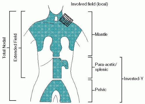
2. Radiation fields (Fig. 21.1)

a. Mantle field encompasses the cervical, supraclavicular, infraclavicular, axillary, hilar, and mediastinal lymph nodes to the level of the diaphragm. Preauricular fields are added for patients with high cervical lymphadenopathy. The lungs and much of the heart are shielded by lead blocks, although many radiotherapists administer some radiation (≤1,500 cGy) to the lung on the involved side, if hilar lymph nodes are enlarged. The whole heart may be treated if the pericardium is involved. A small gap must be left between the inferior border of the mantle field and the superior border of the periaortic field to obviate potential severe spinal cord injury caused by overlap.

b. Inverted-Y field includes the spleen or splenic pedicle and the celiac, periaortic, iliac, inguinal, and femoral lymph nodes. The kidneys, much of the pelvic marrow, and the testes are shielded.

c. Spade and pelvic fields. The inverted-Y field may be divided into a spade field, encompassing the splenic pedicle (or spleen) and periaortic nodes, and a pelvic field, including the iliac, inguinal, and femoral lymph nodes.

d. Subtotal nodal or subtotal lymphoid irradiation consists of mantle and spade fields.

e. Total nodal or total lymphoid irradiation is uncommonly used and consists of mantle and inverted-Y fields.


Figure 21.1. Radiation fields used in Hodgkin lymphoma. Stippled area is the area irradiated. See text for descriptions.

f. Involved-field radiation therapy (IFRT) consists of sites of known disease only and is used with curative intent only in combination with chemotherapy. It has become the most common use of RT in HL with other fields previously described of mostly historical interest. Doses given in combined-modality therapy range from 2,000 to 3,600 cGy.

D. Combination chemotherapy is the mainstay modality for all stages of classical HL and advanced stages of nonclassical HL. Chemotherapy, often in combination with RT, is also preferable for patients with early-stage disease and/or bulky disease. The selection among the available regimens is often guided by the desire to avoid long-term toxicities associated with specific treatments. The advent of the nonleukemogenic, gonadal-sparing ABVD chemotherapy regimen expanded the use of chemotherapy to patients with earlier stages and obviated the need for laparotomy; it has replaced the historic MOPP. More aggressive regimens such as BEACOPP (Appendix D1, Section II) may improve on ABVD (see Section V.D.3), especially in patients with advanced disease. Maintenance therapy is not recommended.

1. Useful chemotherapy regimens for HL are shown in Appendix D1. These regimens must be strictly followed because delays in therapy or reduction in dosages not indicated by the protocol can clearly compromise results. The total dose and dose rate (dose intensity) are important in achieving cure. Regimens used as salvage therapy in HL are shown in Appendix D3.

2. MOPP or COPP regimen (Appendix D1, Section I). The National Cancer Institute (NCI) recommends that vincristine should not be limited to a 2-mg maximum dosage in this regimen, but most clinicians sustain the 2-mg limit. Treatment is administered in 28-day cycles for two additional cycles beyond the attainment of a restaged complete response (CR) and a minimum of six cycles (6 months).


a. The CR rate using the MOPP regimen is between 70% and 80% for stages III and IV HL. About 60% to 70% of CR cases are durable, with relapses rare after 42 months. More than 80% of patients with stage IIIA or IVA disease survive 10 years without recurrence of disease. Histologic subtype appears to have little effect on results with MOPP.

b. The MOPP regimen is particularly emetogenic, and it is associated with myelosuppression neuropathy, leukemogenesis, and infertility. It is believed that COPP (replacing mechlorethamine with cyclophosphamide) may be better tolerated.

3. ABVD regimen (Appendix D1, Section I) is superior to the MOPP regimen and causes much less leukemia and infertility. Potential cardiac toxicity caused by doxorubicin and pulmonary toxicity caused by bleomycin (particularly with the concomitant use of granulocyte growth factors) have been occasional problems using this schedule. The concern is heightened when combined with mediastinal RT. ABVD-based treatment has replaced MOPP as the standard regimen for HL.

a. Generally, the same therapeutic rules as with the MOPP regimen apply: 6 to 8 monthly cycles are usually administered and at least two cycles beyond maximum response.

b. Pulmonary function should be monitored. If dyspnea, pneumonitis, or significant reduction to <40% of predicted lung diffusion capacity is noted, bleomycin should be discontinued. Bleomycin pneumonitis usually responds to corticosteroids and mandates discontinuation of bleomycin. As there is a concern that the use of myeloid growth factors may increase the risk for bleomycin pulmonary toxicity, their use has been discouraged.

c. Cardiac function should be monitored in patients with pre-existing heart disease and in those receiving high cumulative doses of doxorubicin. A baseline measurement of left ventricular ejection fraction is suggested before beginning doxorubicin administration.

4. MOPP and ABVD in alternating cycles and the MOPP/ABV hybrid have both been found to be less satisfactory than ABVD alone. While MOPP/ABV and ABVD were equally efficacious, the hybrid regimen was associated with increased acute toxicity, myelodysplastic syndrome, and leukemia in a randomized trial. While MOPP/ABVD and ABVD were both superior regimens to MOPP alone, ABVD was less myelotoxic than the combined regimen.

5. Dose-intense regimens have been developed with the hope of improving outcome, especially in patients with high-risk HL. The value of these regimens remains unclear.

a. BEACOPP. This aggressive 3-week cycle regimen has been compared favorably to COPP-ABVD in randomized prospective and mature studies. Higher response rates and progression-free survival are reported with dose escalation and mandatory use of growth factors, possibly with a higher risk for secondary leukemia. The effect on sterility is not fully evaluated.

b. Stanford V (Appendix D1, Section II). Excellent results achieved with this weekly regimen in phase II studies at a single institution have not yet been confirmed in multicenter randomized trials.

c. High-dose chemotherapy followed by autologous stem cell transplantation (SCT) for patients in first remission is not generally recommended.

6. Compared effectiveness

a. A large, randomized trial conducted by a cooperative group showed that ABVD alone may be as effective as MOPP plus ABVD and more effective than MOPP alone in the management of most patients with advanced HL.
ABVD is considered the standard first-line treatment for most patients and is superior to MOPP in efficacy and toxicity profile.

b. A three-arm randomized clinical study compared COPP-ABVD with standard-dose BEACOPP and escalated-dose BEACOPP (see Diehl et al. 2003, in “Suggested Reading”). The 5-year relapse-free rates were 69% for COPP-ABVD, 76% for standard BEACOPP, and 87% for escalated BEACOPP. The 5-year survival rate was 83% for the COPP-ABVD arm, 88% for the standard-dose BEACOPP arm, and 91% for the escalated-dose BEACOPP arm. Patients with advanced HL and adverse prognostic factors appear to derive benefit from dose escalation. BEACOPP in one of its forms can clearly be considered the treatment of choice for selected patients with high-risk HL.

7. Combined-modality treatment is becoming popular in the management of early-stage disease. The advantage of this approach is the limitation of radiation to the involved area only (and thus the reduction of the total dose), reducing long-term radiation-related complications.

a. IFRT can complement an abbreviated course of chemotherapy in patients with clinical stage I or II and nonbulky disease.

b. IFRT may be prescribed after a full course of chemotherapy to consolidate previously bulky areas of disease, especially those that respond only partially to chemotherapy. IFRT to prior sites of disease, however, may not be helpful for patients who achieve a CR with chemotherapy.

E. Treatment controversies and recommendations in classical HL (Table 21.4)

1. Stages IA and IIA

a. Supradiaphragmatic disease. Traditionally, most patients used to undergo staging laparotomy and, if found to have pathologic stage I or II disease, would receive subtotal nodal irradiation. This approach resulted in an 80% probability of disease-free survival. Overall survival, on the other hand, may not be affected because most patients who relapse after RT can be salvaged by chemotherapy.

Excellent disease-free survival, however, has been documented after treatment with an abbreviated course of chemotherapy (two to four cycles)
followed by IFRT. ABVD regimen or the Stanford V regimen (Appendix D1, Sections I and II) is often used. In a randomized study using four cycles of ABVD, there was no difference in outcome between groups irradiated with 2,000 or 4,000 cGy, suggesting that the dose of radiation can also be reduced.


Table 21.4 Hodgkin Lymphoma: Recommended Treatment According to Clinical Presentation


Presentation


Recommended Treatment


Early Stages


Classical HL, IA-IIA


ABVD × 4 cycles with IFRT or Stanford V for 2 cycles with IFRT


NLP HL, IA-IIA


IFRT alone; observation (if patient cannot tolerate RT); chemotherapy followed by IFRT can be used in CS IIA


IB, IIB


Full-course chemotherapy


Advanced Stages


Bulky disease stage I-II


ABVD × 6 cycles or Stanford V for 3 cycles with RT to bulky site


Clinical stage III-IV and/or presence of B symptoms


ABVD × 6-8 cycles (or Stanford V or BEACOPP)


HL, Hodgkin lymphoma; IFRT, involved field radiation therapy; NLP, nodular lymphocyte-predominant; RT, radiation therapy; CS, clinical stage.


b. Infradiaphragmatic disease. Generally, similar principles apply for early disease. Most patients could be treated with a combined-modality approach or full-course combination chemotherapy.
Jun 7, 2016 | Posted by in ONCOLOGY | Comments Off on Hodgkin and Non-Hodgkin Lymphoma

Full access? Get Clinical Tree

Get Clinical Tree app for offline access