Miscellaneous Neoplasms
Bartosz Chmielowski
Dennis A. Casciato
I. PRIMARY TUMORS OF THE MEDIASTINUM
A. General features
1. Anatomy. The mediastinum is bounded by the sternum anteriorly, the thoracic vertebral bodies posteriorly, the diaphragm inferiorly, and the first thoracic vertebrae superiorly. Its lateral boundaries are the parietal and pleural surfaces of the lungs. The mediastinum is arbitrarily divided into anterior, middle, and posterior segments by the heart and great vessels.
2. Incidence. The annual incidence of mediastinal tumors is 2/1 million population. Of mediastinal tumors, 75% are benign. Many are detected serendipitously in chest radiographs obtained for other reasons.
a. The most common mediastinal masses are thymoma, teratoma, goiter, and lymphoma. Most mediastinal malignancies represent lymphomas or metastatic cancers from other sites.
b. Lymphomas can involve the anterior, middle, or posterior mediastinum. Hodgkin lymphoma is the most common cause of isolated mediastinal disease among the lymphomas; the nodular sclerosing subtype has a predilection for the anterior mediastinum. Other lymphomas are infrequently limited to the mediastinum at the time of diagnosis. Lymphomas are discussed in Chapter 21.
c. Mediastinal goiters without a cervical component are rare. They usually descend into the left anterosuperior mediastinum. Infrequently, they descend behind the trachea into the middle and posterior mediastinum. Mediastinal goiters contain foci of malignancy rarely.
3. Age and sex. Most of the tumors show no sexual predilection. Mediastinal teratomas usually arise after the age of 30 years. Benign thymomas may occur in any age group. Thymic carcinomas are more common in elderly men. Tumors of nerve tissue origin may occur at any age but are more common in children.
4. Symptoms and signs. Presenting symptoms depend on the tumor location, type, and rate of growth. Symptoms are more likely to be present with rapidly growing, malignant tumors. Hypertrophic osteoarthropathy can occur with any primary mediastinal tumor, particularly sarcomas.
a. Anterior mediastinal tumors can present with retrosternal pain, dyspnea, upper airway obstruction, and development of collateral venous circulation over the chest. Dullness to percussion may be observed over the upper sternum.
b. Posterior mediastinal tumors can cause tracheal compression (cough and dyspnea), phrenic nerve compression (hiccoughs or diaphragm paralysis), involvement of left recurrent laryngeal nerve (hoarseness), esophageal compression (dysphagia), vena cava obstruction, Horner syndrome or pain, or palsies in the brachial or intercostal nerve distribution.
B. Tumors of the anterior and middle mediastinum
1. Thymomas represent 20% of all mediastinal tumors and are the most common cause of anterior mediastinal masses. The peak frequency of the diagnosis is between 40 and 60 years of age. They are composed of cells of both lymphocytic and epithelial origin. Thymomas are benign in 70% of cases and are locally invasive in 30%. Only 1% of thymic tumors are thymic carcinomas. Invasive thymomas involve the pericardium, myocardium, lung, sternum, and large mediastinal vessels. Disseminated metastases are uncommon.
Histologic details have had little bearing on prognosis or evaluation of malignant potential; invasiveness of a thymoma at surgery is the best index of its malignancy. The World Health Organization classification, however, appears to reflect the invasiveness and prognosis of thymic epithelial tumors (see Okumura et al., 2002). As a cautionary note, carcinoid tumors of the mediastinum may be misclassified as thymoma during cytologic interpretation of fine-needle aspirates.
a. Paraneoplastic immunologic syndromes associated with both benign and malignant thymomas occur in 50% to 60% of patients, do not affect prognosis, and may not reverse following thymectomy. These syndromes include the following:
(1) Myasthenia gravis occurs in more than half of patients with thymoma; manifestations are improved in about 70% of patients who undergo thymectomy. About 20% of patients with myasthenia gravis have thymomas. Patients suspected of having thymoma should have an assay of serum anti-acetylcholine-receptor antibody.
(2) Pure red-cell aplasia (PRCA, <5% of thymomas). About 10% of patients with PRCA have a thymoma in contemporary series. The pathophysiology of this complication is poorly understood. Thymectomy results in remission of PRCA in <20% of patients. Various immunosuppressive treatments have been attempted with variable success (cyclosporine, antithymocyte globulin), but can lead to significant morbidity, particularly with pulmonary infections.
(3) Immunodeficiency. Acquired hypogammaglobulinemia with low to absent levels of B cells and CD4+ T lymphocytopenia (Good syndrome) occurs in about 10% of patients with thymoma. Patients experience recurrent sinopulmonary infections secondary to encapsulated organisms, skin or urinary tract infections, and bacterial diarrheas. Therapy with intravenous immunoglobulins (IVIG) should be helpful in reducing the occurrence of infections.
(4) Rare paraneoplastic syndromes associated with thymoma
(a) Ectopic Cushing syndrome
(b) Polymyositis, dermatomyositis, granulomatous myocarditis
(c) Systemic lupus erythematosus
(d) Churg-Strauss syndrome, microscopic polyangiitis, isolated pauci-immune necrotizing crescentic glomerulonephritis
(e) Optic neuritis, limbic encephalitis
(f) Hypertrophic osteoarthropathy
(g) Thymoma-associated multiorgan autoimmunity (TAMA) is a rare complication of thymoma, and it resembles acute graft-versus-host disease with rash, diarrhea, and elevation of liver enzymes.
b. Therapy
(1) Surgical extirpation results in a cure rate that exceeds 95% for encapsulated, noninvasive thymomas. Less than 10% of resected
encapsulated thymomas recur, sometimes years after excision. Surgery alone appears to be insufficient therapy for invasive thymomas.
encapsulated thymomas recur, sometimes years after excision. Surgery alone appears to be insufficient therapy for invasive thymomas.
(2) Radiation therapy, 4,500 to 5,000 cGy given postoperatively for locally invasive or incompletely excised thymomas, reduces the local recurrence rate from about 30% to 5% in 10 years. RT does not appear to be necessary for Masaoka stage II thymoma (microscopic transcapsular invasion or macroscopic invasion into the surrounding fatty tissue). The recurrence rate for locally invasive thymomas treated with RT alone is 20% to 30%.
(3) Combination chemotherapy regimens for locally advanced or metastatic disease usually involve cisplatin, doxorubicin, and cyclophosphamide. Corticosteroid therapy, including high doses, is also beneficial. Reports of chemotherapy efficacy are usually small phase II studies. These combinations consistently result in response rates that are >50%, less than half of those are complete responses. The median duration of complete responses in widespread disease is about 12 months. The 5-year survival rate for these patients is about 30%. For patients with locally advanced disease (for whom there is no standard therapy), it is reasonable to use induction chemotherapy first, followed by resection and RT.
(4) Somatostatin analogs, such as lanreotide (30 mg intramuscular [IM] every 14 days), combined with prednisone is effective therapy in thymic tumors refractory to standard chemotherapeutic agents as long as the tumor is positive on a octreotide scan, which reflects the presence of somatostatin receptors.
(5) Multikinase inhibitors (sunitinib, sorafenib) appear to have activity in thymomas resistant to standard chemotherapy.
2. Thymic carcinomas are obviously malignant histologically and are usually not associated with paraneoplastic syndromes. Neoplasms that are well circumscribed and low grade with a lobular growth pattern have a relatively favorable prognosis for survival (90% 5-year survival rate). High-grade thymic carcinomas are locally invasive; they are frequently associated with pleural or pericardial effusions and frequently metastasize to regional lymph nodes and distant sites. Cisplatin-based chemotherapy plus RT for high-grade tumors is associated with a 5-year survival rate of 15%. Carboplatin plus paclitaxel may also be helpful.
3. Thymic carcinoids are rare. About half have endocrine abnormalities, especially ectopic production of adrenocorticotropic hormone and multiple endocrine neoplasia syndrome, but carcinoid syndrome rarely occurs. Regional lymph node metastases and osteoblastic bone metastases develop in most patients. Metastases are often refractory to therapy.
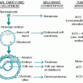
4. Germ cell tumors (see Chapter 12). Teratomas (or dermoids) represent 10% of mediastinal neoplasms. About 10% of these are malignant, usually with a predominant epithelioid component, but occasionally with sarcomatous or endodermal elements. Malignant germ cell tumors of the mediastinum are usually large and solid.
a. Benign (mature) teratoma accounts for about 70% of mediastinal germ cell tumors, especially in children and young adults. They appear as a round, dense mass (often with a calcified capsular shell and occasionally with teeth). They are usually small with multilocular cysts and asymptomatic, but they can attain a large size. The serum of a patient with benign teratoma contains no α-fetoprotein (AFP) or β-human chorionic
gonadotropin (β-hCG). These characteristics often differentiate benign teratoma from germ cell malignancy. The treatment is surgical excision.
gonadotropin (β-hCG). These characteristics often differentiate benign teratoma from germ cell malignancy. The treatment is surgical excision.
b. Seminoma comprises only 2% to 4% of mediastinal masses, but it is the most common malignant germ cell neoplasm of the mediastinum, occurs most frequently in men 20 to 40 years of age. The lesions are rarely calcified. Less than 10% of cases have an elevated β-hCG, and none has an elevated AFP. Treatment of mediastinal seminoma is surgical excision if the tumor is small, followed by irradiation of the mediastinum and the supraclavicular nodes. For locally advanced disease, combination chemotherapy (see Chapter 12, Section VI), followed by resection of residual disease, is preferred. The 5-year survival rate for these patients is >80%.
c. Mediastinal nonseminomatous germ cell tumors are malignant, aggressive, and usually symptomatic. The prognosis is worse than for patients with mediastinal seminoma. They are usually associated with elevations of serum levels of β-hCG, AFP, or lactate dehydrogenase (LDH). Choriocarcinoma in the mediastinum presents with gynecomastia and testicular atrophy in half of all male patients. Embryonal or yolk sac tumors of the mediastinum are highly aggressive cancers that are large and bulky at the time of diagnosis.
Stay updated, free articles. Join our Telegram channel

Full access? Get Clinical Tree


