(1)
Department of Surgery, Dar A lAlafia Medical Company, Qatif, Saudi Arabia
Sickle cell anemia is an inherited disease that existed in Africa for at least 5000 years but there have been no records of its existence till it was discovered in 1904.
It is interesting to note that the deer is the only living creature other than humans known to suffer from sickle cell disease.
In 1840, the British zoologist Gulliver observed under the microscope sickle-shaped red blood cells in the blood of a variety of exotic deer belonging to the London Zoological Garden.
In North America, the white-tailed deer is occasionally encountered dying in sickle cell vaso-occlusive crisis in the forests of Michigan.
The first description of sickle red blood cells in humans was published in 1899 when Dr. Hayem, a French colonial physician, reported his microscopic discovery from Africa. He erroneously explained that the crescent shape of these red blood cells represented a normal variation of erythrocyte morphology.
In 1905, two French colonial physicians, Dr. Sargent and Dr. Sargent, examined the blood of 243 natives of Algiers and published a report describing “half-moon corpuscles” in 5 % of their African patients. Since all these patients suffered from malaria, the authors incorrectly attributed the presence of the sickle erythrocytes to malaria itself.
The first report of the sickle red blood cells in North America was published in 1904 by Dr. Dresbach where in a histology class at Ohio State University, one mulatto student sketched ellyptoid erythrocytes which he observed under his own microscope.
The first clinical description of sickle cell anemia was recorded by James Bryan Herrick in 1910.
James Bryan Herrick was an American cardiologist and professor of medicine at the Chicago Presbyterian Hospital. He was also a major figure in the history of medical science and worked and taught at several hospitals in Chicago. He was born on August 11, 1861, in Oak Park, Illinois, and died in 1954.
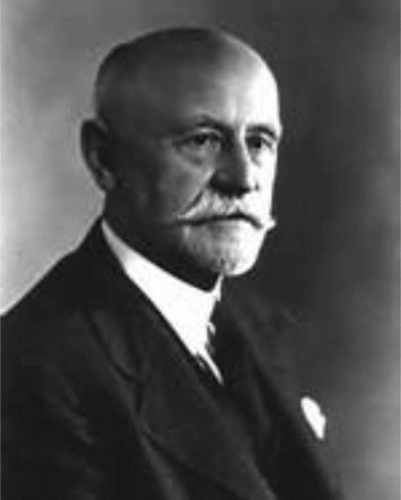
Professor James Bryan Herrick (1861–1954)
In December 1904, a patient named Walter Clement Noel, a 20-year-old first-year dental student from the island of Grenada, was admitted to the Chicago Presbyterian Hospital suffering from painful episodes and symptoms of anemia. Prior to this, Walter Clement Noel suffered from an ulcer on the ankle which was painful and healed with conservative treatment.
Mr. Walter Clement Noel belonged to an affluent African family which owned the Duquesne estate, a large plantation on the island of Grenada.
His father had died at the age of 36 years, reportedly of kidney failure, and his sister also died prematurely at the age of 24 years, presumably of pulmonary tuberculosis.
Mrs. Noel decided to send her 20-year-old son to study in the United States where he was the only African student accepted to the Chicago College of Dental Surgery.
He sailed to the United States in 1904 for the purpose of studying dentistry.
When Mr. Walter Clement Noel disembarked from the ocean liner SS Cearense in New York on September 5, 1904, he already was suffering from chronic ankle ulcers. This was treated by a physician who applied iodine to the skin ulcers, which healed within a week.
He then traveled by train to Chicago and enrolled at the Chicago College of Dental Surgery.
By Christmas time, he became ill with pneumonia or what today would be referred to as acute chest syndrome and presented himself to the Presbyterian Hospital of Chicago. He was also anemic and was examined by Dr. Ernest Irons, an intern who performed a complete blood count including microscopic examination of the red blood cells.
Mr. Walter Clement Noel appeared pale and his red blood cell count as documented on December 31, 1904, was only 2,880,000 cells per cubic milliliter.
By January 22, 1905, he had recovered completely from acute chest syndrome, a complication of sickle cell disease which even today is associated with significant mortality.
During his 3 years of study at the Chicago College of Dental Surgery, Mr. Walter Clement Noel would be treated on several other occasions by Dr. Ernest Irons and Dr. James Herrick for various clinical complications of his sickle cell anemia.
Walter Clement Noel was readmitted several times over the next 3 years for “muscular rheumatism” and “bilious attacks.”
He managed to survive all of his sickle cell crises, completed his studies, graduated from dental school and returned to the capital of Grenada (St. George’s) to practice dentistry in 1907.
Upon return to his native land of Grenada, Mr. Walter Clement Noel established a successful dental practice and remained in more or less satisfactory health for almost 10 years.
He must have suffered several life-threatening crises during his stay in St. George, Grenada. On the day of his death, he went to Grenville on the other side of the island in order to watch a horse race and returned the same day to the capital. He felt very tired, became ill with pneumonia, and died in 1916 at the age of 32 years from what looks like an acute chest syndrome (pneumonia). He was buried in the Catholic cemetery at Sauteurs in the north of Grenada next to his sister and father and their tombs are still intact today in Grenada.
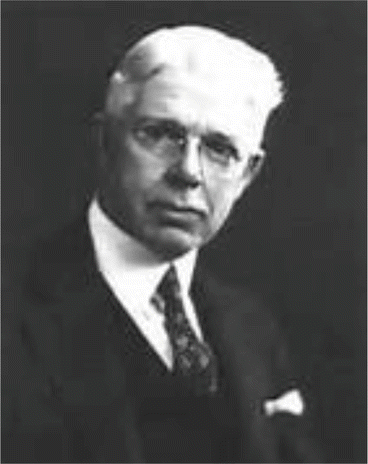
Dr. Ernest Edward Irons (1877–1959)
Professor Herrick had an intern named Ernest Edward Irons (1877–1959). Ernest Irons was looking at Noel’s blood film when he noticed the irregularity in the shape of the blood cells. He called them “peculiar, elongated, and sickle-shaped” red blood cells (Fig. 1.1).
On December 31, 1904, Irons drew a rough sketch of these erythrocytes, the first pictures of sickled cells (Fig. 1.1).
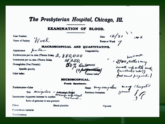
Fig. 1.1
Diagrammatic drawing of the blood film of Mr. Walter Clement Noel as was seen by Dr. Ernest Edward Irons. Note the detailed description of the blood film showing a pale patient with a total red blood cells number of 2,880,000 per cubic mm. The red blood cells appear very irregular and many elongated
In 1910, Dr. James Herrick presented the case of Mr. Walter Clement Noel to the Association of American Physicians in Washington DC and in the same year published a report in the Archives of Internal Medicine.
In 1910, Herrick published the findings with illustration and it was the first time the connection between the irregularities of the shape of red blood cells with sickle cell anemia was discovered and identified.
This was published in the November 1910 issue of “Archives of Internal Medicine,” when James B. Herrick of Chicago reported on “Peculiar Elongated and Sickle-Shaped Red Blood Corpuscles in a Case of Severe Anemia.”
This case is reported because of the unusual blood findings, no duplicate of which I have ever seen describe. (James B. Herrick, M.D.)
1910 is regarded as the year of the discovery of sickle cell anemia.
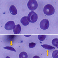
In the beginning and for some time, this condition was called Herrick’s syndrome (Herrick’s anemia) because of his contributions to the discovery of sickle cell anemia (Fig. 1.2).
It is interesting to note that Dr. James Herrick was also the first physician to describe the pathology of heart attack or myocardial infarction and again received no lasting recognition for it. He later became Dean of Rush Medical School and died in 1954.
Dr. Ernest Irons, the house officer who saw the sickle cells and sought no credit or recognition out of respect for his mentor, Dr. James Herrick, also had an illustrious career, becoming President of the American Medical Association in 1949.
Some elements of sickle cell anemia, however, had been recognized earlier than this but were not well documented. In 1846, a paper in the “Southern Journal of Medical Pharmacology” described the absence of a spleen in the autopsy of a runaway slave. The African medical literature also reported this condition in the 1870s, when it was known locally as “ogbanjes” (“children who come and go”) because of the very high infant mortality rate caused by this condition. A history of the condition tracked reports back to 1670 in one Ghanaian family. Also, the practice of using tar soap to cover blemishes caused by sickle cell sores was prevalent in the black community.
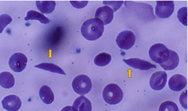
Fig. 1.2
A blood film showing sickle shaped red blood cells. Note also the normal looking red blood cells
In 1922, Verne Rheem Mason (Wapello, Iowa, August 8, 1889 – Miami, Florida, November 16, 1965) an eminent internist named the disease as “sickle cell anemia” based on the description of Ernest Irons. She received a B.S. from the University of California, Berkeley, in 1911, and an M.D. from Johns Hopkins University in 1915. As a medical resident at Johns Hopkins in 1922, Mason gave the disease the name of sickle cell anemia.
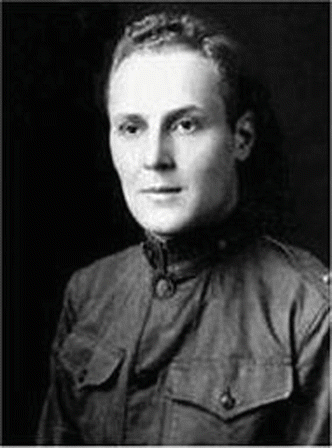
Dr. Verne Rheem Mason (1889–1965)
In 1927, Hahn and Gillespie discovered that red blood cells from persons with the disease could be made to sickle by removing oxygen. There were also people at that time, often relatives of the patients, whose red blood cells were sickling when deprived of oxygen but who had no symptoms. These became known as “sickle trait.” Hahn and Gillespie were the first to associate the red cell sickling to low oxygen and acidic conditions.
The first splenectomy for the treatment of sickle cell anemia was reported by Hahn and Gillespie in 1927.
In the late 1940s and early 1950s, the nature of the disease began to become clearer.
Stay updated, free articles. Join our Telegram channel

Full access? Get Clinical Tree


