(1)
Department of Surgery, Dar A lAlafia Medical Company, Qatif, Saudi Arabia
6.1 Introduction
SCA can affect any part of the body and poses diagnostic and therapeutic dilemmas to the treating physicians.
The hepatobiliary system is one of the common organs to be affected either directly from the sicklening process or indirectly as a result of chronic hemolysis and multiple blood transfusions.
This can manifest in several clinical conditions.
6.2 Cholelithiasis
Cholelithiasis is one of the common complications of SCA (Figs. 6.1 and 6.2).
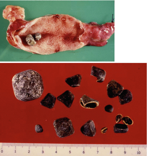
Figs. 6.1 and 6.2
Clinical photographs showing cholelithiasis
These are usually pigment stones that result from chronic hemolysis leading to increased bilirubin production (Fig. 6.3).
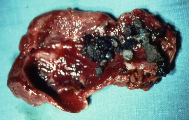
Fig. 6.3
A clinical photograph showing pigmented gallstones in a patient with SCA
The frequency of cholelithiasis in patients with SCA is however variable depending on the age of the patients and the diagnostic method used.
A frequency ranging from 5–55 % has been reported but an overall 70 % of patients with SCA will develop gallstones at one stage of their life.
With better understanding of SCA, improved medical and surgical management of these patients and the development of new drugs such as hydroxyurea have contributed to a better survival and increased life expectancy of these patients.
This will lead to an increased frequency of cholelithiasis in these patients as the frequency of gallstones in patients with SCA increases with age.
An overall of 19.7 % frequency of gallstones was reported in children with SCA. This frequency increased from 8.7 % in those less than 10 years of age to 36 % in those 15–18 years of age.
6.3 Presentation
The presentation of cholelithiasis in patients with SCA is variable.
The classic presentation is that with biliary colic, right upper quadrant abdominal pain and discomfort, and dyspepsia or upper abdominal pain and discomfort.
A small number of these patients present acutely with acute cholecystitis (Figs. 6.4 and 6.5).
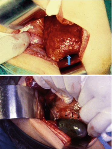
Figs. 6.4 and 6.5
A clinical intraoperative photograph showing acute cholecystitis and acute gangrenous cholecystitis in two patients with SCA
The recent liberal use of abdominal ultrasound as a diagnostic investigation resulted in the discovery of a large number of these patients with asymptomatic gallstones.
6.4 Diagnosis
Abdominal ultrasound is the main diagnostic investigation to diagnose gallstones (Figs. 6.6 and 6.7).
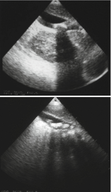
Figs. 6.6 and 6.7
Abdominal ultrasound showing gallstones in patients with SCA
This is a simple, relatively cheap and noninvasive investigation which can accurately diagnose gallstones. Abdominal ultrasound can easily be repeated.
The stones can be single or multiple (Fig. 6.8).
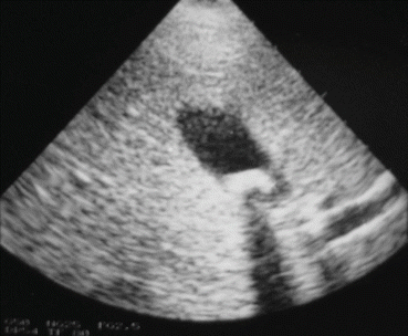
Fig. 6.8
Abdominal ultrasound showing a single gallstone in a patient with SCA
Plain abdominal X-ray may also show these gallstones (Figs. 6.9 and 6.10).
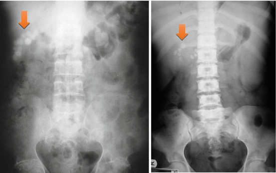
Figs. 6.9 and 6.10
Abdominal X-rays showing multiple gallstones
Abdominal CT scan is rarely used to diagnose gallstones but can be used to diagnose gallstone-related complications such as common bile duct obstruction, live abscess, and pancreatitis.
Endoscopic retrograde cholangiopancreatography (ERCP) is a valuable investigation to diagnose common bile duct stones and delineate the anatomy of the biliary system (Figs. 6.11 and 6.12).
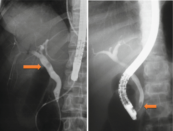
Figs. 6.11 and 6.12
ERCP showing common bile duct stones
It is also valuable as a therapeutic modality to treat common bile duct stones.
6.5 Treatment
The treatment of cholelithiasis in patients with SCA is cholecystectomy.
In the era of minimal invasive surgery, laparoscopic cholecystectomy is now the procedure of choice. This is specially so for patients with SCA where it was shown to be feasible, beneficial, and safe.
The fact that there is no large abdominal wound, laparoscopic cholecystectomy should be advantageous to patients with SCA. Not only is the hospital stay short and cosmetically better but the associated postoperative pain is much less. This makes their early mobilization much easier and will not interfere with their breathing which may prove beneficial in reducing the incidence of postoperative acute chest syndrome.
The treatment of asymptomatic gallstones in patients with SCA is still controversial.
In the past and because of a reported perioperative mortality rate as high as 10 % and a postoperative complication rate up to 50 %, surgery in patients with SCA was not advocated except in symptomatic patients.
This however is not the case nowadays, as with better understanding of SCA and improved perioperative care, surgery is safer.
Many surgeons now advocate cholecystectomy for asymptomatic gallstones in patients with SCA.
The life expectancy of patients with SCA is known to be shorter than the general population and they usually die of SCA-related complications, but as a result of better understanding of SCA and its complications, the use of hydroxyurea, as well as improved medical and surgical care, SCA patients are now living longer. This makes them liable to develop gallstone-related complications including:
Biliary colic
Acute cholecystitis (Figs. 6.13, 6.14 and 6.15)
Choledocholithiasis (Fig. 6.16)
Obstructive jaundice (Fig. 6.17)
Ascending cholangitis
Pancreatitis
Operating on these patients on an emergency basis not fully prepared is definitely associated with a high morbidity and mortality (Fig. 6.2). It is much better to operate on these patients electively when they are well prepared.
Add to this the fact that cholecystectomy should simplify the future management of abdominal crisis as the possibility of acute cholecystitis is eliminated.
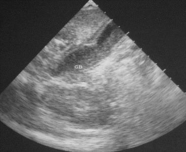
Fig. 6.13
An abdominal ultrasound showing acute cholecystitis with biliary sludge. Note the thickened wall of the gallbladder
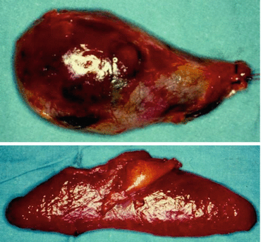
Figs. 6.14 and 6.15
Clinical photographs showing acute cholecystitis in already excised gallbladders
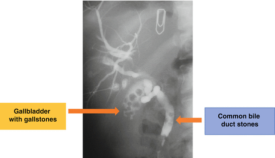
Fig. 6.16
Percutaneous transhepatic cholangiogram showing gallstones and common bile duct stones
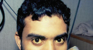
Fig. 6.17
A clinical photograph of a patient with SCA who presented with obstructive jaundice
Complications of Cholelithiasis
Biliary colic and dyspepsia
Acute and chronic cholecystitis
Choledocholithiasis
Obstructive jaundice
Ascending cholangitis
Acute pancreatitis
It has been estimated that about 70 % of patients with SCA will develop gallstones at one stage of their life. This is a high frequency and a question that is being raised nowadays is whether this justifies incidental cholecystectomy for SCA patients who are having abdominal surgery electively for some other reason such as splenectomy or hiatal hernia repair. This is still controversial and most surgeons are not supportive of this.
6.6 Choledocholithiasis
Another relatively common complication of SCA is the development of choledocholithiasis which is usually secondary to cholelithiasis but there are also primary choledocholithiasis (Figs. 6.18, 6.19, 6.20, 6.21, 6.22 and 6.23).
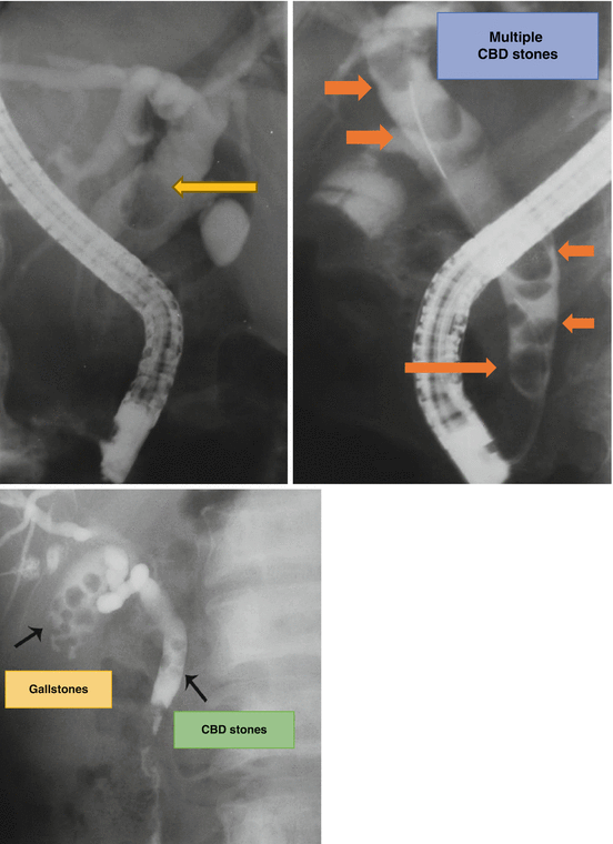
Figs. 6.18, 6.19, and 6.20
ERCP, percutaneous transhepatic cholangiogram (PTC) and ERCP showing single CBD stone, gallstones, and multiple CBD stones
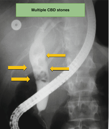
Fig. 6.21
ERCP showing multiple common bile duct stones
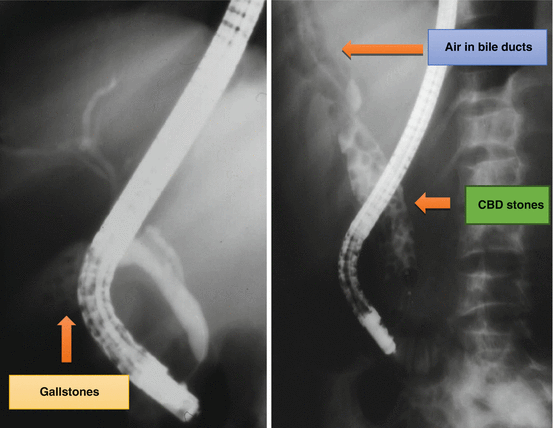
Figs. 6.22 and 6.23
ERCP showing multiple gallstones without common bile duct stones and ERCP showing common bile duct stones complicated by a choledochoduodenal fistula. Note the air in the biliary tree
In the general population with cholelithiasis, the incidence of common bile duct stones has been reported as 10–15 %. In patients with SCA, the frequency of common bile duct stones is 18–30 %.
As a result of this high incidence of choledocholithiasis, routine intraoperative cholangiogram was advocated for those undergoing cholecystectomy (Fig. 6.24).
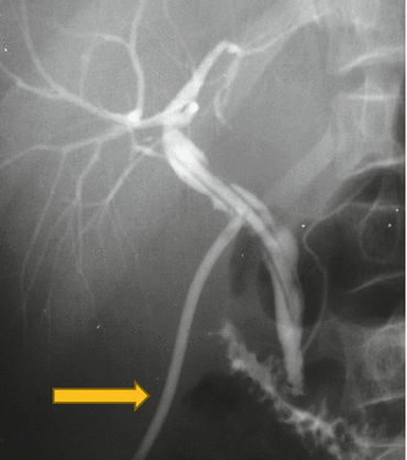
Fig. 6.24
T-tube cholangiogram following intraoperative cholangiogram and CBD exploration for CBD stones. Note the patent biliary ducts and T-tube for temporary drainage of common bile duct
In the era of laparoscopy and endoscopic retrograde cholangiopancreatography (ERCP), the question is whether laparoscopic cholangiography with or without common bile duct exploration is necessary in patients with SCA.
Laparoscopic cholangiography will increase the operative time and makes it difficult to decide when CBD stones are diagnosed whether to convert the operation to open cholecystectomy or wait and do a post-laparoscopic cholecystectomy ERCP. Added to this is a 25 % false-positive rate that may lead to unnecessary CBD exploration or conversion to open cholecystectomy.
Laparoscopic cholangiography and CBD exploration and extraction of CBD stones is feasible and safe in experienced hands.
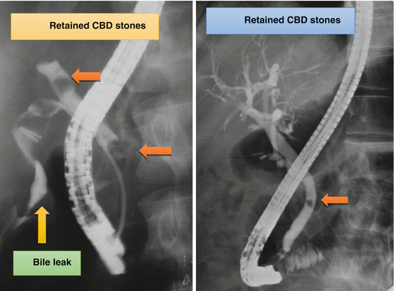
ERCP however is valuable both as a diagnostic and therapeutic procedure for SCA patients with choledocholithiasis both pre- and post-laparoscopic cholecystectomy. This sequential approach of endoscopic sphincterotomy and stone extraction followed by laparoscopic cholecystectomy is a safe and effective approach for the management of SCA patients with cholelithiasis and choledocholithiasis (Figs. 6.25, 6.26, 6.27 and 6.28).
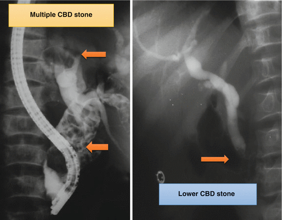
Figs. 6.25 and 6.26
ERCP showing multiple bile duct stones in the first one and a single CBD stone following laparoscopic cholecystectomy in the second one

