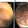
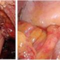
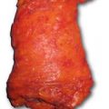
Fig. 17.1
Photomicrograph showing low anterior resection specimen with total mesorectal excision (TME) . a shows posterior surface with good bulk of mesorectum (indicated with an arrow) with smooth surface and small superficial defect (indicated with an arrowhead), indicating a good TME. b shows anterior aspect of the specimen (indicated with an arrow) and highest vascular pedicle (indicated with dark arrow)
Macroscopic Examination of Fixed Specimen
Tumor bed configuration and tumor boundaries are better observed after formalin fixation. Sectioning of the tumor with surrounding tumor is easy and does not produce disruptions or tissue curling after formalin fixation. Tumor dimensions, presence and dimension of other lesions, and status of nonneoplastic colorectal wall and adjoining organ should be assessed in the fixed state.
Sampling for Microscopic Examination
It is recommended that entire tumor bed or scar should be submitted for microscopic examination. In specimens with distinct grossly identifiable tumor, majority of gross tumor should be sampled for microscopic examination. There is more than one approach for sampling tumor for optimal assessment of residual tumor and status and distance of tumor from radial distal and other margins.
The approach of macroscopic examination being followed by majority of the pathology laboratories includes inking, examining, and opening the specimen in the fresh state followed by fixation and sampling in the fixed state. Tumor and adjacent nonneoplastic rectum are submitted by longitudinal or transverse sectioning of tumor to include tumor, rectal wall, mesorectum, and adherent organs. The tumor is serially sectioned in multiple slices, and each slice is divided into multiple sections to accommodate the tissue in a traditional paraffin block . A 5-micron thick section is procured and stained with H&E from each block. This sampling approach identifies discontinuous foci of tumor cells in tumor bed for appropriate pathologic tumor stage. In addition, this method is helpful in accurately measuring distance between tumor and radial or adherent organ margin under a microscope and status of peritumoral lymph nodes . In addition, due to each section derived from the paraffin block that has tissue thickness of <5 mm, the amount of tumor available for microscopic assessment is higher in the H&E-stained sections as compared to having >5 mm thick sections.
Approach for sampling distal margin depends upon the distance between tumor/scar and distal margin . When the distance between the distal edge of the tumor/scar and distal margin is ≤2 cm, perpendicular sections of tumor and corresponding margin should be submitted so that exact distance between tumor cells and/or acellular mucin and distal margin can be measured under the microscope. For specimens with broad distal margin, it is acceptable to submit part of distal margin which is in line with and closest to the tumor/scar in perpendicular sections and peripheral part of the margin as shaved sections with an en face margin. Highest vascular pedicle and proximal colonic margins should be submitted separately as shaved section for en face margins.
Major challenge for pathologic rectal cancer resection specimen evaluation of after neoadjuvant chemoradiation is finding adequate number of lymph nodes. Although processing by chemicals like Bouin’s solution has shown to increase yield of lymph nodes, there is no alternative to meticulous dissection of mesorectal and other soft tissue to identify as many lymph nodes as possible. Not infrequently, the lymph node loses the rounded contour and soft consistency due to radiation, so nodular firm area should be felt and visualized and if suspicious should be sampled. Any lymph node which grossly appears positive should be sampled in its entirety because not infrequently these lymph nodes show reaction to the therapy and/or acellular mucin and not tumor cells. Due to independent prognostic value, inferior mesenteric artery lymph nodes should be identified in advance and sampled separately [5]. It is also essential to make sure that lymph nodes suspected to be positive in the preoperative scans are sampled for microscopic assessment. A review of the preoperative scan by the pathologist is helpful in identifying need of search for specific positive lymph nodes.
Sampling of additional focal lesions such as polyps, ulcers, and diverticula along with random section of nonneoplastic rectum is routinely performed as for any other colorectal resection specimen. Table 17.1 lists the essential components to be included in the gross/macroscopic description of rectal resection specimens .
Table 17.1
Macroscopic features required in a pathology report
Specimen type | Low anterior resection, abdominoperineal resection, reresection of locally recurrent tumor |
Specimen dimension | Length × diameter in cm |
Location/epicenter of tumor | Rectosigmoid, upper/mid and distal rectum, anterior/posterior/lateral wall |
Tumor | Length × width in cm |
Tumor configuration | Ulcerated, exophytic, annular, no definite tumor; scar or ulcer only |
Tumor distance from margins | Proximal, distal, radial, other soft tissue, adherent organ margins |
Macroscopic depth of tumor invasion | Muscularis propria, perimuscular soft tissue, infiltrating the serosa/radial margin, invading into adherent organs |
Other lesions and their distance from primary tumor and margins | Polyps, ulcer, diverticula |
Grossly positive lymph nodes or soft tissue tumor deposits | Distance from primary tumor, radial margin, vascular pedicle margin |
Highest vascular pedicle | Lymph node involvement Possible margin |
Inferior mesenteric artery lymph nodes | Lymph node involvement |
Section codes | Tumor, margins, lymph nodes, other lesions |
A different approach of sampling originally described by Quirke et al. [6] includes whole mount processing of the slices of the tumor and adjoining mesorectal and other soft tissue. In this method, the tumor is serially sliced in a transverse plane into 5–10 mm thick slices. The slice with maximal lateral spread of tumor is identified as “primary slice” and is blocked into multiple paraffin blocks with maintenance of orientation. A 5-micron thick section is procured and stained with H&E from each paraffin block. Remaining slices with tumor and mesorectal soft tissue are obtained, and the slices are whole mounted on paraffin blocks, and a 10-micron thick section is procured by sledge microtome followed by H&E staining . The strength of this approach is ability to consistently measure the distance between deepest extent of the tumor to radial margin and tumor extension beyond muscular layer and correlating the extent of the residual tumor with preoperative imaging findings. The limitations include training of the pathologist grossing these specimens and more demand on histology laboratory due to whole mount of the sections and less sampling of the tumor bed in the vicinity of the tumor epicenter as compared to the method described above, due to the fact that the amount of tissue remaining in the FFPE block and not assessed under microscope is higher than what is described in the first approach.
Histopathologic Assessment and Pathologic Parameters of Response to Neoadjuvant Therapy
Optimal histopathologic examination includes all the histopathology findings which are assessed in any adenocarcinoma of colon or rectal resection specimen including pathologic tumor stage, nodal stage, distant metastases, presence (or absence) of lymphovascular and perineural invasion, and distance of tumor to the margins. In addition, histologic regression of the tumor characterized by reduction of tumor cells and replacement of the tumor bed by fibrosis with or without necrosis, granulation tissue, and inflammation is one of the essential parameters required to be reported in the pathology report.
Pathologic Tumor Stage (ypT)
More than one study have shown that downstaging of tumor stage (ypT < cT) is an independent prognostic factor of disease-free survival in node-positive and node-negative rectal adenocarcinoma [7]. Pathologic T stage is decided based on the depth of invasion of tumor in rectal wall and/or adherent organs. Only difference as compared to the specimens without neoadjuvant therapy is that presence of tumor cells is required in a layer of rectal wall or adjacent organ for appropriate ypT stage designation. Presence of acellular mucin, necrosis, or fibrosis without tumor cells is not considered to be a histologic evidence of tumor for ypT . Radiation-induced injury also includes thinning of the colon wall with partial destruction of submucosa and/or muscularis propria which can lead to an understaging of ypT3 to ypT1 or ypT2.
Pathologic Nodal Stage (ypN)
Pathologic nodal stage is based on the number of lymph nodes involved by the tumor, similar to the specimens without neoadjuvant therapy. Presence of tumor cells is required in a lymph node to identify lymph node as positive for metastatic carcinoma. However, it is important to identify and report the lymph nodes which show features of preoperative therapy like necrosis, fibrosis, calcification, and acellular mucin. These findings help in identifying any lymph node which would have been reported as positive on preoperative imaging and explain cN+ disease. Location of positive lymph node is independently important if it is present at the margin of highest vascular pedicle (R1 resection ) or identified as separate (e.g., inferior mesenteric artery) lymph nodes by the operating surgeon.
Stay updated, free articles. Join our Telegram channel

Full access? Get Clinical Tree


