Hematopoietic Growth Factors


Hematopoietic Growth Factors


Gary H. Lyman

Nicole M. Kuderer


The hematopoietic growth factors (HGFs) are an important class of biologic agents for the support of cancer patients receiving myelosuppressive chemotherapy. They augment the production and functional maturation of hematopoietic cells to reduce hematologic complications while enabling the safe delivery of effective treatment. This chapter focuses on HGFs with known clinical importance for hematopoiesis in the patient with cancer. Myelosuppression and its sequelae represent the most common dose-limiting complications of cancer chemotherapy and are associated with considerable morbidity, mortality, and costs. In addition to direct chemotherapyassociated complications such as neutropenia, anemia, and thrombocytopenia, myelosuppression often results in chemotherapy dose reductions and delays, reducing delivered chemotherapy dose intensity and potentially compromising disease control and long-term survival in patients with responsive and potentially curable malignancies.


Biology and Pharmacology of the Hematopoietic Growth Factors

Endogenous production of HGFs occurs in a wide variety of hematopoietic and nonhematopoietic cells (Table 38-1). Figure 38-1 depicts the hematopoietic lineages derived from the myeloid stem cell and influenced by the various HGFs.


The Myeloid Growth Factors

The term colony-stimulating factor (CSF) relates to early observations that certain glycoproteins were capable of supporting the formation of colonies of hematopoietic elements when bone marrow cells were cultured.1,2 The genes encoding granulocyte colony-stimulating factor (G-CSF), granulocyte-macrophage-colony-stimulating factor (GM-CSF), and interleukin-3 (IL-3) were cloned and produced in large amounts using recombinant DNA technology.3 The currently approved myeloid growth factors for treatment of neutropenia are recombinant human (rhu) G-CSF (filgrastim, lenograstim), rhu GM-CSF (sargramostim, molgramostim, regramostim), and pegylated rhu G-CSF (pegylated filgrastim). Table 38-2 summarizes some of the pharmacokinetic characteristics of the CSFs.


Granulocyte Colony-Stimulating Factor

G-CSF was first identified in 1980 in the serum of mice with an ability to induce granulocyte differentiation of a myelomonocytic leukemia cell line.4,5 G-CSF is an 18.8 kDa glycoprotein with 174 amino acids encoded by a single gene on chromosome 17 (q21-22). G-CSF is produced endogenously by monocytes, macrophages, endothelial cells, fibroblasts, mesenchymal cells, and bone marrow stromal cells. G-CSF is usually absent or present in low concentration but is highly stimulated by inflammatory cytokines and bacterial cell wall products.6,7 The G-CSF receptor is mainly expressed on mature neutrophils and their precursors. It is composed of 813 amino acids and is encoded by a gene located on chromosome 1 (p32-34). The receptor consists of both a cytokine-specific binding subunit and signal-transducing subunit. While lacking intrinsic kinase activity, cytokine binding of G-CSF to its receptor leads to dimerization bringing together signaling proteins associated with the cytoplasmic domains of the receptor. The biologic action of the cytokine is orchestrated by a number of signaling pathways, the most important of which is the Janus kinase-signal transducers and activators of transcription (JAK-STAT) signaling pathway. Activated phosphorylated STAT proteins dissociate from the receptor and translocate to the nucleus where they activate transcription. Other important signaling pathways include ras-mitogen activated protein (MAP) kinases that are necessary for G-CSF-directed cellular proliferation.

G-CSF is necessary to sustain normal neutrophil production and to increase neutrophil numbers in response to stress. Endogenous G-CSF levels rise in neutropenic states and most dramatically with febrile neutropenia (FN). G-CSF acts on late myeloid progenitors leading to increased cell division and reduced transit time through the bone marrow to the peripheral blood and tissues. G-CSF also induces the differentiation and functional maturation of neutrophils increasing chemotaxis, phagocytosis, and antibody-dependent cellular toxicity. G-CSF knockout mice develop chronic neutropenia.6,7 The CSFs are metabolized by binding to the receptor with subsequent internalization and by hepatic enzymes. Unpegylated CSFs are cleared by glomerular filtration and excreted in urine.


Pegylated G-CSF (Pegfilgrastim)

Pegfilgrastim is a recombinant G-CSF that has been bioengineered by covalently binding a 20-kDa polyethylene glycol molecule to the N terminus of filgrastim, increasing the total molecular weight from 18,800 to 39,000 kDa.8 The pegfilgrastim molecule is too large for renal clearance prolonging the half-life while retaining the biological activity of filgrastim leading to an increase in cell division, a shorter transit through the bone marrow, and increasing differentiation
and functional activation of neutrophils. The half-life of pegfilgrastim is approximately 33 hours. Pegfilgrastim has saturable and self-regulating neutrophil-mediated elimination where neutrophil production is stimulated when neutrophil counts are low and rapid clearance of the agent occurs as neutrophil counts recover providing fresh receptors for binding. G-CSF receptor knockout mice exhibit a significantly slower clearance, longer half-life, and greater area under the curve (AUC) than wild-type mice for both filgrastim and pegfilgrastim.9 Similar concentration-time profiles of pegfilgrastim are observed in nephrectomized rats while nephrectomy results in a 60% to 70% decreased clearance of filgrastim.10 In clinical observations, serum clearance of pegfilgrastim decreased with increasing dose, consistent with neutrophil-mediated elimination while the elimination of pegfilgrastim is prolonged after chemotherapy, when the myeloid mass was reduced11 (Fig. 38-2). The apparent neutrophil-dependent clearance of pegfilgrastim results in prolonged circulation and action during recovery from neutropenia. Pegfilgrastim, like filgrastim, binds to cell surface G-CSF receptors and increases differentiation and functional activation of neutrophils.12


TABLE 38.1 Pharmaceutical characteristics of hematopoietic growth factors


Cytokine


Other names


Generic names


Expression vector


Brand names


No. of amino acids


Human chromosome location


Normal endogenous sources


G-CSF


Granulocyte colony-stimulating factor


Filgrastim
Pegfilgrastim
Lenograstim


E. coli
E. coli
CHO


Neupogen (Amgen)
Neulasta (Amgen)
Granocyte (Chugai)


174


17


Monocytes/macrophages, fibroblasts, endothelial cells, keratinocytes


GM-CSF


Granulocyte-macrophage colony-stimulating factor


Sargramostim
Molgramostim
Regramostim


Yeast
E. coli
CHO


Leukine (Immunex)
Leucomax (Schering)


127


5


T lymphocytes, monocytes/macrophages, fibroblasts, endothelial cells, osteoblasts, epithelial cells


EPO


Erythropoietin


Epoetin-α


CHO


Epogen (Amgen)


165


7


Renal cells, hepatocytes


Epoetin-α


CHO


Procrit/Eprex (Ortho)


Epoetin-β


CHO


NeoRecormon (Roche)


Darbepoetin-α


CHO


Aranesp (Amgen)


IL-11


Interleukin-11


Oprelvekin


E. coli


Neumega (Genetics Institute)


178


19


Stromal fibroblasts, trophoblasts


SCF


Stem cell factor, steel factor, mast cell growth factor, c-kit ligand


Ancestim


E. coli


Stemgen (Amgen)


165


12


Endothelial cells, fibroblasts, circulating mononuclears, bone marrow stromal cells


TPO


Thrombopoietin, megakaryocyte growth and development factor


332


3


Liver, kidney


CHO, Chinese hamster ovary cells; CSF, colony-stimulating factor.


Granulocyte-Macrophage Colony-Stimulating Factor

GM-CSF was first identified in 1977 with human GM-CSF purified in 1984.13,14 GM-CSF is a 14- to 35-kDa glycoprotein encoded by a gene located on chromosome 5 (q23-31) close to a cluster of hematopoietic regulatory genes. GM-CSF is produced
by monocytes, macrophages, fibroblasts, endothelial cells, and conditioned lymphocytes.6,7 GM-CSF gene knockout mice exhibit normal hematopoiesis suggesting that it has minimal role in leukocytosis in the steady state.6,7 The GM-CSF receptor is expressed on neutrophils, monocytes, eosinophils, myeloid progenitors, myeloid leukemia cells, T lymphocytes, and dendritic cells. The GM-CSF receptor is composed of two subunits: an a subunit that is GM-CSF specific encoded on chromosome X/Y and a β subunit shared by other cytokines encoded on chromosome 22q31. Binding of GMCSF to its receptor leads to downstream signaling mainly through the JAK-STAT pathway but also through mitogen-activated protein kinase (MAPK).6,7 GM-CSF stimulates the functional activity of
neutrophils, macrophages, monocytes, and eosinophils enhancing phagocytosis. Recombinant GM-CSF is available as sargramostim (yeast derived) and molgramostim (Escherichia coli derived) and is approved by the US Food and Drug Administration (FDA) for use in acute myeloid leukemia (AML), autologous and allogeneic hematopoietic stem cell transplantation (SCT), and stem cell mobilization. It is used experimentally as a vaccine adjuvant.


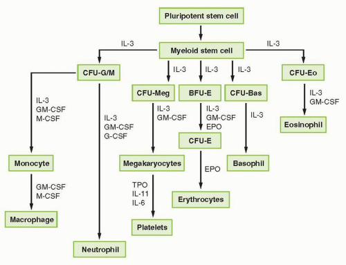
FIGURE 38-1 Representation of myeloid hematopoietic differentiation. Cytokines capable of stimulating specific cells are listed below such cells. See Table 38-2 for other names of cytokines shown here. BFU-E, burst-forming unit, erythroid; CFU-GEMM, colony-forming unit-granulocyte-erythrocyte-megakaryocyte macrophage; CFU-GM, colony-forming unit-granulocyte-macrophage; EPO, erythropoietin; G-CSF, granulocyte colony-stimulating factor; GM-CSF, granulocyte-macrophage colony-stimulating factor; IL, interleukin; M-CSF, macrophage colony-stimulating factor; SCF, stem cell factor; TPO, thrombopoietin.


TABLE 38.2 Pharma cokinetic studies of hematopoietic growth factors


CSF


Route


N


Half-life (h)


Tmax(h)


Cl (mL/min/kg)


G-CSF


SQ


37


2.5-5.8


4-8


19-56


Peg G-CSF


SQ


10


27-47


72-120


0.04-0.68


G-CSF


IV


58


(α: 8a; β: 1.8) 1.3-5.1


NA


4-21


GM-CSF


SQ


55


1.6-5.8


2.7-20


249-312


GM-CSF


IV


63


(α: 5-20a; β: 1.1-2.5) 1.1-2.4


NA


9.9-178


EPO


SQ


125


9-38


12-28


N/A


EPO


IV


135


4-11.2


NA


2.8-6.7


DARBO


SQ


14


33-49


54-86


0.062


DARBO


IV


28


18-25


NA


0.027-0.033


ELTROMOPAG


PO


73


9-12


NA


NA


ROMIPLASTIM


SQ


4


56


24


N/A


IL-11


SQ


18


6.9


3.2


NAb


a Values are in minutes.

b Clearance of IL-11 in infants and children is 1.2- to 1.6-fold higher than in adults or adolescents.


Cl, systemic clearance (values are “apparent“ for SQ route); CSF, colony-stimulating factor; DARBO, darbepoetin alpha; EPO, erythropoietin; G-CSF, granulocyte colony-stimulating factor; GM-CSF, granulocyte-macrophage colony-stimulating factor; IL, interleukin; N, number of patients; NA, not applicable; Peg, pegylated;


Tmax, time of maximal concentration after SQ injection.


Data presented are ranges of mean values in the reviewed studies.38,152, 153, 154


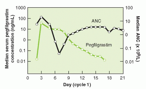
FIGURE 38-2 Pegfilgrastim serum concentrations and absolute neutrophil count (ANC) in patients with breast cancer who received pegfilgrastim as an adjunct to chemotherapy. (Adapted from Green MD, Koelbl H, Baselga J, et al. A randomized double-blind multicenter phase III study of fixed-dose single-administration pegfilgrastim versus daily filgrastim in patients receiving myelosuppressive chemotherapy. Ann Oncol 2003;14:29-35.)

GM-CSF augments the survival and proliferation of cells in the granulocytic and macrophage lineages as well as maintains megakaryocyte progenitors at high concentrations. Increases in granulocyte life span and metabolic functional activity have been noted in vitro. Other alterations of cellular function by GM-CSF include inhibition of neutrophil migration to sterile inflammatory fields.15 GM-CSF is a potent stimulator (in vitro and in vivo) of dendritic cells, which are important initiators of primary immune responses.16 Fever and fluid retention are frequently reported in patients receiving GM-CSF particularly when prepared in E. coli.


The Erythropoiesis-Stimulating Agents

The glycoprotein hormone, erythropoietin (EPO), is the primary regulator of red cell production. The erythropoiesis-stimulating agents (ESAs) available in the United States include epoetin alfa and the hyperglycosylated recombinant EPO, darbepoetin alfa. EPO illustrates how glycosylation can alter the pharmacologic properties of a molecule. Glycoproteins require terminal sialic acid residues on the oligosaccharides to protect against proteolytic attack.17 Darbepoetin results from mutagenesis of the gene encoding ESA adding two N-glycosylation sites yielding a 23% increase in molecular weight and a threefold increase in circulation time through protection from metabolic degradation.18,19 The therapeutic effects of the ESAs include induction, proliferation, and differentiation of erythroid progenitors. The primary effects of EPO are on the erythroid lineage; however, it may also play a role in the stimulation of early multipotent progenitors.20

Multiple factors may account for anemia in patients with cancer including disease stage, radiation and chemotherapy, and renal toxicity since endogenous EPO is mainly produced in the peritubular interstitial cells and regulated by an oxygen sensor. Inappropriately low as well as high EPO concentrations have been reported following chemotherapy due in part to paradoxical elevations of endogenous EPO concentrations immediately following chemotherapy.21, 22, 23 Since the efficacy of EPO is dependent on adequate iron stores, treatment with intravenous iron has been shown to improve the hemoglobin response in cancer patients treated with recombinant EPO.24


The Thrombopoietic Agents

The hematopoietic stem cell gives rise to the early common myeloid progenitor, which then leads to the megakaryocyte-erythroid (MK) progenitor that can then lead to either erythroid or megakaryocyte progenitors. Two to five thousand platelets are released from each mature megakaryocyte regulated by various cytokines working in concert. Stem cell factor or c-kit ligand and IL-3 act at early stages and stimulate proliferation and differentiation of progenitor cells into the MK lineage. Thrombopoietin (TPO) has broad activity, stimulating growth and maturation of MK progenitor cells into mature megakaryocytes. In a normal state, 1011 platelets are produced daily, with platelets lasting about 8 to 9 days in the circulation.25

IL-11 has been shown to synergistically act with early- and later-acting cytokines in various stages of hematopoiesis including megakaryopoiesis, perhaps interacting at a later stage than TPO. Preclinical and in vitro studies indicate that IL-11 directly stimulates megakaryocytes. Oprelvekin (recombinant IL-11) was the first cytokine to reach the market for the prevention of chemotherapy-induced thrombocytopenia. Early clinical trials in patients with cancer demonstrated an ability to increase steady-state platelet counts and to reduce the risk of chemotherapy-induced thrombocytopenia.26,27

TPO was initially identified in 1994 and is the primary regulator of thrombopoiesis while also stimulating platelet adhesion and aggregation.28, 29, 30 TPO is a 332-amino acid glycoprotein with an amino domain essential for thrombopoietic activity and a carboxy domain that increases the half-life. TPO is produced primarily in the liver, and its effects are mediated through the TPO receptor on megakaryocytes and platelets with TPO levels regulated primarily by the amount of receptors available for binding.31 The unbound TPO regulates megakaryocytopoiesis. TPO levels are high in cases of thrombocytopenia due to decreased production while the levels are not sufficiently elevated with increased destruction such as in immune thrombocytopenic purpura (ITP) probably due to the high turnover of TPO with the platelets and their c-Mpl receptors.25 Recombinant human TPO is identical to endogenous TPO and increases platelet counts in a dose-dependent fashion.32 While no neutralizing antibodies have been identified, serial bone marrow biopsies from patients treated with TPO demonstrated hypercellularity, megakaryocytic hyperplasia, and reticulin fibrosis, which resolved within 3 months after rhTPO was stopped.28 Pegylated recombinant human megakaryocyte growth and development factor (PEG-rHuMGDF) consists of the receptor-binding domain of TPO bound to a polyethylene glycol moiety.33 Although early trials showed improvement in the time to platelet recovery and decreased need for platelet transfusions in cancer patients undergoing high-dose chemotherapy, thrombocytopenia associated with cross-reacting
antibody formation that neutralized physiologic TPO occurred in 13 of 325 healthy volunteers and 4 of 650 oncology patients receiving as few as two doses of PEG-rHuMGDF.34 Therefore, alternative TPO agonists have been sought, lacking any sequence homology with endogenous TPO. These new agents include TPO peptide mimetics, nonpeptide mimetics, and agonist antibodies.35

Romiplostim was the first TPO receptor agonist to receive regulatory approval by the US FDA for treatment of thrombocytopenia in patients with chronic ITP poorly responsive to glucocorticoids, immunoglobulins, or splenectomy. The peptide mimetic, romiplostim, is a recombinant fusion protein with two identical subunits consisting of a peptide with two TPO-binding domains covalently bound to the Fc domain of a human IgG molecule.36 The rationale for the use of TPO agonists in ITP is based on the observation that serum TPO levels are inappropriately normal or low in the majority of patients with an apparent defect in platelet production due to immune destruction of platelet precursors.37 Alternatively, eltrombopag is a small molecule biarylhydrazone representing a selective nonpeptide agonist of the TPO receptor that results in receptor phosphorylation and activation of cytoplasmic tyrosine kinases and signal transducers of transcription. Eltrombopag promotes proliferation and differentiation of marrow stem cells into committed megakaryocyte precursors in a dose-dependent fashion. Preclinical data demonstrated that this agent has good oral bioavailability with consistent increases in platelet counts following daily oral administration.38


Clinical Application of the Hematopoietic Growth Factors in Oncology


Granulocyte Colony-Stimulating Factor


Solid Tumor and Lymphoma

The CSFs are the only biological agents used in clinical practice to reduce the risk of neutropenic complications and to maintain chemotherapy dose intensity.39 Primary prophylaxis with G-CSF starting within 3 to 5 days of the initial cycle of chemotherapy is based on evidence that the risk of neutropenic complications including FN is greatest during the first cycle of chemotherapy.40, 41, 42 Multiple randomized controlled trials (RCTs) of primary prophylaxis with G-CSF have been reported in a variety of malignancies and treatment regimens.43 Filgrastim is approved by the US FDA to decrease the incidence of infection, as manifested by FN, in patients with nonmyeloid malignancies receiving myelosuppressive anticancer drugs associated with a significant incidence (>20%) of FN.


Pegfilgrastim

FDA approval of pegfilgrastim was based on two phase III RCTs using filgrastim as an active control due to rates of myelosuppression of 40% associated with the chemotherapy regimen utilized without G-CSF support.44 While otherwise identical, the pegfilgrastim dose was weight based at 100 μg/kg in one trial45 and fixed dose at 6 mg in the other46 (Fig. 38-3). Patients received either a single injection of pegfilgrastim starting 24 hours after chemotherapy followed by daily placebo or daily filgrastim 5 μg/kg until neutrophil recovery or a maximum of 14 days. While equivalent for the duration of severe neutropenia, the incidence of FN was lower in patients who received pegfilgrastim. Combined treatment effect from both studies demonstrated additional relative risk reduction for FN with pegfilgrastim of 44% (95% confidence interval [CI]: 11% to 65%) (P = 0.015).8 The largest RCT of G-CSF reported to date compared pegfilgrastim to placebo in 928 women with breast cancer receiving docetaxel 100 mg/m2 every 3 weeks for four cycles.47 Patients receiving pegfilgrastim experienced a lower incidence of FN (1% versus 17%; P <0.001), FN-related hospitalization (1% versus 14%, P <0.001), and IV antibiotics (2% versus 10%; P <0.001) than placebo control subjects. Pegfilgrastim is approved by the US FDA to decrease the incidence of infection, as manifested by FN, in patients with nonmyeloid malignancies receiving myelosuppressive anticancer drugs associated with an incidence of FN of 17% or greater risk. The recommended dose is 6 mg single SC injection 24 hours after administration of chemotherapy and not <14 days before the next scheduled chemotherapy cycle.


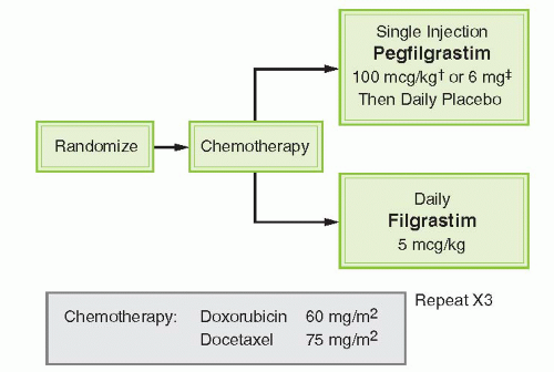
FIGURE 38-3 Design of pegfilgrastim phase III randomized trials in patients receiving chemotherapy for stage II to IV breast cancer. †Holmes FA, Jones SE, O’Shaughnessy J, et al. Comparable efficacy and safety profiles of onceper-cycle pegfilgrastim and daily injection filgrastim in chemotherapy-induced neutropenia: a multicenter dose-finding study in women with breast cancer. Ann Oncol 2002;13:903-909; ‡Green MD, Koelbl H, Baselga J, et al. A randomized double-blind multicenter phase III study of fixed-dose single-administration pegfilgrastim versus daily filgrastim in patients receiving myelosuppressive chemotherapy. Ann Oncol 2003;14:29-35.


Systematic Review of Randomized Controlled Trials

The systematic review of RCTs of primary prophylaxis with G-CSF in patients with solid tumors and lymphoma reported a relative risk for FN with G-CSF prophylaxis of 0.54 (95% CI: 0.43 to 0.67) (P < 0.0001) (Fig. 38-4).43 Likewise, the summary relative risk for infection-related mortality and early all-cause mortality with G-CSF was 0.55 (95% CI: 0.33 to 0.90) (P = 0.018) and 0.60 (95% CI: 0.43 to 0.83) (P = 0.002) (Fig. 38-4), respectively. Finally, median relative dose intensity (RDI) among control subjects was 88.5% compared to 95.5% in patients receiving G-CSF in these trials. Increasing the RDI of chemotherapy utilizing abbreviated treatment schedules (dose dense) with G-CSF support has improved clinical outcomes.48, 49, 50

Only gold members can continue reading. Log In or Register to continue

Stay updated, free articles. Join our Telegram channel

May 27, 2016 | Posted by in ONCOLOGY | Comments Off on Hematopoietic Growth Factors

Full access? Get Clinical Tree

Get Clinical Tree app for offline access