Chapter 71 Hematologic Manifestations of HIV/AIDS
Peripheral Blood Smear and Bone Marrow Morphology in HIV/AIDS
The peripheral blood smear of a patient with HIV/AIDS might show anisocytosis, poikilocytosis, and rouleaux formation. Anemia when present is usually normocytic and normochromic. Sometimes macrocytic anemia can be seen even in the absence of zidovudine therapy. Lymphopenia is seen in advanced disease. Hypogranular neutrophils and Pelger-Huët forms are rarely present. Platelets can be normal or hypogranular. In cases of thrombocytopenia the platelets can be normal-sized or large when thrombocytopenia is due to immune destruction with preserved marrow function.
The bone marrow is usually hypercellular but can be normocellular or hypocellular. Interstitial and perivascular polyclonal plasmacytosis is usually present. HIV-associated stromal changes include edema, gelatinous transformation, and increased reticulin fibers (dense collagen fibers are not a feature of HIV). Normal bone marrow architecture is often disturbed, and dysplastic changes can be seen, including dyserythropoiesis, dysgranulopoiesis, and abnormal megakaryocytes (including clusters and bare megakaryocytic nuclei). However, the following features distinguish the bone marrow morphology in HIV from that of myelodysplastic syndrome (MDS): Dysplasia is less severe in HIV. Dyserythropoiesis occurs mainly in patients on highly active antiretroviral therapy (HAART). Megaloblastic changes are associated with zidovudine therapy. Whereas erythropoiesis is usually hyperplastic in MDS, myeloid/erythroid ratio is usually normal in HIV. Increased blasts can be seen in MDS but never in HIV. Lastly, in contrast to MDS, the basement membrane in HIV often shows eosinophilia, lymphohistiocytic infiltrates, and plasmacytosis.
AIDS, Acquired immunodeficiency syndrome; HIV, human immunodeficiency virus.

Figure 71-1 TYPICAL PERIPHERAL BLOOD AND BONE MARROW FINDINGS IN HIV.
The peripheral smear not uncommonly shows anemia, which is sometimes macrocytic (A), but can be normochromic and normocytic. There frequently is a neutrophilia with left shift, toxic granulation, and some mild dysplastic change in the granulocytes (B). In some patients, particularly those with severe disease, some of the segmented neutrophils show cytoplasmic inclusions similar to Howell-Jolly bodies seen in red cells (B, top, right cell). These are nuclear in origin and are not microorganisms. The bone marrow can show granulocytic hyperplasia with left shift and megaloblastoid change in the myeloid and erythroid cell lines (C). Typically, there is also a reactive plasmacytosis (D). The biopsy specimen can be hypocellular, normocellular, or hypercellular (E) and commonly shows cellular atypia/dysplasia (insert) and atypical reactive lymphoid infiltrates (F), poorly formed (G) or well-formed granuloma (H), and increased plasma cells (I).

Figure 71-2 ACID-FAST ORGANISMS IN GRANULOMA.
Large poorly formed granuloma in the bone marrow (A), is composed of loosely aggregated histiocytes, lymphocytes, and plasma cells (B), with occasional giant cells (C). The acid-fast stain shows rare elongated, slightly beaded organisms, typical of Mycobacterium tuberculosis (D, top). In Mycobacterium avium complex, the organisms frequently stuff histiocytes (D, bottom).

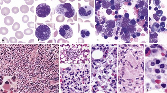
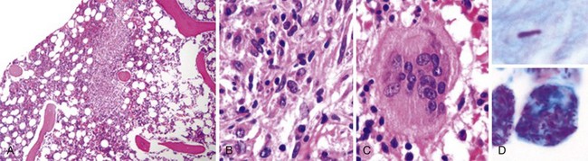
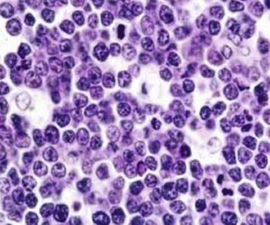
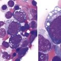
Figure 71-3 BURKITT LYMPHOMA INVOLVING THE BONE MARROW OF A PATIENT WITH ACQUIRED IMMUNODEFICIENCY SYNDROME (AIDS).
(Zhao X, Sun NC, Witt MD, et al: Changing pattern of AIDS: A bone marrow study. Am J Clin Pathol 121:393, 2004.)

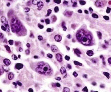
Figure 71-4 CLASSIC HODGKIN LYMPHOMA INVOLVING THE BONE MARROW OF A PATIENT WITH ACQUIRED IMMUNODEFICIENCY SYNDROME (AIDS).
(Zhao X, Sun NC, Witt MD, et al: Changing pattern of AIDS: A bone marrow study. Am J Clin Pathol 121:393, 2004.)
Table 71-1 Surveillance Case Definition for HIV Infection in Adults and Adolescents (Age Over 13 Years)
Stage | Laboratory Evidence | Clinical Evidence |
---|---|---|
Stage1 | Laboratory confirmation of HIV infection and CD4+ T-lymphocyte count of ≥500 cells/µL or CD4+ T-lymphocyte percentage of ≥29* | No AIDS-defining condition (see Table 71-2) |
Stage 2 | Laboratory confirmation of HIV infection and CD4+ T-lymphocyte count of 200-499 cells/µL or CD4+ T-lymphocyte percentage of 14-28* | No AIDS-defining condition (see Table 71-2) |
Stage 3 | Laboratory confirmation of HIV infection and CD4+ T-lymphocyte count of <200 cells/µL or CD4+ T-lymphocyte percentage of <14* | Documentation of an AIDS-defining condition with laboratory confirmation of HIV infection (see Table 71-2) |
Stage unknown | Laboratory confirmation of HIV infection and no information on CD4+ T-lymphocyte count or percentage | No information on presence of an AIDS-defining condition |
AIDS, Acquired immunodeficiency syndrome; HIV, human immunodeficiency virus.
*The CD4+ T-lymphocyte percentage is the percentage of the total lymphocyte count.
Table 71-2 Surveillance Definitions of AIDS-Defining Conditions
Pneumocystis jiroveci (carinii) Candidiasis: esophageal and systemic ![]() Stay updated, free articles. Join our Telegram channel![]() Full access? Get Clinical Tree![]() ![]() ![]() |