Hairy Cell Leukemia


Hairy Cell Leukemia


James B. Johnston

Michael R. Grever


Hairy cell leukemia (HCL), or leukemic reticuloendotheliosis, is a chronic B-cell disorder that was initially described by Bouroncle et al. in 1958.1 The disease is characterized by the presence of typical hairy cells in the peripheral blood and marrow, pancytopenia, and a variable degree of splenomegaly.1, 2, 3, 5, 6 The disease has always aroused interest, initially over the unique morphologic and clinical features of this disorder. The tumor cells are B-cells that typically express CD11c, CD25, CD103, and CD123, are usually IgVH-mutated and are unique memory B-cells in expressing multiple immunoglobulin (Ig) isotypes. More recently, it has been demonstrated that most hairy cells contain a mutated active form of the BRAF gene (V600E) which may be used as a diagnostic tool and a potential target for therapy. Most cases of HCL are very sensitive to interferon (IFN)-α and the nucleoside analogs pentostatin (2′-deoxycoformycin, dCF) and cladribine (2-chlorodeoxyadenosine CdA).5, 6 Less responsive patients generally respond to monoclonal antibodies, such as rituximab (against CD20) or to immunotoxin-labeled antibodies to CD22 or CD25. Prior to the development of these treatments the median survival for HCL patients was 4 years whereas now the survival of these patients is no different from an age- and sex-matched control population.


INCIDENCE AND ETIOLOGY

HCL is a rare disorder, accounting for 2% of all leukemias, and occurs more frequently in men, with an incidence in the United States of 2.9 per million per year for men and 0.6 per million per year for women.5, 7 The etiology of HCL is unknown. Several case-controlled studies have identified possible relationships to radiation exposure,7, 8 exposure to benzene,9 to farm animals, and to commercial herbicides and pesticides.9, 10 A familial predisposition is suggested by reports of the disorder in 15 families where HCL occurred among first-degree relatives. In these families, disease is usually associated with the following haplotypes; A2, B7, and Bw4/6; A1/3 and B7/8; A2, B51, and DR52/53.11


PATHOGENESIS

Immunologic,12, 13, 14 molecular genetic,15, 16, 17, 18, 19, 20, 21 chromosomal,22, 23 and gene expression24 studies have demonstrated that the hairy cell is of B-cell origin. Although some T-cell features have been reported,25, 26, 27, 28, 29, 30 including two cases in which the retrovirus human T-cell leukemia virus type II was isolated,31, 32 no instances of T-cell receptor gene rearrangements have been found. Fluctuations in the expression of T- and B-cell markers have been demonstrated both in vivo and under various in vitro culture conditions.33, 34, 35, 36 In most instances, the alteration noted was between a surface-immunoglobulin-positive (SIg+) CD2phenotype and a SIgCD2+ phenotype. Other B-cell antigens, however, such as CD20, were retained, even with the loss of SIg and the appearance of CD2.35

The cell of origin of the hairy cell has been a subject of much debate and has been discussed in several recent reviews.37, 38, 39 Gene expression profiling has demonstrated that the hairy cell is most similar to a memory B-cell, although it differs in that the hairy cell has increased expression of adhesion and chemokine receptor genes.24 The profile is extremely homogeneous, quite different from other lymphoid malignancies, and the abnormal expression of specific genes explains the properties of this disease which include unique cell structure, marrow fibrosis, marrow suppression, and the tendency of tumor cells to home to spleen and marrow, rather than to lymph nodes. As hairy cells do not express CD27, which is normally expressed on memory B-cells, it has been suggested that the normal counterpart is a memory cell which lacks CD27 but has somatic mutations of the the IgVH genes.40 However, the profile also demonstrated expression of many genes associated with macrophages suggesting that these cells are unique in having phagocytic properties.24 Consistent with being a memory B-cell, the majority of cases with HCL show mutations of the variable chain of the Ig gene, demonstrating that the hairy cell has passed through the germinal center (Fig. 91.1).15, 16, 17, 18, 19, 20, 21, 41 However, 10% to 20% of cases have unmutated IgVH and in general these patients have very poor prognosis and are resistant to chemotherapy.17, 18 The IgVH gene usage is shown in Figure 91.2 and demonstrates that in classical HCL IgVH 3-23 (17%), 3-30 (8%), and 4-34 (7%) are most commonly used. IgVH 1-69, 3-11, 3-48, and 4-39 are each used in 5% of cases The major difference between the HCL variant and classical HCL, is the usage of IgVH V4-34, which is present in 54% of variant cases but only 17% of classical cases.20 The majority (94%) of IgVH4-34 cases are unmutated. As expected, the IgVH is more likely to be unmutated in the HCL variant (54%) as compared to classical HCL (17%). It is interesting that, as discussed later, patients with unmutated IgVH or those that use IgVH4-34 have more aggressive and drug-resistant disease.17, 18 The patterns of IgVH gene family usage and the incidence of IgVH mutations is different in HCL and chronic lymphocytic leukemia (CLL).20 In CLL, the most commonly used IgVH genes are IgVH1-69, 3-21, 3-30, 4-34, and 1-02 and whereas most IgVH 1-69 cases are unmutated, most IgVH4-34 cases are mutated.42 Overall, approximately 60% of CLL cases have mutated IgVH and 40% are unmutated.

Hairy cells are unique in expressing multiple Ig isotypes that are related to RNA splicing. Thus, individual hairy cells may express IgG alone or in combination with IgM and/or IgD and/or IgA.15, 16, 41 Similarly, IgVH mutational analysis has demonstrated intraclonal variation in gene expression, both in the IgVH unmutated and mutated varieties. These studies have suggested that the hairy cell is in maturation arrest before deletional recombination within the heavy chain locus.


Cytogenetics

Cytogenetic studies have been difficult to carry out in HCL, because the number of circulating hairy cells is small, and attempts at marrow aspiration are usually unsuccessful. In addition, until recently it has been difficult to induce hairy cells to proliferate and to obtain hairy cells in metaphase. In early reports, the abnormalities observed in HCL tended to be nonclonal.43, 44 However, more recently, 15 of 19 patients were found to have an abnormal karyotype, and a 14q+marker involving the breakpoint q32 at the Ig heavy chain locus was the most frequent clonal abnormality.22 Abnormalities involving the short arm of chromosome 12 have been observed in four patients.13 Haglund et al.45 observed deletions and inversions in 67% of patients. Clonal abnormalities of chromosome 5 were found in 40% of cases (most commonly trisomy 5, pericentric inversions, and interstitial deletions involving 5q13). The frequency with which these abnormalities occur is unique and is not found in other B-cell malignancies. Abnormalities in 5q13 thus occur in one third of patients, and subsequent studies have identified three expressed sequences as candidates for a putative tumor-suppressor gene at 5q13.3.46 Cytogenetic analysis has been carried out using monoclonal antibodies to CD40, because this
appears to be the most effective way of inducing hairy cell proliferation.47 This technique allowed cytogenetic analysis in 42 of 43 cases and demonstrated that clonal abnormalities were present in 19% of cases and involved numeric or structural abnormalities in chromosomes 5, 7, and 14. In contrast, abnormalities of chromosome 5 were not observed in the HCL variant, and translocations more frequently involved either chromosome 2 or 14.48


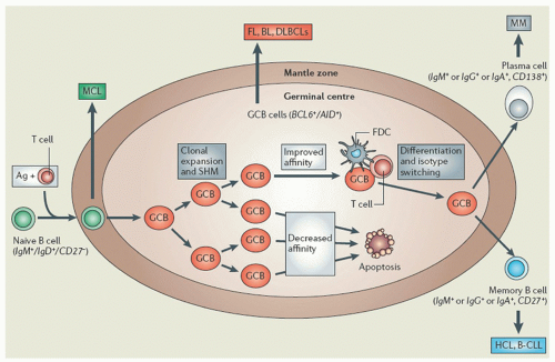
FIGURE 91.1. The germinal center (GC) reaction is a key event in adaptive immunity to T-cell-dependent antigens. In lymph nodes and the spleen, antigen (Ag)-activated naive B-cells (immunoglobulin [Ig] M+/IgD+) are driven into primary follicles where, aided by T-cells, they undergo clonal expansion, form the GC, and displace nonproliferating naive B-cells to the periphery or mantle zone (shown in diagram). Within the GC, somatic hypermutation (SHM) mutates the Ig variable region of proliferating B-cells to increase their affinity for the stimulating Ag. Although most GC B-cells (GCB) acquire unfavorable mutations and die by apoptosis, a few improve their Ig affinity and are selected for further differentiation through interactions with T-cells and follicular dendritic cells (FDC). A proportion of these selected B-cells then undergo switch recombination of the Ig constant region, which changes the isotype and, consequently, the antibody-effector function. B-cells of the GC express BCL6, which is required for GC formation, and activation-induced cytidine deaminase (AID) which is essential for SHM and switch recombination. Finally, selected B-cells differentiate into memory B-cells (CD27+) or antibody-secreting plasma cells (CD138+). Follicular lymphoma (FL), Burkitt lymphoma (BL), and some diffuse large B-cell lymphomas (DLBCLs) are thought to derive from B-cells of the GC, mantle cell lymphoma (MCL) mainly from mantle-zone naive B-cells, B-cell chronic lymphocytic leukemia (B-CLL) and hairy-cell leukemia (HCL) from memory B-cells, and multiple myeloma (MM) from plasma cells. The majority of cases of CLL and HCL have undergone SHM, but a proportion have not and these cases have more aggressive disease. (From Tiacci E, Liso A, Piris M, and Falini B. Evolving concepts in the pathogenesis of hairy-cell leukaemia. Nature Rev 2006;6:437-448, with permission).

High-density genomewide DNA profiling in HCL showed that, in contrast with CLL, HCL has a remarkably stable genome.49, 50 Using a high-density single nucleotide polymorphism array, it has been shown that only 25% of patients had gross copy number abnormalities.49 Alterations in the genes for FGF12 and FGF receptor were observed, and increased expression of these genes is typically observed in HCL, which is responsible for the marrow fibrosis seen in this disease.49 Although a high incidence of TP53 mutations was initially reported in HCL, subsequent studies have suggested that this is uncommon and occurs primarily in patients with unmutated IgVH.17, 51, 52


Oncogenes and Signal Transduction

To date, there has been little information regarding the role of oncogenes in HCL. It has been shown that CSF1R, which encodes the receptor for macrophage colony-stimulating factor and is normally expressed in macrophages/monocytes, is highly expressed in hairy cells.53 The addition of macrophage colony-stimulating factor to hairy cells causes enhanced mobility of the cells, the extent of which depends on the underlying stratum, and this effect is mediated through alterations in the integrin αvβ4.54 More recently, it has been demonstrated that, compared with normal lymphocytes, hairy cells have markedly increased levels and activities of the protein tyrosine kinase pp60c-src, which is the product of the proto-oncogene CSK.55 This increase was not observed in CLL cells and was unrelated to the amplification or structural rearrangements of the gene.55 Cyclin D1 messenger RNA and protein levels are increased in HCL, but the increase is less than is seen in mantle cell lymphoma and is not related to t11, 14 (q13;q32), which is typically observed in mantle cell lymphoma.56, 57 Finally, because of the high frequency of abnormalities of chromosome 5 in classic HCL, it has been suggested that a transforming oncogene might be located at 5q13.3.46

Gene expression profiling identifies a unique homogeneous signature for HCL distinct from other B-cell malignancies. In HCL samples, decreased expression was noted for CXCR5, TNFRAF5, CD40, CD27, and CCR7, whereas increased expression was noted for GAS7, FGFR1, FGF2, FLT3, TIMP1, TIMP4, RECK, ANXA1,
Syndecan3, Cyclin D1, IGFBP, and NUDT6.24 The proposed effects of these changes on the pathophysiology of HCL are outlined in Table 91.1. More recently, a heterozygous mutation of GTG to GAG at codon 600 of the BRAF gene has been observed in typical HCL producing a V600E variant protein, in which glutamate substitutes for valine at position 600.58, 59, 60, 61, 62 BRAF is a serine-threonine kinase and the variant protein (V600E) demonstrates increased activity resulting in enhanced phosphorylation and activation of mitogen-activated protein-ERK kinase (MEK) and extracellular signal-regulated kinase (ERK), which increase cell survival and proliferation by increasing the expression cyclin D1 and decreasing p27.58 The functionality of the variant protein was demonstrated in primary hairy cells by showing the presence of phosphorylated MEK and ERK in these cells, and demonstrating that the phosphorylation was decreased using the BRAF inhibitor, PLX-4720.58 Initial studies suggested that all patients with classical HCL contained a V600E; however, a more recent study demonstrated that 82% of patients with classical HCL express BRAF, whereas the V4-34 subtype and HCL variant do not express BRAF.21 Thus, BRAF inhibitors may not be useful in the treatment of the aggressive V4-34 HCL subtype of the HCL variant.


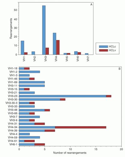
FIGURE 91.2. Immunoglobulin (Ig) VH family and gene usage in hairy-cell leukemia (HCL). Number of IgVH rearrangements in each family (A) and for genes used by more than one patient (B) for HCLc (black) and HCLv (gray). (From Arons E, Roth L, Sapolsky J, et al. Evidence of canonical somatic hypermutation in hairy cell leukemia. Blood 2011;117:4844-4851, with permission.)

It has also been demonstrated that the activities of MEK and ERK are increased in HCL through the epsilon form of protein kinase C (PKCε), which in turn is activated by tyrosine nitration (Fig. 91.3).63 Tyrosine nitration is produced by the high levels of reactive oxygen species (ROS) and nitric oxide (NO) in hairy cells.63 In addition, apart from its role in cell survival, activated PKCε is also involved in causing the unusual hairy cell structure by increasing the Rho guanosine triphosphatases (GTPases), Rac1 and Cdc42.63 The Rho GTPases are members of the Ras family of GTP-binding proteins and can either be active (GTP-bound) or inactive (GDP-bound).63 The GTPases are important for actin structure within the cell and controlling cell shape. It has been shown that three groups of Rh0 GTPases, RhoA, Rac1, and Cdc42, are overexpressed in HCL and are responsible for the filamentous projections and growth of these cells.64


TABLE 91.1 MECHANISMS FOR HAIRY CELL LEUKEMIA FEATURES


Hairy Cell Feature


Mechanism


Hairy cell projections


Alteration of actin structure through up-regulation of the Rho GTPase Rac 1.


Prolonged survival of hairy cells


Autocrine stimulation by TNF-α, increased cellular levels of bcl-2.


Ability of hairy cell to phagocytose and expression of macrophage markers, for example, CD11c


Up-regulation of annexin1 mediates phagocytic function and c-Maf transcription factor for macrophage differentiation.


Marrow fibrosis and monocytopenia


Up-regulation of FGF2 which activates FGFR1 on the hairy cell producing fibronectin and TGFβ, which causes the adjacent fibroblasts to secrete collagen.


TNF-α and TGF-β cause marrow suppression and monocytopenia.


Infliltration of red pulp of spleen by hairy cells with the formation of “pseudosinuses”


Up-regulation of TIMP1, TIMP4, RECK inhibitors of matrix metalloproteinases.


Lack of lymph node involvement by hairy cells


Hairy cells lack L-selectin which is required for binding to venule endothelium and chemokine receptor 7 (CCR7) which is required for transendothelial migration.


GTPases, guanosine triphosphatases; TGF-β, transforming growth factor-β; TNF-α, tumor necrosis factor.


Swerdlow SH, Campo E, Harris NL, et al. WHO classification of tumours of haematopoietic and lymphoid tissues, vol. 2, 4th ed. WHO 2008; Basso K, Liso A, Tiacci E, et al. Gene expression profiling of hairy cell leukemia reveals a phenotype related to memory B cells with altered expression of chemokine and adhesion receptors. J Exp Med 2004;199:59-68 and Cawley JC, Hawkins SF. The biology of hairy-cell leukaemia. Curr Opin Hematol 2010;17:341-349.


Cytokines

There has been great interest in the role of cytokines in the pathogenesis of HCL.37, 38, 39 Identified factors that may be involved include interleukin-2 (IL-2), tumor necrosis factor (TNF)-α, IL-4, IL-6, B-cell growth factor, IFN-α, transforming growth factor-β (TGF-β), and basic fibroblast growth factor (bFGF, FGF2).65 Some of these factors may be produced by the hairy cells themselves or by normal T-cells.65, 66 T-cell clones have been shown to produce cytokines that stimulate the growth of hairy cells, but not normal B-cells; this effect is prevented by the addition of IFN-α.66

A feature of classical HCL is the reactivity of the hairy cells with anti-Tac (CD25), which detects the α-chain of the IL-2 receptor.67, 68, 69, 70 Increased serum levels of IL-2 receptor have been found in untreated HCL patients, and there is evidence that the leukemic cells release the receptor.70 The serum levels of IL-2 receptor correlate with the extent of disease and decrease after effective therapy with IFN-α.71, 72, 73 Paradoxically, even though the IL-2 receptor is present on hairy cells, these cells do not respond to stimulation by IL-2, and IL-2 probably does not play a major role in the pathogenesis of HCL.65

It has been demonstrated that TNF-α, but not TNF-β, stimulates the growth of hairy cells, whereas in CLL, both forms of TNF stimulate leukemic cell growth (Fig. 91.4).74, 75 Hairy cells can also produce TNF-α,76, 77 and the serum level of TNF-α is increased in HCL, the level correlating with tumor burden.77 The TNF-α receptor can also be detected in the serum of these patients, and the level of this receptor also decreases after treatment with IFN-α.78, 79 These findings suggest that TNF-α production by the hairy cells may play an important role in the pathogenesis of HCL by stimulating further growth of hairy cells and producing pancytopenia through the inhibition of normal marrow function.74, 76, 77


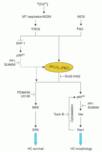
FIGURE 91.3. Proposed pathways of constitutive signaling in hairy cells (HCs). This figure shows the schematic representation of the possible associations into two parallel pathways of active signaling components detected in HCs. Protein kinase C (PKCε) plays a central role, controlling the activation of p60src, Rac1, and extracellular signal-regulated kinase (ERK). PKCε is itself activated through nitration of tyrosine residues by reactive oxygen species (ROS) and nitric oxide (NO) which are increased in HCs. (From Slupsky JR, Kamiguti AS, Harris RJ, et al. Central role of protein kinase Cε in constitute activation of ERK1/2 and Rac 1 in malignant cells in hairy cell leukemia. Am J Path 2007;170:745-754, with permission.) MEK, mitogen-activated protein-ERK kinase; MT, mitochondrial; NOX5, NADPH oxidase 5; iNOS, inducible nitric-oxide synthetase.

Hairy cells secrete low levels of IL-6, and the serum level of this cytokine is increased in HCL.65, 80 The production of IL-6 messenger RNA and IL-6 secretion is markedly increased by incubating HCL cells with TNF.80 IL-6 antisense oligonucleotide can inhibit the effect of TNF on IL-6 secretion and DNA synthesis, suggesting that IL-6 mediates the activity of TNF in HCL.80

Hairy cells also produce bFGF and TGF-β and express the bFGF receptor.81, 82 Adhesion of the hairy cells to hyaluronan via CD44 induces the secretion of bFGF, but not TGF-β.81 The secreted bFGF then feeds back on the hairy cell to secrete fibronectin, which is a major component of the marrow fibrosis in this disease. As the spleen does not have hyaluronic acid, this explains the lack of bFGF and fibrosis in this organ despite abundant hairy cells.81 TGF-β has been shown to be present in increased
quantities in the plasma and marrow of patients with HCL, and this also contributes to the marrow fibrosis by inducing the production of collagen and reticulin by adjacent fibroblasts.82


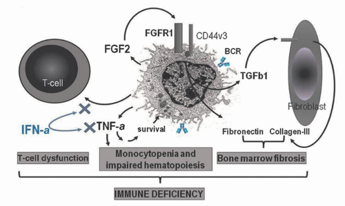
FIGURE 91.4. Immune suppression in hairy-cell leukemia (HCL). Immune suppression is a result of T-cell dysfunction and impaired hematopoiesis with pancytopenia, bone marrow fibrosis, and hypersplenism. T-cell activation, decreased numbers of memory T-cells, restricted T-cell repertoire, and opportunistic infections are the result of inappropriate activation and suppression of T-cell responses directly by cytokines produced by the neoplastic B-cells. Cytopenia with severe monocytopenia is caused by the secretion of tumor necrosis factor (TNF)-α by the hairy cells. TNF-α also has autocrine prosurvival effects on the tumor clone. Treatment of HCL with IFN-α is able to restore the abnormal T-cell repertoire and hematopoiesis by inhibiting cytokine (including TNF-α) mediated effects on the T-cells and tumor cell. Fibrosis is caused by production of fibroblast growth factor-2 (FGF-2) and overexpression of its receptor FGFR1. The FGF2-FGFR1 interaction increases with CD44v3 co-receptor and syndecan family members. FGFR1 signals secretion of autocrine fibronectin and of transforming growth factor-β (TGFβ) by hairy cells. TGFβ stimulates adjacent fibroblasts to produce fibronectin and collagen type III. (From Forconi F. Hairy cell leukaemia: biological and clinical overview from immunogenetic insights. Haematol Oncol 2011;29:55-66, with permission.)


CLINICAL FINDINGS

The median age in HCL is between 50 and 55 years (range, 22 to 85 years), with the male-to-female ratio being 4:1.1, 2, 3, 4, 5, 6, 83 Classically, these patients present with weakness and fatigue, and the symptoms can usually be related to pancytopenia and splenomegaly (Table 91.2).1, 2, 3, 4, 5, 6, 83 Ninety percent of patients have a palpable spleen, which may be massive, and one third of patients have hepatomegaly.1, 2, 3, 4, 5, 6, 83 Peripheral lymphadenopathy is uncommon,5 but with computed tomography scanning, significant abdominal lymphadenopathy may be observed.84, 85The degree of abdominal lymphadenopathy correlates with the duration and extent of disease. The incidence at presentation is 17%, but increases to 56% at relapse after chemotherapy.84 Less frequently, mediastinal node enlargement may also be observed.86 Compared to 30 years ago, patients are now being diagnosed earlier and have less marked splenomegaly and anemia at presentation.6

Most patients have pancytopenia, the etiology of which is multifactorial but is primarily related to marrow failure and splenomegaly. Marrow failure is related to replacement of marrow by hairy cells and the associated reticulin fibrosis, and to the inhibition of myelopoiesis by cytokines, such as TNF-α, released by the hairy cells.37, 38, 39 Splenomegaly in HCL may produce pancytopenia by three mechanisms. First, the major factor responsible for the cytopenias is pooling (sequestration, margination) of normal peripheral blood cells in the enlarged spleen.87, 88, 89, 90, 91, 92 As much as 90% of the peripheral platelet mass,88 30% of the red cell mass,91 and 65% of the granulocytes89 may pool in a massively enlarged spleen. In HCL, apparently because of the formation of blood-filled pseudosinuses by the hairy cells, a greater proportion of the peripheral red cell volume (as much as 48%) may be pooled in a massively enlarged spleen.91 A second mechanism responsible for cytopenias with splenomegaly is the increased destruction of cells in the spleen. Finally, an expanded plasma volume contributes to the appearance of cytopenia with splenomegaly; this is particularly true for the observed anemia.87, 90


TABLE 91.2 HAIRY CELL LEUKEMIA: CLINICAL MANIFESTATIONS


Manifestation


Incidence (%)


Weakness, easy fatigue


80


Fever, sweats, weight loss, anorexia


20-35


Infection


20-30


Easy bruising, bleeding


20-30


Left upper quadrant abdominal discomfort


25


Autoimmune disorders


15-30


Splenomegaly


80-90


Hepatomegaly


30-40


Ecchymoses, petechiae


20-30


Infections

Prior to the development of effective treatments for HCL, infections were a major problem and the leading cause of death.83, 93, 94, 95, 96 Nowadays, approximately one third of patients will develop an infection during the course of their disease and two thirds of these will be serious.96 Fever is rarely a manifestation of the underlying HCL; when present, it should prompt a careful search for an infectious process. Approximately 30% of patients present with infection, and 70% have either documented or suspected infections during the course of the disease.93 Pyogenic infections account for ˜50% of infectious episodes; Gram-negative and Gram-positive bacteria are identified with approximately equal frequency.83, 93 However, nonpyogenic infections occur in >30% of patients,83, 93 and infections can occur with unusual organisms such as atypical mycobacteria, including Mycobacterium kansasii, which is a unique feature of HCL, in comparison to other lymphoproliferative disorders.83, 94, 97 Other organisms include Toxoplasma gondii, Legionella, Listeria monocytogenes, and Pneumocystis jirovecii, as well as various fungi and viruses.83, 98, 99, 100

The high infection rate can be ascribed primarily to neutropenia, although several other immune defects have been described. Monocytopenia is often a prominent feature,101, 102 and functional abnormalities of monocytes and granulocytes may occur.99, 101, 103, 104, 105 The defects in monocyte production and function may account for the unusual susceptibility of these patients to atypical mycobacterial and fungal infections.95, 96 The T-cells are also highly abnormal in HCL, demonstrating an inversion in the CD4:CD8 ratio, and they have a poor antigen response which is likely related to absence of CD28.106, 107 Moreover, there is clonogenic expansion of CD8+ cytotoxic lymphocytes but the target for these cells has not yet been identified.108 These abnormalities can resolve with IFN-α therapy, although it may take up to 2 years to see this effect.109 A depressed helper to suppressor T-cell ratio associated with a decrease in the number of T-helper cells and an increase in the number of T-suppressor cells also has been demonstrated.109 As expected, lymphocyte functional studies reveal impaired delayed-type hypersensitivity to recall antigens, as well as near-absent antibody-dependent cellular cytotoxicity.106 In contrast to CLL, the serum Ig levels are normal.110


Autoimmune Disorders

Clinical manifestations secondary to various autoimmune disorders are being recognized with increasing frequency in patients
with HCL.83, 111, 112, 113, 114, 115 In one series of patients, these complications were second only to infection as a cause of morbidity.83, 112 The onset may occur any time during the course of the disease and is not related to the tumor burden. Most frequently, patients present with arthritis, arthralgias, palpable purpura, or nodular skin lesions resulting from cutaneous vasculitis, and low-grade fever.83, 112, 114 Occasionally, patients may have involvement of the lung, liver, intestine, and kidney, with a clinical picture that resembles polyarteritis nodosa.83, 112, 113, 115 These patients often have fever, malaise, and weight loss, and a co-existent infection must be ruled out.83, 112 If skin lesions are present, the diagnosis can be confirmed by biopsy, which usually shows changes compatible with a diagnosis of polyarteritis nodosum or leukocytoclastic vasculitis; occasionally, a vasculitis related to the invasion of the vessel wall by hairy cells occurs; this may appear very similar to polyarteritis nodosa with the presence of aneurysms.114 In some organs, such as the lung, a granulomatous vasculitis may be found.112 Angiography may reveal peripheral aneurysms.112 Antinuclear antibodies, rheumatoid factor, immune complexes, and hepatitis B antigen are variably positive.111 Cryoglobulinemia has been detected in some patients.116, 117 It has been postulated that the increased incidence of vasculitis in HCL may be related to infections with hepatitis B and other viruses, cross-reactivity of antibodies against hairy cells with epitopes on endothelial cells, and decreased clearance of immune complexes by the impaired immune system.114

These autoimmune manifestations may be self-limited, but if therapy is required, a short course of corticosteroids is usually effective.114 Remissions have also been observed after splenectomy, IFN-α, and pentostatin therapy.112, 114, 115


Unusual Manifestations


Lytic Bone Lesions

Although the immunophenotypic profile of hairy cells closely resembles that of B-cells at a developmental stage just before terminal differentiation to plasma cells, lytic bone lesions are distinctly unusual (Table 91.3).118, 119, 120, 121 In some patients with osteolytic lesions, HCL and multiple myeloma were thought to coexist.122, 123 However, several patients with classic HCL and without any evidence of plasma cell proliferation have been reported to develop osteolytic lesions.119, 120, 121 These lesions have a predilection for the proximal femur and usually are associated with extensive bone marrow infiltration by hairy cells.120, 121 The lesions, as with those seen in association with multiple myeloma, respond well to radiotherapy. The administration of corticosteroids can produce prompt relief of bone pain.118


Skin Involvement

Cutaneous lesions referable to thrombocytopenia (ecchymoses, petechiae), infection, or vasculitis are common during the course of HCL, but lesions caused by infiltration of the skin by hairy cells are unusual.124, 125 In a retrospective review of 600 cases, skin lesions thought to be due to infiltration by hairy cells were reported in 8.0% of cases, but histopathologic verification was present in only 1.3%.124 Infiltrative lesions usually are widely disseminated and consist of erythematous maculopapules. Biopsy shows the infiltrates to be perivascular, involving the dermis but not the epidermis.126


Splenic Rupture

Surprisingly, even with massive splenomegaly, spontaneous splenic rupture is rare in HCL, occurring in ˜2% of cases.1, 127


Other Organ Dysfunction

Although hairy cell infiltration of multiple organs and tissues is a frequent finding at autopsy, clinically significant organ dysfunction is unusual.128 Infiltration of connective tissue and fat surrounding organs is common.128 Central nervous system involvement is unusual, and only a rare case of meningeal involvement has been documented.128, 129 Infection is by far the most frequent cause for neurologic complications.129 Pleural effusions, ascites, proteinlosing enteropathy, and spinal cord compression may occur rarely in HCL and result from tissue infiltration by hairy cells.118


LABORATORY FINDINGS

The relative incidence of the most characteristic laboratory findings is listed in Table 91.4. In a series of 725 cases studied by the Italian Cooperative Group, 80% of patients had pancytopenia at presentation, with one third of all patients having a hemoglobin level <8.5 g/dl, neutrophils <0.5 × 10⁹/L, and platelets <50 × 109/L.6 A careful inspection of the peripheral blood smear demonstrates the presence of typical hairy cells in >85% of patients,1, 6 and in 13% of cases, there are >5 × 10⁹/L hairy cells.6 In Bouroncle’s series of 82 patients, hairy cells accounted for ≥10% of the leukocytes in 80% of patients and ≥50% in 43% of patients.2 The total leukocyte count was elevated in 20%, but in only 4% did the count exceed 50 × 109/l.2 Monocytopenia also typically occurs in active HCL.101, 102

Abnormal liver function studies, usually with an isolated elevation of the serum alkaline phosphatase level, are obtained in 10% to 19% of individuals with HCL.4 The leukocyte alkaline phosphatase scores are high in most cases. The Ig levels are normal in HCL and monoclonal gammopathy is rare, occurring in 1% to 3% of patients.3, 4 It has been difficult to prove that the hairy cells are secreting the paraprotein130; in other cases, the M band was thought to be related to a co-existing myeloma.120, 122, 123


TABLE 91.3 HAIRY CELL LEUKEMIA: UNUSUAL CLINICAL MANIFESTATIONS


Manifestation


Incidence (%)


Peripheral lymphadenopathy


<5


Lytic bone lesions


3


Skin involvement


5


Splenic rupture


<5


Other organ dysfunction


<5


TABLE 91.4 HAIRY CELL LEUKEMIA: LABORATORY MANIFESTATIONS


Manifestation


Incidence (%)


Pancytopenia


70


Neutropenia


80


Thrombocytopenia


80


Anemia


75


Monocytopenia


98


Leukocytosis


15


Hairy cells in peripheral blood


85


Hairy cells in bone marrow


99


Hairy Cells

The characteristic morphologic appearance of hairy cells on a Wright-stained peripheral blood smear is the single most important diagnostic finding (Fig. 91.5).12 Other B-cell disorders that may have hairy projections on the cells include B-cell prolymphocytic leukemia, atypical HCL, and splenic lymphoma with villous lymphocytes (SLVL), although careful analysis and flow cytometry can differentiate these disorders.131 Typical hairy cells are mononuclear with relatively abundant cytoplasm and a cell diameter in the range of 10 to 25 µm. The cytoplasm is pale blue-gray and agranular with a variable number of elongated (hairy) projections. The mechanism for the hairy cell villi has been related to up-regulation of the expression of the Rho GTPases in the hairy cells.64 The nuclei are round, oval, reniform, or dumbbell-shaped with a nuclear chromatin pattern that is homogeneous and less clumped and lighter staining than that of normal mature lymphocytes and those seen in classic CLL and prolymphocytic leukemia.6, 25, 131 A prominent nucleolus is rarely seen. On both transmission and scanning electron microscopy, the cytoplasmic projections appear as elongated slender microvilli or broad-based ruffles or pseudopods (Fig. 91.6).132 Other ultrastructural features include numerous mitochondria, polyribosomes, strands of rough endoplasmic reticulum, intermediate filaments, and some lysosomal granules.25 In 50% of cases, cytoplasmic inclusions known as ribosome-lamellar complexes are found.6, 133 These complexes appear as rod-shaped structures under light microscopic analysis.133 Although seen most frequently in individuals with HCL, they are not pathognomonic, having been reported in association with other chronic B-cell lymphoproliferative disorders, acute monocytic leukemia, and even nonhematologic neoplasms.134 The characteristic filamentous projections can be seen readily in living hairy cells; using supravital dyes and phase-contrast microscopy, one can observe the cytoplasmic projections protruding and then retracting constantly.6


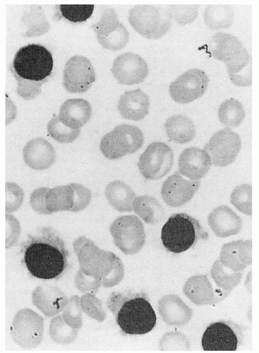
FIGURE 91.5. Hairy cells from a peripheral blood smear (light microscopy). Cells have abundant agranular cytoplasm with multiple cytoplasmic projections. Nucleus is round, oval, or reniform with light-staining homogeneous nuclear chromatin (Wright stain, × 1,500).

A useful diagnostic test for HCL is the presence of acid phosphatase isoenzyme 5 in the cytoplasm of hairy cells, but this can also be positive in other disorders.6, 135, 136, 137, 138, 139 Cytochemical staining procedures demonstrate that this isoenzyme, unlike other acid phosphatase isoenzymes, is not inhibited by tartrate.135, 136, 137, 138, 139 The enzyme concentrates primarily in the Golgi area and nuclear membrane.135 Even though a positive tartrate-resistant acid phosphatase (TRAP) stain is not pathognomonic for the diagnosis of HCL, it has stood the test of time as an important tool in the differential diagnosis of this disease.137 The test is positive in more than 95% of cases.4 In one report,137 the TRAP stain was positive in 76%, intermediate in 21%, and negative in only 3% of 29 patients with HCL. In contrast, in 37 patients with CLL and other lymphoproliferative disorders, the test was positive in only 3%, intermediate in 32%, and negative in 65%.137 The TRAP stain can now be carried out on paraffin-embedded tissue using 9C5, a monoclonal antibody to TRAP.138, 139 The majority of cases of HCL are detected using this antibody, although only 50% of the cells stain positively.139

Gene expression profiling has identified ANXA1 as one of the most up-regulated genes in HCL.24 ANXA1 codes for annexin1, and is not expressed in normal B-cells. By using an immunocytochemical assay staining for the presence of annexin A1 on either paraffin-fixed bone marrow specimens or acetone-fixed cytospins of peripheral blood, 100% sensitivity and specificity has been shown in differentiating HCL from other B-cell malignancies, including variant HCL.140 This is now considered one of the best tests.

More recently it has been shown that the majority of patients with classical HCL have GTG to GAG mutations at codon 600 of the BRAF gene, the (V600E) allowing glutamic acid to be substituted for valine at position 600 of the BRAF protein.58, 59, 60, 61, 62 A recent study by Kreitman et al21 has demonstrated that all classical HCL cases have the mutation apart from the aggressive and drug-resistant IgVH4-34 subtypes and the HCL variant. Several recent reports have described highly sensitive polymerase chain reaction (PCR) assays to detect the V600E mutation, which should be useful in the diagnosis of HCL and monitoring for minimal residual disease (MRD) following therapy.60, 61, 62

Immunophenotyping has proved very useful in assisting in the diagnosis of HCL and in differentiating it from variants and other B-cell malignancies.1, 131, 141, 142, 143 Using flow cytometry, very small numbers of abnormal cells can be detected and the diagnosis of HCL can be made on the peripheral blood of the majority of patients. The hairy cells are monoclonal and, in contrast with most other lymphoid malignancies, cases with κ or λ expression are equally represented or there is a predominance of cases with κ expression.142 Table 91.5 shows the immunophenotype of typical HCL in comparison to other chronic B-cell disorders with which it can be confused. In HCL, the leukemia cells have SIg (IgM+/-, IgG, IgD, or IgA) and B-cell-associated antigens (CD19, CD20, and CD22); typically, the cells are positive for CD25, the IL-2 receptor. CD11c (leu-M5), which reacts with the α subunit of p150/95, a member of the lymphocyte functionassociated antigen subfamily of integrin molecules, is present on virtually all hairy cells, but it is also present on monocytes and neutrophils and on the cells of other lymphoid malignancies.6, 141 Several antibodies have been raised against hairy cells, although none is entirely specific for this leukemia. B-ly-7 reacts with a 144-kd antigen (mucosal lymphocyte antigen; CD103) on hairy cells and on a small number of normal lymphocytes, which have been postulated to be the normal hairy cell counterpart.142 Like
B-ly 7, the antibodies HML-1, Ber-ACT8, and LF61 can also recognize CD103. Two further antibodies, RAB-I and anti-HC2, are also relatively specific for hairy cells.6 CD123 (α chain of the IL-3 receptor) is present in 95% of cases with classical HCL and is only rarely detected in other chronic lymphoid malignancies.142, 143, 144 Hairy cells are also typically strongly FMC7 positive (an epitope of CD20), negative for CD79b (an epitope of B-cell receptor β chain), and CD27, CD10, and CD38 are rarely positive.142 In summary, the diagnostic immunophenotypic features for HCL are monoclonal B-cells that are positive for CD11c, CD25, CD103, and CD123.142

Only gold members can continue reading. Log In or Register to continue

Stay updated, free articles. Join our Telegram channel

Oct 21, 2016 | Posted by in HEMATOLOGY | Comments Off on Hairy Cell Leukemia

Full access? Get Clinical Tree

Get Clinical Tree app for offline access