Genetic Mouse Models of Cancer


Figure 9-1 Non–genetically engineered mouse models of cancer (A,B) Mice develop tumors spontaneously or in response to carcinogen exposure. (C,D) Transplantation of human or mouse tumor cells into recipient mice provides a rapid method to study cell growth and progression in vivo.


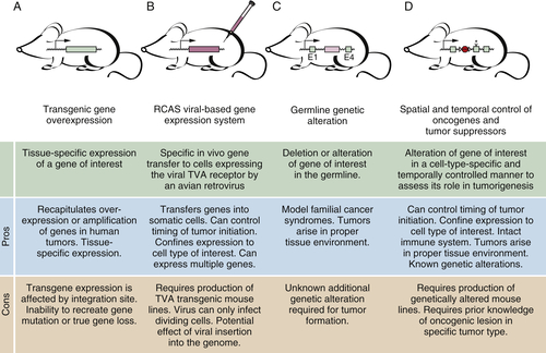
Techniques to alter the germline of mice allow the deletion or alteration of genomic loci (see Figure 9-2, C and D). These alterations can also be induced in cell-type and temporally regulated fashions. Such powerful approaches allow mouse models to be created that mimic the loss of tumor suppressor genes and activation of oncogenes that occur in different human cancers, resulting in mouse models that closely resemble the human disease. These genetically engineered mouse models are being used in a myriad of research settings to further our understanding of tumor biology.


Techniques to Modify the Mouse Genome


Different genetic strategies can be used to overexpress, alter, or reduce the expression of genes that affect tumor incidence or progression. Genetic mouse models begin to recapitulate the selected human cancer when the genetic alterations are consistent with those detected in human cancers and when those alterations produce a tumor lesion that appears histologically similar to the human disease. Transgenic overexpression, induced and germline gene deletion, and conditional expression of activated oncogenes allow most of the genetic alterations found in human cancers to be modeled in mice.


Transgenic Mice


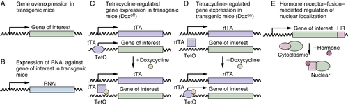
Transgenic mice have an extra copy of the gene of interest controlled by a ubiquitous or tissue-specific promoter (Figure 9-3 , A). The use of a cell-type–specific promoter provides spatial control over the expression of the transgene. A normal or mutant form of a gene can be overexpressed to ascertain its effect on tumor development. In addition to gene overexpression, transgenic mice can also be used to reduce gene expression or protein function. The expression of dominant negative or viral proteins that interfere with endogenous protein function has been used to assess the effect of disrupting certain pathways on in vivo tumorigenesis. In addition, RNA interference (RNAi) can be used to reduce the expression of a gene of interest in mice (see Figure 9-3, B). 17,18

Traditional transgenic mice constitutively express the transgene in the chosen cell type, potentially disrupting organ development or tissue homeostasis. Therefore, systems have been developed to allow the temporal control of transgene expression or function. Two complementary systems rely on a tetracycline-dependent transactivation to control the spatial and temporal expression of the gene of interest 19–21 (see Figure 9-3, C and D). The tetracycline transactivator (tTA) drives the expression of genes under the control of the bacterial tetracycline-dependent operator (tetO). The transactivation function of the tTA is blocked when tetracycline derivatives, often doxycycline, are present (see Figure 9-3, C). The reverse tTA (rtTA) works analogously to tTA, except that the expression of the tetO-controlled gene is induced only in the presence of doxycycline (see Figure 9-3, D). Exposure of mice with cell-type–specific expression of the tTA or rtTA transgene and a tetO-controlled gene of interest to doxycycline can be used to turn gene expression on and off. These systems have allowed investigators to control tumor initiation and evaluate the requirement for continued oncogene expression during tumor maintenance and progression. 22–28


image

Figure 9-2 Genetically modified mouse models of cancer (A,B) Transgenic gene expression and (C,D) the alteration of endogenous loci allow induction of tumors in mice with genetic alterations analogous to those in human cancer.


image

Figure 9-3 The toolbox for the transgenic control of gene expression and protein function in genetically modified mice (A,B) The use of tissue-specific promoters allows the expression of a gene or interfering RNA of interest in the desired tissue. (C,D) Regulation of gene expression by the tetracycline system adds a level of temporal control based on a change in conformation of the tetracycline transactivator (tTA) or reverse tetracycline transactivator (rtTA) in the presence of doxycycline. (E) The expression of hormone receptor (HR)-fusion proteins allows the nuclear translocation of proteins of interest only in the presence of the hormone.

The fusion of oncogenes and tumor suppressors to hormone receptors has also been used to regulate protein function by controlling their subcellular localization (see Figure 9-3, E). In-frame fusion of a gene of interest to the estrogen receptor (ER) or a truncated progesterone receptor (APR) creates a fusion protein that is sequestered in the cytoplasm until the cell is exposed to the appropriate hormone that induces its nuclear import (see Figure 9-3, E). Modified ERs (ER(TAM) and ER(T2)) have been created that translocate to the nucleus in the presence of 4-hydroxytamoxifen but not natural ER ligands, thus reducing background translocation. 29,30 These acutely switchable protein alleles have been used to determine the execution point for various nuclear proteins, including oncogenes and tumor suppressors. 31–33


Gene-Targeted Mice


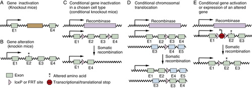
The ability to alter endogenous loci within the mouse genome has dramatically affected every field of biology. 34 Homologous recombination in embryonic stem cells allows the specific deletion or alteration of genomic loci (Figure 9-4 , A and B). This technique was initially used by cancer biologists to make germline deletions of several genes implicated in human cancer. 35–38 These conventional “knockout mice” lack the gene of interest in every cell in the animal. Germline deletion of some genes results in embryonic lethality, necessitating the analysis of heterozygous mutant mice or the use of conditional deletion strategies. Several tumor suppressor genes are mutated in the germlines of families, predisposing them to cancer, and mice with heterozygous deletion or mutation of these genes can serve as useful models to study tumor development under these sensitizing genetic conditions. 39–43

The ability to delete genes specifically in a chosen cell type is comparable to the use of tissue-specific promoters to drive transgene expression (see Figure 9-4, C). Using bacteriophage-derived Cre recombinase, it is possible to delete genomic regions flanked by loxP nucleotide sequences (these loci are referred to as floxed). 34,44 FLPe recombinase is used less frequently but can also be used to recombine loci flanked by FRT sequences (see Figure 9-4, C).

The development of mice that express Cre recombinase in defined cell types and the creation of floxed alleles of many important cancer genes have allowed researchers to investigate the role of these genes in the development of various types of tumors in a highly controlled manner. Cre recombinase can also be used to induce chromosomal translocations analogous to the translocations that are pathognomonic of certain hematopoietic cancers 45,46 (see Figure 9-4, D).


image

Figure 9-4 The toolbox for the deletion or genetic modification of endogenous genes in mice Genetic alteration of endogenous loci to inactivate (A), alter (B) or conditionally activate (D,E), or inactivate (C) genes. Homologous recombination allows the deletion or alteration of gene coding sequences. (C-E) The expression of a recombinase (Cre or FLPe) from a tissue-specific promoter or virus allows the spatially restricted deletion (C), translocation (D), or induced expression (E) of a targeted allele.

The expression of activated oncogenes is an important aspect of mouse models of human cancer. To express a mutated oncogene at its physiologic level from its endogenous promoter (as is the case in most human cancers), mice have been engineered with a floxed transcription/translation stop cassette in the first exon of a chosen mutant oncogene. These oncogenes remain silent until Cre-recombinase removes the stop cassette, allowing the expression of the mutant oncogene in the chosen cell type (see Figure 9-4, E).

Specific promoters direct the expression or deletion of genes to a desired cell lineage, and sophisticated systems can also allow the timing of gene alteration to be controlled. In these situations, however, every cell of the chosen cell type undergoes the same oncogenic event, which is in stark contrast to the initiation of human tumors where a single cell likely incurs the oncogenic alteration. Although inducing these genetic changes in a single cell may not be the most appropriate approach in experimental research, the use of viruses to deliver Cre recombinase to a subset of cells may be an acceptable medium. In these systems, viruses (often adenoviral or lentiviral vectors) are used to deliver Cre to a fraction of the cells in the organ of interest in mice that are genetically poised to express or delete genes of interest. These viral vectors have been used to initiate multifocal non–small-cell and small-cell lung cancer, hepatocellular carcinoma, ovarian cancer, and various brain tumors. 47–51

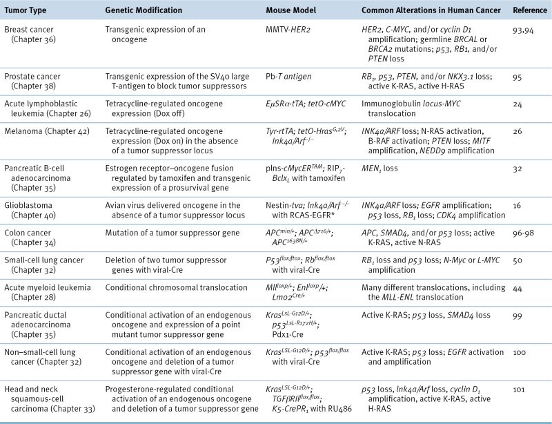
Rational creativity may be the underlying theme of these mouse models. Table 9-1 contains a selection of mouse models that use a variety of different genetic techniques to model different tumor types. As our knowledge of the genetic alterations in human cancers increases, our ability to control their expression in mice will also expand with the application of additional orthogonal systems.

Applications of Mouse Models to Cancer Biology


Combinations of the methods described in the preceding sections have been used to address several important questions in cancer biology, including oncogene addiction and the cooperation and interdependence of various oncogenes and tumor suppressors.


Cross-Species Comparisons


The comparison of tumors from different species has highlighted the central role for several oncogenic and tumor suppressor pathways. Mutations in p53 are found in about half of human tumors, but p53 is also mutated in tumors in the soft-shell clam, Mya arenaria, underscoring the importance of this tumor suppressor and the conservation of critical alterations across diverse phyla. 52,53 Cross-species comparison of gene expression and genomic changes in tumors from mice and humans has also yielded valuable insight into the important genetic changes in cancer. 54–58 The genetic changes in human tumors are often complex and are overlaid on the considerable allelic variation among individuals. Although possible, pinpointing the important somatic changes or genomic alterations can become unwieldily complex. 59 By comparing the overlapping genomic and genetic changes in mouse and human tumors of the same type (and even tumors containing several of the same oncogenic events), the minimal critical genetic changes can be established. In addition, these changes can be functionally validated in the mouse models that aided in their identification.


Table 9-1


Examples of Genetically Modified Mouse Models of Cancer

image


Oncogene Addiction


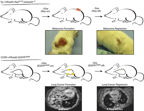
Mutation or overexpression of oncogenes can initiate tumor development. The use of tetracycline-regulated expression systems has documented the requirement for continued oncogene expression for growth and survival of established tumors and metastases. 22–28 Although most tumors undergo dramatic cell death and regress after oncogene inactivation, the regression is not always complete, and tumor subclones that escape the requirement for the initiating oncogene can recur (Figure 9-5 ). 55 Interestingly, in a model of MYC-induced hepatocellular carcinoma, MYC reactivation after tumor regression results in the development of tumors that are clonally related to the initial primary tumor, indicating that dormancy can also be a result of oncogene inactivation. 25 These dramatic results validate the future use of these models to predict the outcome of altering specific pathways predicted to influence tumor survival or progression. Clinically, pharmacologic oncogene inactivation can successfully reduce tumor growth, supporting the concept of oncogene addiction. In particular, a subset of non–small-cell lung cancer with mutant EGFR, 60,61 gastrointestinal stromal tumors with active/mutant c-Kit, 62 and chronic myeloid leukemia with the BCR-ABL translocation 63 have been successfully treated with small molecules targeting these driving oncogenes.


Oncogene Cooperation and Codependence


The hypothesis of a multistep model of tumorigenesis mediated by multiple genetic alterations raises the interesting question of how these genes cooperate to promote tumor development. In vitro studies in immortalized cell lines and primary fibroblasts were initially used to show the cooperativity of different oncogenes. 64 The tumor suppressor networks, the relationship between oncogenes and their target genes, and the cooperation of different genetic changes in promoting tumor initiation and progression have also been studied in vivo using genetic methods. 65–67 Genetic epistasis experiments in mice have identified several critical targets of specific oncogenes that mediate different aspects of tumorigenesis. 33,65,68 Moreover, genes that enhance or reduce the effect of tumor suppressor gene mutation and oncogene expression have also been identified. 66,67,69


image

Figure 9-5 Oncogene addiction of mouse tumors predicts the outcome of therapeutic blockade of oncogene expression or activity Melanocyte-specific oncogenic Ras expression leads to the formation of melanoma, which regresses when Ras is no longer expressed. Lung epithelial expression of an active point mutant of EGFR produces lung adenocarcinoma development. The maintenance of these lung tumors relies on the continued expression of the oncogene. (Melanoma images are courtesy Joseph Gans and Lynda Chin, Dana-Farber Cancer Institute, Harvard University. Lung adenocarcinoma images are courtesy Katerina Politi and Harold Varmus, Memorial Sloan-Kettering Cancer Center.)

Future Directions of Cancer Models


The most advanced genetically engineered mouse models of human cancer reflect their human counterparts at the genetic and histologic levels. These mouse models are now poised to lead the way to the discovery of new genes and pathways dysregulated in cancer and aid in the development and screening of potential therapeutics.


In Vivo Screens


Transposon and retroviral insertional mutagenesis, short-interfering RNA (siRNA) libraries, and advances in the analysis of gene expression and genomic alteration allow mouse models to be used as tools in the discovery of new cancer genes and pathways. Each of these approaches has been used to identify genes that promote tumorigenesis. 70–79 Unlike chemical or physical mutagens, insertional mutagens allow the identification of mutated genes. By using these mutagens in sensitized backgrounds (e.g., loss of a tumor suppressor or expression of an oncogene), the genes that regulate tumor initiation, invasion, or metastasis can be identified. Whole-genome or focused siRNA and shRNA library screens for genes that influence transformation have been conducted in vitro 74–76 and in vivo. 80–82 Genes discovered by these methods can be confirmed in the same tumor model in which they are found, and these unbiased approaches have begun to contribute to the identification of genes and pathways that are potential therapeutic targets.


Validation of Pharmaceutical Targets and Preclinical Trials


The development of new therapeutics requires carefully designed preclinical studies in models that most closely approximate human disease. Xenograft tumor models are the mainstay of current preclinical testing. Although several obstacles must be overcome before genetic mouse models can fully reach their potential in pharmacologic and biotechnological settings, these models may more accurately reflect the therapeutic response of patients. 7,8,83,84 The use of genetically defined mouse models may prioritize potential therapeutic compounds and stratify patients based on the mutational status of their tumors. 85 These efforts should accelerate the translation of novel therapies into the clinic.


Biomarkers for Early Tumor Detection


The detection of cancer at an early stage is of paramount importance, as patients diagnosed with early-stage disease invariably have a better prognosis. Unfortunately, there is a dearth of sensitive and reliable screening tests for most solid tumors. Here again, mouse models on inbred backgrounds with controllable and reproducible disease, coupled with advances in proteomic and molecular imaging technologies, may allow new diagnostic markers to be identified. 86


Identification of the Cell of Origin


Spatial and temporal restriction of genetic alterations in mice also allow the initial events that are triggered by oncogene expression to be investigated and the cells that respond to these initial genetic lesions to be identified. Specific genetic manipulation in defined cell types can identify the cell type in a given tissue that is susceptible to oncogenic transformation. 87,88 Alternatively, analyzing the cells that respond after in vivo oncogene activation may identify the cells of origin. 89 The appeal of these approaches is not solely to identify tumor-initiating cells but also to allow their subsequent manipulation and the identification of critical pathways dysregulated in these cells.


Recruitment and Function of Immune, Vascular, and Stromal Cells in the Tumor Environment


It has become increasingly clear that tumor growth and progression are greatly influenced by surrounding nontumor cells, including various immune cell types, vascular cells, stromal fibroblasts, and myofibroblasts. 90,91 Mouse models in which each of these tumor cell populations can be manipulated independently allow the function of each cell type to be identified. 92 Moreover, molecules that regulate the recruitment, survival, and function of these cells within the tumor can be characterized in mouse models in vivo. The secreted and cell-surface molecules used by these cells to communicate with each other and with the tumor cells will lead to the identification of important regulators of tumor growth, angiogenesis, invasion, and metastasis.

Conclusions


Genetically engineered mouse models of human cancers are an important component of the arsenal of experimental systems that will allow the in vivo dissection of tumor biology over the next several decades. The versatility of mouse models that recapitulate human cancer will lead to timely identification and validation of therapeutic targets that will ultimately influence human health.


References


1. Linzer D.I. , Levine A.J. Characterization of a 54K Dalton cellular SV40 tumor antigen present in SV40-transformed cells and uninfected embryonal carcinoma cells . Cell . 1979 ; 17 : 43 – 52 .


2. Lane D.P. , Crawford L.V. T antigen is bound to a host protein in SV40-transformed cells . Nature . 1979 ; 278 : 261 – 263 .


3. Sheiness D. , Bishop J.M. DNA and RNA from uninfected vertebrate cells contain nucleotide sequences related to the putative transforming gene of avian myelocytomatosis virus . J Virol . 1979 ; 31 : 514 – 521 .


4. Stehelin D. , Varmus H.E. , Bishop J.M. et al. DNA related to the transforming gene(s) of avian sarcoma viruses is present in normal avian DNA . Nature . 1976 ; 260 : 170 – 173 .


5. Van Dyke T. , Jacks T. Cancer modeling in the modern era: progress and challenges . Cell . 2002 ; 108 : 135 – 144 .


6. Hirst G.L. , Balmain A. Forty years of cancer modelling in the mouse . Eur J Cancer . 2004 ; 40 : 1974 – 1980 .


7. Sharpless N.E. , Depinho R.A. The mighty mouse: genetically engineered mouse models in cancer drug development . Nat Rev Drug Discov . 2006 ; 5 : 741 – 754 .


8. Singh M. , Johnson L. Using genetically engineered mouse models of cancer to aid drug development: an industry perspective . Clin Cancer Res . 2006 ; 12 : 5312 – 5328 .


9. Berghmans S. , Jette C. , Langenau D. et al. Making waves in cancer research: new models in the zebrafish . Biotechniques . 2005 ; 39 : 227 – 237 .


10. Poulin G. , Nandakumar R. , Ahringer J. Genome-wide RNAi screens in Caenorhabditis elegans: impact on cancer research . Oncogene . 2004 ; 23 : 8340 – 8345 .


11. Vidal M. , Cagan R.L. Drosophila models for cancer research . Curr Opin Genet Dev . 2006 ; 16 : 10 – 16 .


12. Pagliarini R.A. , Xu T. A genetic screen in Drosophila for metastatic behavior . Science . 2003 ; 302 : 1227 – 1231 .


13. Adams J.M. , Harris A.W. , Pinkert C.A. et al. The c-myc oncogene driven by immunoglobulin enhancers induces lymphoid malignancy in transgenic mice . Nature . 1985 ; 318 : 533 – 538 .


14. Hanahan D. Heritable formation of pancreatic beta-cell tumours in transgenic mice expressing recombinant insulin/simian virus 40 oncogenes . Nature . 1985 ; 315 : 115 – 122 .


15. Brinster R.L. , Chen H.Y. , Messing A. et al. Transgenic mice harboring SV40 T-antigen genes develop characteristic brain tumors . Cell . 1984 ; 37 : 367 – 379 .


16. Holland E.C. , Hively W.P. , DePinho R.A. , Varmus H.E. A constitutively active epidermal growth factor receptor cooperates with disruption of G1 cell-cycle arrest pathways to induce glioma-like lesions in mice . Genes Dev . 1998 ; 12 : 3675 – 3685 .


17. Dickins R.A. , McJunkin K. , Hernando E. et al. Tissue-specific and reversible RNA interference in transgenic mice . Nat Genet . 2007 ; 39 : 914 – 921 .


18. Premsrirut P.K. , Dow L.E. , Kim S.Y. et al. A rapid and scalable system for studying gene function in mice using conditional RNA interference . Cell . 2011 ; 145 : 145 – 158 .


19. Gossen M. , Bujard H. Tight control of gene expression in mammalian cells by tetracycline-responsive promoters . Proc Natl Acad Sci U S A . 1992 ; 89 : 5547 – 5551 .


20. Gossen M. , Freundlieb S. , Bender G. , Müller G. , Hillen W. , Bujard H. Transcriptional activation by tetracyclines in mammalian cells . Science . 1995 ; 268 : 1766 – 1769 .


21. Furth P.A. , St Onge L. , Böger H. et al. Temporal control of gene expression in transgenic mice by a tetracycline-responsive promoter . Proc Natl Acad Sci U S A . 1994 ; 91 : 93029306 .


22. Fisher G.H. , Wellen S.L. , Klimstra D. et al. Induction and apoptotic regression of lung adenocarcinomas by regulation of a K-Ras transgene in the presence and absence of tumor suppressor genes . Genes Dev . 2001 ; 15 : 3249 – 3262 .


23. Ji H. , Li D. , Chen L. et al. The impact of human EGFR kinase domain mutations on lung tumorigenesis and in vivo sensitivity to EGFR-targeted therapies . Cancer Cell . 2006 ; 9 : 485 – 495 .


24. Felsher D.W. , Bishop J.M. Reversible tumorigenesis by MYC in hematopoietic lineages . Mol Cell . 1999 ; 4 : 199 – 207 .


25. Shachaf C.M. , Kopelman A.M. , Arvanitis C. et al. MYC inactivation uncovers pluripotent differentiation and tumour dormancy in hepatocellular cancer . Nature . 2004 ; 431 : 1112 – 1117 .


26. Chin L. , Tam A. , Pomerantz J. et al. Essential role for oncogenic Ras in tumour maintenance . Nature . 1999 ; 400 : 468 – 472 .


27. Politi K. , Zakowski M.F. , Fan P.D. , Schonfeld E.A. , Pao W. , Varmus H.E. Lung adenocarcinomas induced in mice by mutant EGF receptors found in human lung cancers respond to a tyrosine kinase inhibitor or to down-regulation of the receptors . Genes Dev . 2006 ; 20 : 1496 – 1510 .


28. Moody S.E. , Sarkisian C.J. , Hahn K.T. et al. Conditional activation of Neu in the mammary epithelium of transgenic mice results in reversible pulmonary metastasis . Cancer Cell . 2002 ; 2 : 451 – 461 .


29. Littlewood T.D. , Hancock D.C. , Danielian P.S. , Parker M.G. , Evan G.I. A modified oestrogen receptor ligand-binding domain as an improved switch for the regulation of heterologous proteins . Nucleic Acids Res . 1995 ; 23 : 1686 – 1690 .


30. Indra A.K. , Warot X. , Brocard J. et al. Temporally-controlled site-specific mutagenesis in the basal layer of the epidermis: comparison of the recombinase activity of the tamoxifen-inducible Cre-ER(T) and Cre-ER(T2) recombinases . Nucleic Acids Res . 1999 ; 27 : 4324 – 4327 .


31. Christophorou M.A. , Ringshausen I. , Finch A.J. , Swigart L.B. , Evan G.I. The pathological response to DNA damage does not contribute to p53-mediated tumour suppression . Nature . 2006 ; 443 : 214 – 217 .


32. Pelengaris S. , Khan M. , Evan G.I. Suppression of Myc-induced apoptosis in beta cells exposes multiple oncogenic properties of Myc and triggers carcinogenic progression . Cell . 2002 ; 109 : 321 – 334 .


33. Finch A. , Prescott J. , Shchors K. et al. Bcl-xL gain of function and p19 ARF loss of function cooperate oncogenically with Myc in vivo by distinct mechanisms . Cancer Cell . 2006 ; 10 : 113 – 120 .


34. Capecchi M.R. Gene targeting in mice: functional analysis of the mammalian genome for the twenty-first century . Nat Rev Genet . 2005 ; 6 : 507 – 512 .


35. Jacks T. , Fazeli A. Schmitt EM, Bronson RT, Goodell MA, Weinberg RA. Effects of an Rb mutation in the mouse . Nature . 1992 ; 359 : 295 – 300 .


36. Jacks T. , Shih T.S. , Schmitt E.M. , Bronson R.T. , Bernards A. , Weinberg R.A. Tumour predisposition in mice heterozygous for a targeted mutation in Nf1 . Nat Genet . 1994 ; 7 : 353 – 361 .


37. Donehower L.A. , Harvey M. , Slagle B.L. et al. Mice deficient for p53 are developmentally normal but susceptible to spontaneous tumours . Nature . 1992 ; 356 : 215 – 221 .


38. Serrano M. , Lee H. , Chin L. , Cordon-Cardo C. , Beach D. , DePinho R.A. Role of the INK4a locus in tumor suppression and cell mortality . Cell . 1996 ; 85 : 27 – 37 .


39. Wallace M.R. , Marchuk D.A. , Andersen L.B. et al. Type 1 neurofibromatosis gene: identification of a large transcript disrupted in three NF1 patients . Science . 1990 ; 249 : 181 – 186 .


40. Malkin D. , Li F.P. , Strong L.C. et al. Germ line p53 mutations in a familial syndrome of breast cancer, sarcomas, and other neoplasms . Science . 1990 ; 250 : 1233 – 1238 .


41. Srivastava S. , Zou Z.Q. , Pirollo K. , Blattner W. , Chang E.H. Germ-line transmission of a mutated p53 gene in a cancer-prone family with Li-Fraumeni syndrome . Nature . 1990 ; 348 : 747 – 749 .


42. Groden J. , Thliveris A. , Samowitz W. et al. Identification and characterization of the familial adenomatous polyposis coli gene . Cell . 1991 ; 66 : 589 – 600 .


43. Olive K.P. , Tuveson D.A. , Ruhe Z.C. et al. Mutant p53 gain of function in two mouse models of Li-Fraumeni syndrome . Cell . 2004 ; 119 : 847 – 860 .


44. Gu H. , Marth J.D. , Orban P.C. , Mossman H. , Rajewsky K. Deletion of a DNA polymerase beta gene segment in T cells using cell type-specific gene targeting . Science . 1994 ; 265 : 103 – 106 .


45. Forster A. , Pannell R. , Drynan L.F. et al. Engineering de novo reciprocal chromosomal translocations associated with Mll to replicate primary events of human cancer . Cancer Cell . 2003 ; 3 : 449 – 458 .


46. Drynan L.F. , Pannell R. , Forster A. et al. Mll fusions generated by Cre- loxP-mediated de novo translocations can induce lineage reassignment in tumorigenesis . EMBO J . 2005 ; 24 : 3136 – 3146 .


47. Jackson E.L. , Willis N. , Mercer K. et al. Analysis of lung tumor initiation and progression using conditional expression of oncogenic K-ras . Genes Dev . 2001 ; 15 : 3243 – 3248 .


48. Kalamarides M. , Niwa-Kawakita M. , Leblois H. et al. Nf2 gene inactivation in arachnoidal cells is rate-limiting for meningioma development in the mouse . Genes Dev . 2002 ; 16 : 1060 – 1065 .


49. Dinulescu D.M. , Ince T.A. , Quade B.J. , Shafer S.A. , Crowley D. , Jacks T. Role of K-ras and Pten in the development of mouse models of endometriosis and endometrioid ovarian cancer . Nat Med . 2005 ; 11 : 63 – 70 .


50. Meuwissen R. , Linn S.C. , Linnoila R.I. , Zevenhoven J. , Mooi W.J. , Berns A. Induction of small cell lung cancer by somatic inactivation of both Trp53 and Rb1 in a conditional mouse model . Cancer Cell . 2003 ; 4 : 181 – 189 .


51. DuPage M. , Dooley A.L. , Jacks T. Conditional mouse lung cancer models using adenoviral or lentiviral delivery of Cre recombinase . Nat Protoc . 2009 ; 4 : 1064 – 1072 .


52. Vogelstein B. , Lane D. , Levine A.J. Surfing the p53 network . Nature . 2000 ; 408 : 307 – 310 .


53. Barker C.M. , Calvert R.J. , Walker C.W. et al. Detection of mutant p53 in clam leukemia cells . Exp Cell Res . 1997 ; 232 : 240 – 245 .


54. Lu J. , Getz G. , Miska E.A. et al. Micro RNA expression profiles classify human cancers . Nature . 2005 ; 435 : 834 – 838 .


55. Kim M. , Gans J.D. , Nogueira C. et al. Comparative oncogenomics identifies NEDD9 as a melanoma metastasis gene . Cell . 2006 ; 125 : 1269 – 1281 .


56. Zender L. , Spector M.S. , Xue W. et al. Identification and validation of oncogenes in liver cancer using an integrative oncogenomic approach . Cell . 2006 ; 125 : 1253 – 1267 .


57. Sweet-Cordero A. , Mukherjee S. , Subramanian A. et al. An oncogenic KRAS2 expression signature identified by cross-species gene-expression analysis . Nat Genet . 2005 ; 37 : 48 – 55 .


58. Winslow M.M. , Dayton T.L. , Verhaak R.G. et al. Suppression of lung adenocarcinoma progression by Nkx2-1 . Nature . 2011 ; 473 : 101 – 104 .


59. Sjoblom T. , Jones S. , Wood L.D. et al. The consensus coding sequences of human breast and colorectal cancers . Science . 2006 ; 314 : 268 – 274 .


60. Lynch T.J. , Bell D.W. , Sordella R. et al. Activating mutations in the epidermal growth factor receptor underlying responsiveness of non-small-cell lung cancer to gefitinib . N Engl J Med . 2004 ; 350 : 2129 – 2139 .


61. Paez J.G. , Janne P.A. , Lee J.C. et al. EGFR mutations in lung cancer: correlation with clinical response to gefitinib therapy . Science . 2004 ; 304 : 1497 – 1500 .


62. Heinrich M.C. , Corless C.L. , Demetri G.D. et al. Kinase mutations and imatinib response in patients with metastatic gastrointestinal stromal tumor . J Clin Oncol . 2003 ; 21 : 4342 – 4349 .


63. Druker B.J. , Talpaz M. , Resta D.J. et al. Efficacy and safety of a specific inhibitor of the BCR-ABL tyrosine kinase in chronic myeloid leukemia . N Engl J Med . 2001 ; 344 : 1031 – 1037 .


64. Land H. , Parada L.F. , Weinberg R.A. Tumorigenic conversion of primary embryo fibroblasts requires at least two cooperating oncogenes . Nature . 1983 ; 304 : 596 – 602 .


65. Batlle E. , Bacani J. , Begthel H. et al. EphB receptor activity suppresses colorectal cancer progression . Nature . 2005 ; 435 : 1126 – 1130 .


66. Ma L. , Teruya-Feldstein J. , Behrendt N. et al. Genetic analysis of Pten andTsc2 functional interactions in the mouse reveals asymmetrical haploinsufficiency in tumor suppression . Genes Dev . 2005 ; 19 : 1779 – 1786 .


67. Chen M.L. , Xu P.Z. , Peng X.D. et al. The deficiency of Aktl is sufficient to suppress tumor development in Pten mice . Genes Dev . 2006 ; 20 : 1569 – 1574 .


68. Weng A.P. , Millholland J.M. , Yashiro-Ohtani Y. et al. c-Myc is an important direct target of Notch1 in T-cell acute lymphoblastic leukemia/lymphoma . Genes Dev . 2006 ; 20 : 2096 – 2109 .


69. Jacobs J.J. , Scheijen B. , Voncken J.W. , Kieboom K. , Berns A. , van Lohuizen M. Bmi-1 collaborates with c-Myc in tumorigenesis by inhibiting c-Myc-induced apoptosis via INK4a/ARF . Genes Dev . 1999 ; 13 : 2678 – 2690 .


70. Mikkers H. , Allen J. , Knipscheer P. et al. High-throughput retroviral tagging to identify components of specific signaling pathways in cancer . Nat Genet . 2002 ; 32 : 153 – 159 .


71. Ding S. , Wu X. , Li G. , Han M. , Zhuang Y. , Xu T. Efficient transposition of the piggyBac (PB) transposon in mammalian cells and mice . Cell . 2005 ; 122 : 473 – 483 .


72. Collier L.S. , Carlson C.M. , Ravimohan S. , Dupuy A.J. , Largaespada D.A. Cancer gene discovery in solid tumours using transposon-based somatic mutagenesis in the mouse . Nature . 2005 ; 436 : 272 – 276 .


73. Dupuy A.J. , Akagi K. , Largaespada D.A. , Copeland N.G. , Jenkins N.A. Mammalian mutagenesis using a highly mobile somatic Sleeping Beauty transposon system . Nature . 2005 ; 436 : 221 – 226 .


74. Kolfschoten I.G. , van Leeuwen B. , Berns K. et al. A genetic screen identifies PITX1 as a suppressor of RAS activity and tumorigenicity . Cell . 2005 ; 121 : 849 – 858 .


75. Westbrook T.F. , Martin E.S. , Schlabach M.R. et al. A genetic screen for candidate tumor suppressors identifies REST . Cell . 2005 ; 121 : 837 – 848 .


76. Ngo V.N. , Davis R.E. , Lamy L. et al. A loss-of-function RNA interference screen for molecular targets in cancer . Nature . 2006 ; 441 : 106 – 110 .


77. Haupt Y. , Alexander W.S. , Barri G. , Klinken S.P. , Adams J.M. Novel zinc finger gene implicated as myc collaborator by retrovirally accelerated lymphomagenesis in E mu-myc transgenic mice . Cell . 1991 ; 65 : 753 – 763 .


78. van Lohuizen M. , Verbeek S. , Scheijen B. , Wientjens E. , van der Gulden H. , Berns A. Identification of cooperating oncogenes in E mu-myc transgenic mice by provirus tagging . Cell . 1991 ; 65 : 737 – 752 .


79. Suzuki T. , Shen H. , Akagi K. et al. New genes involved in cancer identified by retroviral tagging . Nat Genet . 2002 ; 32 : 166 – 174 .


80. Bric A. , Miething C. , Bialucha C.U. et al. Functional identification of tumor-suppressor genes through an in vivo RNA interference screen in a mouse lymphoma model . Cancer Cell . 2009 ; 16 : 324 – 335 .


81. Meacham C.E. , Ho E.E. , Dubrovsky E. , Gertler F.B. , Hemann M.T. In vivo RNAi screening identifies regulators of actin dynamics as key determinants of lymphoma progression . Nat Genet . 2009 ; 41 : 1133 – 1137 .


82. Zender L. , Xue W. , Zuber J. et al. An oncogenomics-based in vivo RNAi screen identifies tumor suppressors in liver cancer . Cell . 2008 ; 135 : 852 – 864 .


83. Olive K.P. , Jacobetz M.A. , Davidson C.J. et al. Inhibition of Hedgehog signaling enhances delivery of chemotherapy in a mouse model of pancreatic cancer . Science . 2009 ; 324 : 1457 – 1461 .


84. Singh M. , Lima A. , Molina R. et al. Assessing therapeutic responses in Kras mutant cancers using genetically engineered mouse models . Nat Biotechnol . 2010 ; 28 : 585 – 593 .


85. Chen Z. , Cheng K. , Walton Z. et al. A murine lung cancer co-clinical trial identifies genetic modifiers of therapeutic response . Nature . 2012 ; 483 : 613 – 617 .


86. Taguchi A. , Politi K. , Pitteri S.J. et al. Lung cancer signatures in plasma based on proteome profiling of mouse tumor models . Cancer Cell . 2011 ; 20 : 289 – 299 .


87. Passegue E. , Wagner E.F. , Weissman I.L. JunB deficiency leads to a myeloproliferative disorder arising from hematopoietic stem cells . Cell . 2004 ; 119 : 431 – 443 .


88. Sutherland K.D. , Proost N. , Brouns I. , Adriaensen D. , Song J.Y. , Berns A. Cell of origin of small cell lung cancer: inactivation of Trp53 and Rb1 in distinct cell types of adult mouse lung . Cancer Cell . 2011 ; 19 : 754 – 764 .


89. Kim C.F. , Jackson E.L. , Woolfenden A.E. et al. Identification of bronchioalveolar stem cells in normal lung and lung cancer . Cell . 2005 ; 121 : 823 – 835 .


90. Hanahan D. , Folkman J. Patterns and emerging mechanisms of the angiogenic switch during tumorigenesis . Cell . 1996 ; 86 : 353 – 364 .


91. Bhowmick N.A. , Neilson E.G. , Moses H.L. Stromal fibroblasts in cancer initiation and progression . Nature . 2004 ; 432 : 332 – 337 .


92. Kraman M. , Bambrough P.J. , Arnold J.N. et al. Suppression of antitumor immunity by stromal cells expressing fibroblast activation protein-alpha . Science . 2010 ; 330 : 827 – 830 .


93. Muller W.J. , Sinn E. , Pattengale P.K. , Wallace R. , Leder P. Single-step induction of mammary adenocarcinoma in transgenic mice bearing the activated c-neu oncogene . Cell . 1988 ; 54 : 105 – 115 .


94. Bouchard L. , Lamarre L. , Tremblay P.J. , Jolicoeur P. Stochastic appearance of mammary tumors in transgenic mice carrying the MMTV/c-neu oncogene . Cell . 1989 ; 57 : 931 – 936 .


95. Greenberg N.M. , DeMayo F. , Finegold M.J. et al. Prostate cancer in a transgenic mouse . Proc Natl Acad Sci U S A . 1995 ; 92 : 3439 – 3443 .


96. Su L.K. , Kinzler K.W. , Vogelstein B. et al. Multiple intestinal neoplasia caused by a mutation in the murine homolog of the APC gene . Science . 1992 ; 256 : 668 – 670 .


97. Oshima M. , Oshima H. , Kitagawa K. , Kobayashi M. , Itakura C. , Taketo M. Loss of Apc heterozygosity and abnormal tissue building in nascent intestinal polyps in mice carrying a truncated Apc gene . Proc Natl Acad Sci U S A . 1995 ; 92 : 4482 – 4486 .


98. Fodde R. , Edelmann W. , Yang K. et al. A targeted chain-termination mutation in the mouse Apc gene results in multiple intestinal tumors . Proc Natl Acad Sci U S A . 1994 ; 91 : 8969 – 8973 .


99. Hingorani S.R. , Wang L. , Multani A.S. et al. Trp53R172H and KrasG12D cooperate to promote chromosomal instability and widely metastatic pancreatic ductal adenocarcinoma in mice . Cancer Cell . 2005 ; 7 : 469 – 483 .


100. Jackson E.L. , Olive K.P. , Tuveson D.A. et al. The differential effects of mutant p53 alleles on advanced murine lung cancer . Cancer Res . 2005 ; 65 : 10280 – 10288 .


101. Lu S.L. , Herrington H. , Reh D. et al. Loss of transforming growth factor-beta type II receptor promotes metastatic head-and-neck squamous cell carcinoma . Genes Dev . 2006 ; 20 : 1331 – 1342 .

Only gold members can continue reading. Log In or Register to continue

Stay updated, free articles. Join our Telegram channel

Feb 15, 2017 | Posted by in ONCOLOGY | Comments Off on Genetic Mouse Models of Cancer

Full access? Get Clinical Tree

Get Clinical Tree app for offline access