Inflammation and Cancer


Figure 19-1 Hematopoiesis Hematopoietic progenitor cells (HPCs) can self-renew or differentiate into two multipotent progenitor cell types, myeloid and lymphoid progenitors, which further differentiate to give rise to all cells of the immune system. Myeloid cells differentiate into megakaryocytes, erythrocytes, mast cells, and myeloblasts, which further differentiate into macrophages and dendritic cells. Lymphoid cells give rise to natural killer (NK) cells, and small lymphocytes including T cells (including helper, cytotoxic, and regulatory T cells) and B cells. Together, these cell types work in concert to coordinate both innate and adaptive inflammatory responses.


Table 19-1


Inflammatory Conditions and Infectious Agents That Are Associated with Specific Types of Cancers


Condition/Infection Associated Neoplasm(s)
Asbestos Lung cancer
Bronchitis Lung cancer
Gingivitis Oral cancer
Inflammatory bowel disease Colorectal cancer
Skin inflammation (UV) Skin cancer
Hepatitis Liver cancer
AIDS Non-Hodgkin’s lymphoma
Chronic pancreatitis Pancreatic cancer

AIDS, Acquired immunodeficiency syndrome; UV, ultraviolet.
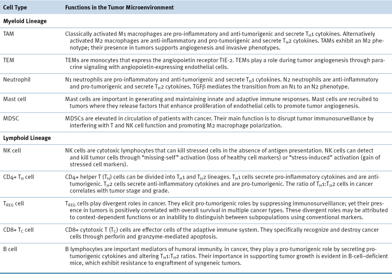

Another piece of evidence that deregulated inflammation contributes to tumorigenesis is the correlation between chronic viral infection and cancer initiation. In 1911, the discovery of a tumor virus in chickens by Peyton Rous, later termed the Rous sarcoma virus (RSV), was a pivotal discovery in molecular cancer biology that led to the discovery of src, the first oncogene. 6,7 Decades later, Bissell and colleagues demonstrated a clear connection between inflammation and tumorigenesis, when they showed that chickens systemically infected with RSV only developed tumors at the site of initial injection or a subsequent inflicted wound. 8,9 It is also known that people infected with hepatitis B or C virus are prone to developing cirrhosis of the liver, which increases the risk of hepatocellular carcinoma by 100-fold. 10 In fact, it has recently been estimated that approximately 2 million cancer cases worldwide, representing 16% of total cases, are caused by infectious agents every year. 11 For a list of infectious agents, inflammatory conditions, and associated cancers, refer to Table 19-1. 1

Recent attempts to understand the connection between infection, inflammation, and cancer have led to the Human Microbiome Project, which was initiated by the National Institutes of Health (NIH) Roadmap for Medical Research. 12 The project was launched in an effort to gain insight into how microorganisms influence health and disease, given that the human body contains 10 times more microbial cells than human cells, and 100 times more microbial genes (i.e., the microbiome) than human genes. 13 Indeed, it is estimated that in humans, the distal gut contains up to 1000 species and 7000 strains of microbes. It is currently known that microorganisms contribute to the development of diseases including cancer, despite often maintaining symbiotic relationships with the human body. 1315 For example, it has been shown that stomach cancer can arise from chronic gastric inflammation caused by Helicobacter pylori infection. Inflammatory bowel disease, comprising ulcerative colitis and Crohn’s disease, is also associated with recurrent bacterial infection and can predispose to colorectal cancer. 16,17 However, the specific mechanisms and intercellular interactions that disrupt microbial homeostasis, leading to inflammation-induced cancer, remain elusive and an area of active investigation.


Table 19-2


Stromal Cell Populations in the Tumor Microenvironment Are Defined by Various Cell Surface Markers and Have Distinct Functions During Tumorigenesis


image


Inflammation and the Metastatic Cascade


In each of the cases just described, the onset of tumorigenesis is supported by an unresolved inflammatory response that contributes to a pro-tumorigenic niche, characterized by a plethora of different stromal cell types, growth factors, and cytokines (Figure 19-2 ). 2,5 For example, studies in breast cancer have shown that the type of inflammatory response is an important predictor of tumor development. In particular, acute inflammation involving cytolytic CD8+ T lymphocytes, CD4+ T helper (TH1) cells, or classically activated M1 macrophages is generally anti-tumorigenic, whereas chronic inflammation involving B lymphocytes, CD4+ T helper (TH2) cells, or alternatively activated M2 macrophages is frequently pro-tumorigenic (Table 19-2 ). 2,18,19 These findings demonstrate a complex relationship between tumor cells and their microenvironment and suggest that the development of a pro-tumorigenic niche is highly dependent on the type of immune response that ensues.

The metastatic cascade begins at the primary tumor site, when tumor cells recruit a vascular supply (angiogenesis), invade through the extracellular matrix (ECM), intravasate into the circulation, disseminate through the body, extravasate at secondary sites, and self-renew to sustain secondary tumor growth. Bone marrow–derived cells have diverse effects on each step of the metastatic cascade and render the microenvironment susceptible or resistant to tumorigenic growth. 2 For example, studies in breast cancer have shown that tumor-associated macrophages (TAMs) are critical for promoting angiogenesis and tumor cell invasion and helping cancer cells to cross blood vessel walls. Within the circulation, platelets can enhance the survival of tumor cells by protecting them from NK cell-mediated death and promoting their adhesion to the endothelium at the site of metastasis. 20 Furthermore, myeloid-derived suppressor cells play a role in suppressing immune surveillance of cancer cells, promoting tumor growth. 21 In an elegant study by Lyden and colleagues, hematopoietic progenitor cells (HPCs) positive for vascular endothelial growth factor receptor 1 (VEGFR1) and endothelial progenitor cells positive for VEGFR2 were both shown to be required for mediating neovascularization at sites of future metastasis: the premetastatic niche. 22 Another study reported that recruited CD11c+ DC precursors were capable of assembling tumor-associated neovessels in a model of ovarian carcinoma. 23 As illustrated by these examples, the diverse effects of all these different immune cell types during multiple steps of the metastatic cascade underscore the complexity of tumor-microenvironment relationships in cancer. In the following section, various immune cell types and their roles during tumorigenesis and metastasis are reviewed.


image

Figure 19-2 The primary tumor environment Cells within a tumor are supported by a complex and dynamic microenvironment composed of multiple infiltrating cell types, including endothelial cells (which line blood and lymphatic vessels), cancer-associated fibroblasts (CAFs), and a variety of bone marrow–derived cells (BMDCs). Infiltrating BMDCs mediate inflammatory responses during cancer progression and can have negative or positive consequences. Major BMDCs within the tumor niche include tumor-associated macrophages (TAMs), myeloid-derived suppressor cells (MDSCs), TIE2-expressing monocytes (TEMs), mesenchymal stem cells (MSCs), and various other cell types from lymphocyte and monocyte lineages. This tumor-associated cellular cocktail largely dictates the evolution of the surrounding environment and, ultimately, the outcome of disease. (Image adapted from Joyce JA, Pollard JW. Microenvironmental regulation of metastasis. Nat Rev Cancer. 2009;9: 239-252).

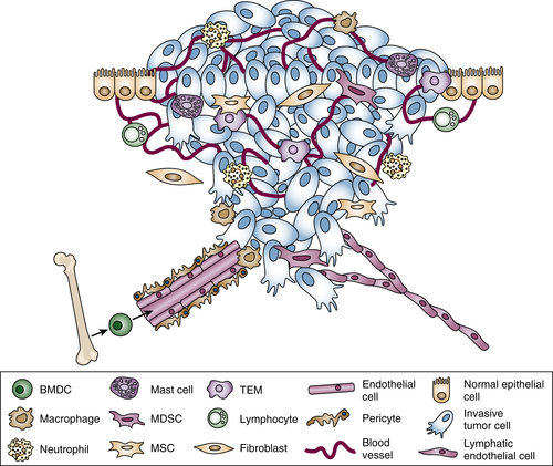
Immune Cells in Cancer
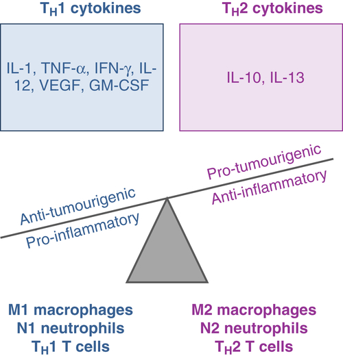


Myeloid Lineage


Macrophages


In normal physiological contexts, macrophages defend against infection, clear debris, and remodel injured tissue to maintain homeostasis. In cancer, normal macrophage function is hijacked by tumor cells to support tumor progression. In fact, in 80% of epithelial cancers, it has been shown that high macrophage infiltration is associated with poor patient prognosis. 24 TAMs typically represent the major immune cell type infiltrating tumors, and in some cancers, such as gliomas and breast cancer, TAMs can constitute up to 30% of the total tumor mass. TAM progenitors are largely recruited from the bone marrow and, once in the tumor mass, represent a critical source of secreted growth factors, proteases, and cytokines that participate in paracrine signaling loops with tumor cells to support invasive phenotypes. 25,26 One important function of TAMs is that they help tumor cells enter blood vessels, a process called intravasation. Condeelis and colleagues have published seminal studies using sophisticated multiphoton intravital imaging techniques to observe intimate macrophage–tumor cell interactions during metastatic dissemination in live animals. 27 These studies have shown that macrophages are primarily localized in perivascular areas, where they help tumor cells intravasate into the circulation (Figure 19-3 ). 2730


image

Figure 19-3 Live images of the tumor microenvironment of metastasis (TMEM) in a mouse breast carcinoma TMEM, the direct interaction of a macrophage, migratory tumor cell, and vascular endothelial cell, are sites of intravasation of tumor cells into the circulation. TMEM density correlates with increased risk of distant metastasis in breast cancer patients. 29,30 TMEM are detected in this live image, from a mouse model of breast cancer, as pairs of macrophages (green) and tumor cells (blue) attached to blood vessels (red), as visualized with a custom-built high-resolution multiphoton microscope. (Image courtesy of Drs. Allison Harney and John Condeelis. Microscope details obtained from Entenberg D, et al. Setup and use of a two-laser multiphoton microscope for multichannel intravital fluorescence imaging. Nat Protoc. 2011;6:1500-1520).

One explanation for the divergent functions of macrophages during normal tissue homeostasis versus tumorigenesis lies in their polarization state. Macrophages are phenotypically plastic. They can alter their polarization status to rapidly accommodate for the needs of different physiological contexts. At the extremes of their phenotypic continuum, macrophages range from M1 to M2 polarization states. 31 “Classically activated” (M1-polarized) macrophages produce type I pro-inflammatory cytokines and participate in antigen presentation, and they play an anti-tumorigenic role in cancer. 2,32 On the other hand, “alternatively activated” (M2-polarized) macrophages produce type II cytokines and anti-inflammatory responses, and they play a pro-tumorigenic role in cancer (Figure 19-4 ). 2,32 Of note, M2-polarized TAMs have been shown to promote tumorigenesis by providing a major source of proteases and chemokines that support tumor invasion and therapeutic resistance in multiple cancer types. 3336 For example, it has been shown that TAM-derived cathepsin proteases B and S promote breast cancer growth and metastasis by blunting chemotherapy-induced apoptosis. 34 Furthermore, tumor-secreted cytokines, such as interleukin-4 (IL-4), hijack macrophages in the tumor niche, activating them toward a pro-tumorigenic state. 33,37 Additional characterization of bidirectional interactions between tumor cells and TAMs will likely provide valuable information about how to manipulate the tumor niche in a therapeutic context.


image

Figure 19-4 The balance of pro- and anti-inflammatory cytokines and cellular states govern cancer outcome Tumor and stromal cells produce a variety of cytokines and chemokines that either contribute to or disrupt the development of a pro-tumorigenic niche. These factors can also reprogram infiltrating immune cells to adopt a pro- or anti-inflammatory state. Generally, M1, N1, and TH1 cell types produce and respond to TH1 cytokines and exhibit anti-tumorigenic phenotypes, whereas M2, N2, and TH2 cell types produce and respond to TH2 cytokines and exhibit pro-tumorigenic phenotypes. It is likely that this inherent plasticity is important for these cells to reestablish normal homeostasis during inflammation; however, their ability to adapt to different types of microenvironments is hijacked in the tumor niche. (Image adapted from Coussens LM, Werb Z. Inflammation and cancer. Nature. 2002;420:860-867).


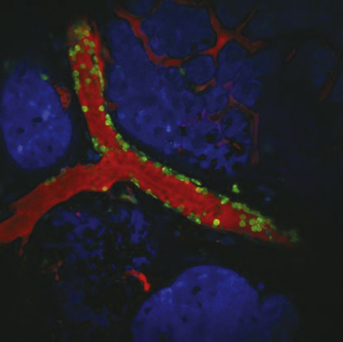
TIE-2–Expressing Monocytes


TEMs are monocytes that express TIE-2, which is a tyrosine kinase receptor for the angiogenic growth factor Angiopoietin. In healthy individuals, TEMs are present at very low levels in the bloodstream and are rarely detected in normal tissue. In contrast, in cancer patients, TEMs are present in higher numbers in the bloodstream and infiltrate neoplastic tissue. 38 TEMs have been implicated in various aspects of tumorigenesis, but they are best known for their role in promoting tumor angiogenesis. 38,39 TEMs regulate angiogenesis by participating in a paracrine signaling loop with Angiopoietin-expressing endothelial cells. This signaling loop is thought to contribute to the inefficacy of anti-angiogenic therapies, such as VEGF-targeted agents. Studies have shown that interfering with the TIE-2/Angiopoietin signaling axis in spontaneous breast or pancreatic neuroendocrine tumor models significantly inhibits tumor vascularization and blocks tumor growth. 40 These findings suggest that interfering with the TIE-2/Angiopoietin axis may have clinical benefits in reducing tumor vascularization; however, this has not yet been explored in humans.


Neutrophils


Neutrophil granulocytes are the most abundant circulating leukocyte population in humans and play an early role during inflammation by rapidly defending against microorganisms at the site of an infection. Likewise, neutrophils are recruited to tumor sites in response to cytokines and chemoattractants, and the neutrophil:lymphocyte ratio is used as a prognostic indicator of survival and therapeutic outcome in a variety of cancer types. 4145 At the tumor site, neutrophils have quite diverse roles. For example, it has been shown that neutrophils stimulated with granulocyte colony-stimulating factor (G-CSF) enhance the capacity of circulating tumor cells to seed and grow in secondary sites such as lung. 46 Infiltrating neutrophils were implicated in driving the initial angiogenic switch in pancreatic islet cancers through their ability to activate matrix metalloproteinase (MMP)-9. 47 In contrast, in a recent report on breast cancer by Benezra and colleagues, primary tumor-entrained neutrophils were shown to colonize the premetastatic lung, where they prevented metastatic colonization of disseminated tumor cells via H2O2-mediated cell death. 48 In another study, in renal cell carcinoma, it was demonstrated that tumor-secreted chemokines induce recruitment of neutrophils to the premetastatic lung, where they establish a barrier that blunts metastatic colonization. 49 Interestingly, microarray expression analysis revealed that poorly metastatic tumor cells expressed higher levels of these neutrophil chemokines compared to highly metastatic tumor cells, providing a possible explanation for their metastatic inefficiency. 49

In light of these divergent findings, the role of neutrophils during tumor progression remains unclear. However, similar to the different polarized states of macrophages, it has been shown that neutrophils can acquire N1 or N2 phenotypes, which have very different effects on cancer 50 (see Figure 19-4). The N1 phenotype is pro-inflammatory and elicits an anti-tumoral response by recruiting CD8+ T cells and secreting pro-inflammatory TH1 cytokines such as tumor necrosis factor-α (TNFα) or IL-12 (see Figure 19-4). In contrast, the N2 phenotype promotes tumorigenesis and plays a role in immune suppression mediated by transforming growth factor (TGF)-β. Indeed, studies have shown that blocking TGFβ signaling can revert pro-tumorigenic N2 neutrophils to an anti-tumorigenic N1 phenotype. 51 This inherent plasticity may explain why neutrophils have been reported to have both pro- and anti-tumorigenic effects in animal models and in patients. 50

Interestingly, a common side effect of patients undergoing chemotherapy is neutropenia, which is characterized by a significant deficiency in the population of neutrophil white blood cells. This deficiency underlies the extreme risk of infection in cancer patients, as the body loses a major rapid defensive mechanism against invading microorganisms. Recombinant G-CSF protein is frequently used in combination with chemotherapy in cancer patients in order to stimulate the bone marrow to produce more neutrophils. 52 Despite the fact that neutropenia is dangerous for patients because of increased risk of infection, retrospective analyses have reported that neutropenia in response to chemotherapy is correlated with improved overall survival, suggesting that a depletion of systemic neutrophils may be important for optimizing the anti-tumoral effects of therapy. 52,53


Mast Cells


Mast cells are also derived from myeloid progenitors and act as cellular barriers of infection. Mast cells contain histamine and are best known for their roles during allergic responses and autoimmune diseases. As mediators of inflammation, mast cells become activated in response to tissue injury and release their granules containing histamine, proteases, heparin, prostaglandins, and various cytokines into the microenvironment, where they induce proliferation of nearby endothelial cells. Of note, as tumors hijack inflammatory responses to tissue injury, it is not surprising that mast cells have also been implicated during tumor angiogenesis. 54,55 In patients, studies have shown that mast cells are abundant in the tumor microenvironment and that the degree of mast cell infiltration correlates with microvascular density and poor prognosis. 54,55 Moreover, in animal models of squamous-cell carcinoma and pancreatic islet cancer, oncogene activation was shown to rapidly recruit mast cells, which were essential for induction of angiogenesis via activation of different proteolytic cascades. 56,57


Myeloid-Derived Suppressor Cells


Myeloid-derived suppressor cells (MDSCs) are immunosuppressive precursors of dendritic cells, macrophages, and granulocytes and play a role in maintaining normal tissue homeostasis in response to various systemic insults. MDSCs are elevated in the circulation in response to bacterial and parasitic infection 5860 and are also elevated in the blood of tumor-bearing hosts. Mobilization of MDSCs into the bloodstream is mediated by cytokines that are secreted by tumor cells. For example, studies in breast cancer have shown that TGFβ signaling in tumor cells increases secretion of tumor-derived chemokine CXCL5, which acts as a chemoattractant for CXCR2-expressing MDSCs. 61 Interestingly, the accumulation of MDSCs is a common response to cancer therapy and is thought to contribute to lack of therapeutic efficacy. For instance, MDSCs have been shown to accumulate in the blood of breast cancer patients receiving doxorubicin-cyclophosphamide chemotherapy and are present in highest numbers in stage IV patients with metastatic disease. 62

Once MDSCs arrive at the tumor site, their main function is to disrupt major mechanisms of tumor immune surveillance, including antigen presentation and cell-mediated anti-tumor immunity. 21 They achieve immune suppression in multiple ways: First, they have been reported to directly inhibit proliferation and activation of CD4+ T cells and CD8+ T cells. 63,64 Several studies have demonstrated that the immunosuppressive effects of MDSCs on T cells are largely mediated by the production of nitric oxide, among other factors. 6466 They are also associated with promoting M2-polarization of TAMs to yield a pro-tumorigenic phenotype, as described earlier. 67 MDSCs have also been shown to participate in a paracrine loop with TAMs, whereby MDSC-derived IL-10 (a type II cytokine) causes inhibition of macrophage-derived IL-12 (a type I cytokine), which feeds back to amplify the production of IL-10 from MDSCs. 68 Last, they are capable of impairing NK cell function. Specifically, MDSCs inhibit cytotoxicity of NK cells by reducing production of NK-derived interferon γ (IFNγ) in a TGF-β–dependent manner. 69,70 In light of these findings, it is not surprising that increased peripheral MDSC levels correlate with advanced disease. 62


Lymphoid Lineage


NK Cells


Natural killer (NK) cells are cytotoxic lymphocytes that play an important role in both innate and adaptive immune responses. NK cells are unique compared to other lymphoid cell types in that they are capable of distinguishing between healthy cells and stressed cells (such as tumor cells) in the absence of antigen presentation. 71 Therefore, it is generally recognized that NK cells provide an anti-tumorigenic immune response. Indeed, reduced NK cell levels in patients with cancer have been shown to correlate with decreased overall survival. 7274 One way that NK cells can recognize and kill cancer is through “missing-self” activation, whereby MHC class I molecules, which characterize healthy cells and inhibit NK cytotoxicity, are lost from tumor cells. Alternatively, NK cells can recognize cancer through “stress-induced activation,” whereby stress-induced ligands are upregulated on tumor cells to activate receptors on adjacent NK cells. 71

Despite the many anti-tumor roles that NK cells have, it has been recently shown that following surgical removal of tumors in cancer patients, NK cell cytotoxicity is significantly reduced, largely attributed to surgical tissue stress. 75 This effect is correlated with increased metastatic incidence in mouse models, demonstrating that the anti-tumorigenic functions of NK cells can be disrupted with standard-of-care treatment regimens. Therefore, therapies that opt to enhance NK populations following surgery may provide an opportunity for improving therapeutic efficacy at early stages of treatment.


CD4+ Helper T Cells


CD4+ helper T (TH) cells play diverse roles in cancer. Like polarized macrophages, they can be subdivided into phenotypically divergent TH1 and TH2 lineages, directed by secretion of IL-12 and IL-4, respectively. 76 Specifically, TH1 cells secrete pro-inflammatory cytokines, including IFN-γ, TNFα, IL-2, and IL-12 (see Figure 19-4) to induce antigen presentation on MHC molecules by antigen-presenting cells (APCs), and CD8+ T cell cytotoxicity. They also exhibit direct cytotoxic functions by releasing granules that directly kill tumor cells in their microenvironment. 77 On the other hand, TH2 cells play a role in humoral immunity. They secrete high levels of anti-inflammatory cytokines, such as IL4-6, IL-10, and IL-13 (see Figure 19-4), and elicit pro-tumorigenic effects through inhibition of CD8+ T-cell cytotoxicity and immunosuppression to promote tumor growth. 77

Despite the finding that TH2 cells are generally associated with pro-tumorigenic functions, they may retain some inherent plasticity that allows them to provide beneficial anti-tumor effects. For instance, it has been shown that TH2 cell infiltrates are positively correlated with disease-free survival in patients with Hodgkin’s lymphoma. 78 In other types of cancers, the TH cell balance seems to be more important for disease outcome. For example, in breast cancer, the ratio of TH1 to TH2 cells within the tumor microenvironment correlates with tumor stage and grade, suggesting that the balance of TH cell types, rather than the degree of infiltration of any one TH subset, may be useful for predicting prognosis in patients. 79


Regulatory T Cells (TREGS)


Regulatory T cells, or TREG cells, are a unique subtype of CD4+FoxP3+ T cells that play divergent roles during tumorigenesis. On the one hand, increased TREG cell infiltrate has been correlated with reduced overall survival in various cancers such as breast cancer and hepatocellular carcinoma. 80,81 In these pro-tumorigenic roles, TREG cells function by releasing immunosuppressive factors, such as TGF-β and TH2 cytokines, which disrupt antigen presentation by APCs and impair the anti-tumorigenic effects of CD8+ cytotoxic T cells and NK cells. 82,83 Furthermore, TREG cells have been shown to impair M1-polarization of macrophages and support immunosuppressive myeloid cells. 84


On the other hand, it has been shown that infiltration of TREG cells is correlated with improved overall survival in cancers as diverse as colorectal cancer, bladder cancer, and head and neck cancer. 8587 The mechanisms of these divergent roles remain elusive in the literature; it is not clear whether TREG cells exhibit context-dependent functionality or whether they encompass multiple subpopulations of cell types, with distinct functions, that are not differentiated using conventional markers. 76


CD8+ Cytotoxic T Cells


Cytotoxic CD8+ T (TC) cells are lymphocytes that kill cells infected with viruses and also have the capacity to kill tumor cells. They express T-cell receptors (TCRs) on their surfaces, which recognize antigens presented on MHC molecules on APCs. In cancer, infiltration of TC cells is associated with an anti-tumorigenic capacity across a wide range of cancer types, including melanoma, breast cancer, ovarian cancer, gliomas, and hepatocellular carcinoma, and is associated with prolonged overall survival in patients. 76,88,89 On activation, TC cells release granules containing cytotoxic factors, for example, perforin and granzyme proteases, which induce apoptosis of infected cells. 83 It has been reported that TC cells are abundant at the invasive edge of tumors, where they improve patient response to chemotherapy and prognosis. 88


B Cells


B cells play an important role in humoral immunity and mediate both antibody production and activation of T cells through antigen presentation. They are most abundant at the tumor invasive edge and in adjacent tertiary lymphoid structures. 76 Although infiltration of B cells in tumors tends to be correlated with better disease outcome and prolonged survival in patients, it remains largely unclear whether B cells have positive or negative roles in disease progression based on confounding literature. 90 For instance, it has been reported that B cells elicit pro-tumorigenic effects by secreting tumorigenic cytokines, altering the ratio of TH1:TH2 cell infiltrates within tumors, and mediating immune cell recruitment. 91 In addition, studies in breast cancer have shown that B cells influence phenotypic transitions between T-cell states and mediate the conversion of CD4+ TH cells into TREG cells to promote metastatic dissemination. 92 In contrast, other studies have demonstrated anti-tumorigenic effects of tumor-infiltrating B cells. For example, B cells can release cytotoxic factors to directly kill tumor cells, such as TNFα and the protease granzyme B, and can induce immune suppression through secretion of IL-10. 93 Taken together, these studies suggest that further classification of B-cell subpopulations may explain their diverse functions and may suggest novel therapeutic avenues for disease intervention.

Nonhematopoietic Stromal Cell Types in the Tumor Microenvironment


In addition to the inflammatory cells that constitute the primary tumor and metastatic microenvironment, additional accessory cells in the tumor stroma, such as cancer-associated fibroblasts (CAFs) and mesenchymal stem cells (MSCs), engage in a dynamic interplay between immune cells and tumor cells. In the following section, we discuss the role of CAFs and MSCs during tumor progression and how immune cells modulate their function.


Cancer-Associated Fibroblasts


Fibroblasts are a predominant cell type in connective tissue and are responsible for depositing ECM and basement membrane components, regulating differentiation events in associated epithelial cells, modulating immune responses, and mediating wound healing. 94,95 In the tumor microenvironment, fibroblasts are present in aberrantly high numbers and are generally regarded as genomically stable compared to tumor cells. 96 However, CAFs are quite different from normal fibroblasts; it has been reported that normal prostate epithelial cells resemble intraepithelial neoplasia in mice when co-injected with CAFs, but not when co-injected with normal fibroblasts. 97 Once CAFs accumulate in the tumor microenvironment, they are activated by growth factors and cytokines such as TGF-β, platelet-derived growth factor (PDGF), and various proteases. 95,98,99 Following activation, CAFs provide a major source of oncoproteins and growth factors that fuel the growing tumor and modulate immune function. 100,101 For example, CAFs are a significant source of hepatocyte growth factor (HGF) in the tumor microenvironment, which supports angiogenesis, acts as a chemotactic agent for monocytes, and interferes with normal function and maturation of B cells. 102104


Mesenchymal Stem Cells


Bone marrow–derived MSCs play an important role during tissue remodeling and repair. They are mobilized into the circulation in response to cytokines released by tissue injury and have a multipotent capacity to give rise to osteoblasts, adipocytes, and chondrocytes. 105 Given that tumorigenesis shares many similarities with the process of wound healing, tumors are likewise able to mobilize and recruit bone marrow–derived MSCs to support remodeling events. 106 Indeed, it has been shown that mobilization of MSCs into circulation is pronounced in patients with advanced breast cancer and is associated with metastasis and chemoresistance. 107,108 Similar to the divergent roles that immune cells play in cancer, MSCs likewise have seemingly distinct functions. For example, it has been shown that glioma cells co-injected with MSCs into BALB/c-nu/nu mice exhibit a marked reduction of tumor vascularization compared to glioma cells injected with normal astrocytes. 109 In contrast, other studies have demonstrated a positive regulatory relationship between MSCs and tumor angiogenesis, whereby co-injection of MSCs and colon cancer cells into BALB/c-nu/nu mice induces a significant increase in tumor volume and microvascular density. 110 The differential effects of MSCs may be due to their capacity to modulate immune cells and thereby change the landscape and secretome of the tumor microenvironment. 105 Indeed, MSCs have been shown to alter both innate and adaptive arms of the immune system. For example, tumor-associated MSCs have been shown to suppress the pro-inflammatory function of dendritic cells, by blunting their ability to produce TNFα. 101 Furthermore, MSCs can interfere with NK activation and cytotoxicity. 111 Finally, MSCs have been shown to negatively regulate CD8+ TC cell–mediated cytotoxicity and have a capacity to switch pro-inflammatory TH1 cells to adopt an anti-inflammatory phenotype. 101,112 Taken together, in addition to the complex relationship between tumor cells and immune cells, additional accessory cells in the tumor-associated stroma contribute to disease progression by influencing tumor-immune interactions.

Summary


Despite the classical role of the immune system in defending against infection and systemic insult, many studies have shown that immune cell functions in the tumor niche often correlate with disease progression. This correlation is largely attributed to the reprogramming effects of the tumor microenvironment, which contains a plethora of growth factors and cytokines that hijack immune cells to play oncogenic roles. Although many immune cells are receptive to tumor-secreted factors and can adopt pro-tumorigenic functions, these cells have also been reported to retain many defensive functions in the tumor microenvironment, speaking to the complexity of tumor–stroma interactions during disease. It is likely that the diverse outcomes of immune cell function are highly context dependent, involving a reprogramming decision that is based on a large variety of factors, including tissue type, tumor secretome, tumor landscape, oxygen availability, tissue pH, and ECM architecture.

The therapeutic implications of tumor microenvironment research are vast. For example, disrupting key tumor–stroma interactions that play known roles during disease progression may interfere with the tumor’s ability to exploit immune cells in the microenvironment. Furthermore, attempts have been made to deplete specific immune-cell populations in the tumor environment; however, this type of an approach has been reported to lead to compensatory infiltration of alternative cell types that completely alter the tumor landscape in a way that is unpredictable and often unfavorable. 113,114 Of note, rather than depleting immune cell populations in tumors, it may be advantageous to develop therapies that opt to “re-educate” immune cells in the microenvironment, in order to take advantage of the valuable defense functions of these cell types. For instance, inducing anti-tumorigenic activation states in plastic immune-cell types, such as forcing classical activation of TAMs, may be a unique way to manipulate the tumor microenvironment in a way that re-establishes normal mechanisms of tissue homeostasis. Many of these therapies have been studied or are currently being explored in the laboratory; it will be interesting to observe the evolution of these novel approaches in patients in the future, with the ultimate goal of maximizing positive responses to therapy.


References


1. Coussens L.M. , Werb Z. Inflammation and cancer . Nature . 2002 ; 420 : 860 867 .


2. Joyce J.A. , Pollard J.W. Microenvironmental regulation of metastasis . Nat Rev Cancer . 2009 ; 9 : 239 252 .


3. Orkin S.H. Diversification of haematopoietic stem cells to specific lineages . Nat Rev Genet . 2000 ; 1 : 57 64 .


4. Balkwill F. , Mantovani A. Inflammation and cancer: back to Virchow? Lancet . 2001 ; 357 : 539 545 .


5. Medzhitov R. Origin and physiological roles of inflammation . Nature . 2008 ; 454 : 428 435 .


6. Murphy J.B. , Rous P. The behavior of chicken sarcoma implanted in the developing embryo . J Exp Med . 1912 ; 15 : 119 132 .


7. Rous P. A transmissible avian neoplasm. (Sarcoma of the common fowl.) . J Exp Med . 1910 ; 12 : 696 705 .


8. Martins-Green M. , Boudreau N. , Bissell M.J. Inflammation is responsible for the development of wound-induced tumors in chickens infected with Rous sarcoma virus . Cancer Res . 1994 ; 54 : 4334 4341 .


9. Dolberg D.S. , Hollingsworth R. , Hertle M. , Bissell M.J. Wounding and its role in RSV-mediated tumor formation . Science . 1985 ; 230 : 676 678 .


10. Bruix J. , Llovet J.M. Hepatitis B virus and hepatocellular carcinoma . J Hepatol . 2003 ; 39 ( suppl 1 ) : S59 S63 .


11. de Martel C. , Ferlay J. , Franceschi S. et al. Global burden of cancers attributable to infections in 2008: a review and synthetic analysis . Lancet Oncol . 2012 ; 13 : 607 6215 .


12. Proctor L.M. The Human Microbiome Project in 2011 and beyond . Cell Host Microbe . 2011 ; 10 : 287 291 .


13. Cho I. , Blaser M.J. The human microbiome: at the interface of health and disease . Nat Rev Genet . 2012 ; 13 : 260 270 .


14. Howitt M.R. , Garrett W.S. A complex microworld in the gut: gut microbiota and cardiovascular disease connectivity . Nat Med . 2012 ; 18 : 1188 1189 .


15. Schwabe R.F. , Wang T.C. Cancer. Bacteria deliver a genotoxic hit . Science . 2012 ; 338 : 52 53 .


16. Manichanh C. , Borruel N. , Casellas F. , Guarner F. The gut microbiota in IBD . Nat Rev Gastroenterol Hepatol . 2012 ; 9 : 599 608 .


17. Lampe J.W. The Human Microbiome Project: getting to the guts of the matter in cancer epidemiology . Cancer Epidemiol Biomarkers Prev . 2008 ; 17 : 2523 2524 .


18. DeNardo D.G. , Brennan D.J. , Rexhepaj E. et al. Leukocyte complexity predicts breast cancer survival and functionally regulates response to chemotherapy . Cancer Discov . 2011 ; 1 : 54 67 .


19. DeNardo D.G. , Coussens L.M. Inflammation and breast cancer. Balancing immune response: crosstalk between adaptive and innate immune cells during breast cancer progression . Breast Cancer Res . 2007 ; 9 : 212 .


20. Gay L.J. , Felding-Habermann B. Contribution of platelets to tumour metastasis . Nat Rev Cancer . 2011 ; 11 : 123 134 .


21. Ostrand-Rosenberg S. , Sinha P. Myeloid-derived suppressor cells: linking inflammation and cancer . J Immunol . 2009 ; 182 : 4499 4506 .


22. Lyden D. , Hattori K. , Dias S. et al. Impaired recruitment of bone-marrow-derived endothelial and hematopoietic precursor cells blocks tumor angiogenesis and growth . Nat Med . 2001 ; 7 : 1194 1201 .


23. Conejo-Garcia J.R. , Benencia F. , Courreges M.C. et al. Tumor-infiltrating dendritic cell precursors recruited by a beta-defensin contribute to vasculogenesis under the influence of Vegf-A . Nat Med . 2004 ; 10 : 950 958 .


24. Bingle L. , Brown N.J. , Lewis C.E. The role of tumour-associated macrophages in tumour progression: implications for new anticancer therapies . J Pathol . 2002 ; 196 : 254 265 .


25. Pollard J.W. Tumour-educated macrophages promote tumour progression and metastasis . Nat Rev Cancer . 2004 ; 4 : 71 78 .


26. Gocheva V. , Joyce J.A. Cysteine cathepsins and the cutting edge of cancer invasion . Cell Cycle . 2007 ; 6 : 60 64 .


27. Wyckoff J.B. , Wang Y. , Lin E.Y. et al. Direct visualization of macrophage-assisted tumor cell intravasation in mammary tumors . Cancer Res . 2007 ; 67 : 2649 2656 .


28. Entenberg D. , Wyckoff J. , Gligorijevic B. et al. Setup and use of a two-laser multiphoton microscope for multichannel intravital fluorescence imaging . Nat Protoc . 2011 ; 6 : 1500 1520 .


29. Condeelis J. , Pollard J.W. Macrophages: obligate partners for tumor cell migration, invasion, and metastasis . Cell . 2006 ; 124 : 263 266 .


30. Robinson B.D. , Sica G.L. , Liu Y.F. et al. Tumor microenvironment of metastasis in human breast carcinoma: a potential prognostic marker linked to hematogenous dissemination . Clin Cancer Res . 2009 ; 15 : 2433 2441 .


31. Mosser D.M. , Edwards J.P. Exploring the full spectrum of macrophage activation . Nat Rev Immunol . 2008 ; 8 : 958 969 .


32. Mantovani A. , Sica A. , Sozz ani S. et al. The chemokine system in diverse forms of macrophage activation and polarization . Trends Immunol . 2004 ; 25 : 677 686 .


33. Gocheva V. , Wang H.W. , Gadea B.B. et al. IL-4 induces cathepsin protease activity in tumor-associated macrophages to promote cancer growth and invasion . Genes Dev . 2010 ; 24 : 241 255 .


34. Shree T. , Olson O.C. , Elie B.T. et al. Macrophages and cathepsin proteases blunt chemotherapeutic response in breast cancer . Genes Dev . 2011 ; 25 : 2465 2479 .


35. Bell-McGuinn K.M. , Garfall A.L. , Bogyo M. et al. Inhibition of cysteine cathepsin protease activity enhances chemotherapy regimens by decreasing tumor growth and invasiveness in a mouse model of multistage cancer . Cancer Res . 2007 ; 67 : 7378 7385 .


36. Joyce J.A. , Baruch A. , Chehade K. et al. Cathepsin cysteine proteases are effectors of invasive growth and angiogenesis during multistage tumorigenesis . Cancer Cell . 2004 ; 5 : 443 453 .


37. Wang H.W. , Joyce J.A. Alternative activation of tumor-associated macrophages by IL-4: priming for protumoral functions . Cell Cycle . 2010 ; 9 : 4824 4835 .


38. Venneri M.A. , De Palma M. , Ponzoni M. et al. Identification of proangiogenic TIE2-expressing monocytes (TEMs) in human peripheral blood and cancer . Blood . 2007 ; 109 : 5276 5285 .


39. De Palma M. , Venneri M.A. , Galli R. et al. Tie2 identifies a hematopoietic lineage of proangiogenic monocytes required for tumor vessel formation and a mesenchymal population of pericyte progenitors . Cancer Cell . 2005 ; 8 : 211 226 .


40. Mazzieri R. , Pucci F. , Moi D. et al. Targeting the ANG2/TIE2 axis inhibits tumor growth and metastasis by impairing angiogenesis and disabling rebounds of proangiogenic myeloid cells . Cancer Cell . 2011 ; 19 : 512 526 .


41. Walsh S.R. , Cook E.J. , Goulder F. et al. Neutrophil-lymphocyte ratio as a prognostic factor in colorectal cancer . J Surg Oncol . 2005 ; 91 : 181 184 .


42. Cho H. , Hur H.W. , Kim S.W. et al. Pre-treatment neutrophil to lymphocyte ratio is elevated in epithelial ovarian cancer and predicts survival after treatment . Cancer Immunol Immunother . 2009 ; 58 : 15 23 .


43. Sarraf K.M. , Belcher E. , Raevsky E. et al. Neutrophil/lymphocyte ratio and its association with survival after complete resection in non-small cell lung cancer . J Thorac Cardiovasc Surg . 2009 ; 137 : 425 428 .


44. Shimada H. , Takiguchi N. , Kainuma O. et al. High preoperative neutrophil-lymphocyte ratio predicts poor survival in patients with gastric cancer . Gastric Cancer . 2010 ; 13 : 170 176 .


45. Chua W. , Charles K.A. , Baracos V.E. et al. Neutrophil/lymphocyte ratio predicts chemotherapy outcomes in patients with advanced colorectal cancer . Br J Cancer . 2011 ; 104 : 1288 1295 .


46. Kowanetz M. , Wu X. , Lee J. et al. Granulocyte-colony stimulating factor promotes lung metastasis through mobilization of Ly6G+Ly6C+ granulocytes . Proc Natl Acad Sci U S A . 2010 ; 107 : 21248 21255 .


47. Nozawa H. , Chiu C. , Hanahan D. Infiltrating neutrophils mediate the initial angiogenic switch in a mouse model of multistage carcinogenesis . Proc Natl Acad Sci U S A . 2006 ; 103 : 12493 12498 .


48. Granot Z. , Henke E. , Comen E.A. et al. Tumor entrained neutrophils inhibit seeding in the premetastatic lung . Cancer Cell . 2011 ; 20 : 300 314 .


49. Lopez-Lago M.A. , Posner S. , Thodima V.J. et al. Neutrophil chemokines secreted by tumor cells mount a lung antimetastatic response during renal cell carcinoma progression . Oncogene . 2013 ; 32 : 1752 1760 .


50. Fridlender Z.G. , Albelda S.M. Tumor-associated neutrophils: friend or foe? Carcinogenesis . 2012 ; 33 : 949 955 .


51. Fridlender Z.G. , Sun J. , Kim S. et al. Polarization of tumor-associated neutrophil phenotype by TGF-beta: “N1” versus “N2” TAN . Cancer Cell . 2009 ; 16 : 183 194 .


52. Epstein R.J. The CXCL12-CXCR4 chemotactic pathway as a target of adjuvant breast cancer therapies . Nat Rev Cancer . 2004 ; 4 : 901 909 .


53. Mayers C. , Panzarella T. , Tannock I.F. Analysis of the prognostic effects of inclusion in a clinical trial and of myelosuppression on survival after adjuvant chemotherapy for breast carcinoma . Cancer . 2001 ; 91 : 2246 2257 .


54. Lin E.Y. , Pollard J.W. Role of infiltrated leucocytes in tumour growth and spread . Br J Cancer . 2004 ; 90 : 2053 2058 .


55. Ribatti D. , Vacca A. , Nico B. et al. The role of mast cells in tumour angiogenesis . Br J Haematol . 2001 ; 115 : 514 521 .


56. Soucek L. , Lawlor E.R. , Soto D. et al. Mast cells are required for angiogenesis and macroscopic expansion of Myc-induced pancreatic islet tumors . Nat Med . 2007 ; 13 : 1211 1218 .


57. Coussens L.M. , Raymond W.W. , Bergers G. et al. Inflammatory mast cells up-regulate angiogenesis during squamous epithelial carcinogenesis . Genes Dev . 1999 ; 13 : 1382 1397 .


58. Haile L.A. , von Wasielewski R. , Gamrekelashvili J. et al. Myeloid-derived suppressor cells in inflammatory bowel disease: a new immunoregulatory pathway . Gastroenterology . 2008 ; 135 : 871 881 881 e1–e5 .


59. Delano M.J. , Scumpia P.O. , Weinstein J.S. et al. MyD88-dependent expansion of an immature GR-1(+)CD11b(+) population induces T cell suppression and Th2 polarization in sepsis . J Exp Med . 2007 ; 204 : 1463 1474 .


60. Cuervo H. , Guerrero N.A. , Carbajosa S. et al. Myeloid-derived suppressor cells infiltrate the heart in acute Trypanosoma cruzi infection . J Immunol . 2011 ; 187 : 2656 2665 .


61. Yang L. , Huang J. , Ren X. et al. Abrogation of TGF beta signaling in mammary carcinomas recruits Gr-1+CD11b+ myeloid cells that promote metastasis . Cancer Cell . 2008 ; 13 : 23 35 .


62. Diaz-Montero C.M. , Salem M.L. , Nishimura M.I. et al. Increased circulating myeloid-derived suppressor cells correlate with clinical cancer stage, metastatic tumor burden, and doxorubicin-cyclophosphamide chemotherapy . Cancer Immunol Immunother . 2009 ; 58 : 49 59 .


63. Gabrilovich D.I. , Velders M.P. , Sotomayor E.M. , Kast W.M. Mechanism of immune dysfunction in cancer mediated by immature Gr-1+ myeloid cells . J Immunol . 2001 ; 166 : 5398 5406 .


64. Mazzoni A. , Bronte V. , Visintin A. et al. Myeloid suppressor lines inhibit T cell responses by an NO-dependent mechanism . J Immunol . 2002 ; 168 : 689 695 .


65. Young M.R. , Wright M.A. , Matthews J.P. et al. Suppression of T cell proliferation by tumor-induced granulocyte-macrophage progenitor cells producing transforming growth factor-beta and nitric oxide . J Immunol . 1996 ; 156 : 1916 1922 .


66. Mundy-Bosse B.L. , Lesinski G.B. , Jaime-Ramirez A.C. et al. Myeloid-derived suppressor cell inhibition of the IFN response in tumor-bearing mice . Cancer Res . 2011 ; 71 : 5101 5110 .


67. Sinha P. , Clements V.K. , Ostrand-Rosenberg S. Reduction of myeloid-derived suppressor cells and induction of M1 macrophages facilitate the rejection of established metastatic disease . J Immunol . 2005 ; 174 : 636 645 .


68. Sinha P. , Clements V.K. , Bunt S.K. et al. Cross-talk between myeloid-derived suppressor cells and macrophages subverts tumor immunity toward a type 2 response . J Immunol . 2007 ; 179 : 977 983 .


69. Liu C. , Yu S. , Kappes J. et al. Expansion of spleen myeloid suppressor cells represses NK cell cytotoxicity in tumor-bearing host . Blood . 2007 ; 109 : 4336 4342 .


70. Li H. , Han Y. , Guo Q. et al. Cancer-expanded myeloid-derived suppressor cells induce anergy of NK cells through membrane-bound TGF-beta 1 . J Immunol . 2009 ; 182 : 240 249 .


71. Vivier E. , Ugolini S. , Blaise D. et al. Targeting natural killer cells and natural killer T cells in cancer . Nat Rev Immunol . 2012 ; 12 : 239 252 .


72. Qiu H. , Xiao-Jun W. , Zhi-Wei Z. et al. The prognostic significance of peripheral T-lymphocyte subsets and natural killer cells in patients with colorectal cancer . Hepatogastroenterology . 2009 ; 56 : 1310 1315 .


73. Takeuchi H. , Maehara Y. , Tokunaga E. et al. Prognostic significance of natural killer cell activity in patients with gastric carcinoma: a multivariate analysis . Am J Gastroenterol . 2001 ; 96 : 574 578 .


74. Villegas F.R. , Coca S. , Villarrubia V.G. et al. Prognostic significance of tumor infiltrating natural killer cells subset CD57 in patients with squamous cell lung cancer . Lung Cancer . 2002 ; 35 : 23 28 .


75. Tai L.H. , de Souza C.T. , Bélanger S. et al. Preventing post-operative metastatic disease by inhibiting surgery-induced dysfunction in natural killer cells . Cancer Res . 2013 ; 73 : 97 107 .


76. Fridman W.H. , Pagès F. , Sautès-Fridman C. , Galon J. The immune contexture in human tumours: impact on clinical outcome . Nat Rev Cancer . 2012 ; 12 : 298 306 .


77. DeNardo D.G. , Andreu P. , Coussens L.M. Interactions between lymphocytes and myeloid cells regulate pro- versus anti-tumor immunity . Cancer Metastasis Rev . 2010 ; 29 : 309 316 .


78. Schreck S. , Friebel D. , Buettner M. et al. Prognostic impact of tumour-infiltrating Th2 and regulatory T cells in classical Hodgkin lymphoma . Hematol Oncol . 2009 ; 27 : 31 39 .


79. Chin Y. , Janseens J. , Vandepitte J. et al. Phenotypic analysis of tumor-infiltrating lymphocytes from human breast cancer . Anticancer Res . 1992 ; 12 : 1463 1466 .


80. Bates G.J. , Fox S.B. , Han C. et al. Quantification of regulatory T cells enables the identification of high-risk breast cancer patients and those at risk of late relapse . J Clin Oncol . 2006 ; 24 : 5373 5380 .


81. Fu J. , Xu D. , Liu Z. et al. Increased regulatory T cells correlate with CD8 T-cell impairment and poor survival in hepatocellular carcinoma patients . Gastroenterology . 2007 ; 132 : 2328 2339 .


82. Feuerer M. , Hill J.A. , Mathis D. , Benoist C. Foxp3+ regulatory T cells: differentiation, specification, subphenotypes . Nat Immunol . 2009 ; 10 : 689 695 .


83. Yang Z.Z. , Novak A.J. , Ziesmer S.C. et al. Attenuation of CD8(+) T-cell function by CD4(+)CD25(+) regulatory T cells in B-cell non-Hodgkin’s lymphoma . Cancer Res . 2006 ; 66 : 10145 10152 .


84. Tiemessen M.M. , Jagger A.L. , Evans H.G. et al. CD4+CD25+Foxp3+ regulatory T cells induce alternative activation of human monocytes/macrophages . Proc Natl Acad Sci U S A . 2007 ; 104 : 19446 19451 .


85. Badoual C. , Hans S. , Rodriguez J. et al. Prognostic value of tumor-infiltrating CD4+ T-cell subpopulations in head and neck cancers . Clin Cancer Res . 2006 ; 12 : 465 472 .


86. Winerdal M.E. , Marits P. , Winerdal M. et al. FOXP3 and survival in urinary bladder cancer . BJU Int . 2011 ; 108 : 1672 1678 .


87. Frey D.M. , Droeser R.A. , Viehl C.T. et al. High frequency of tumor-infiltrating FOXP3(+) regulatory T cells predicts improved survival in mismatch repair-proficient colorectal cancer patients . Int J Cancer . 2010 ; 126 : 2635 2643 .


88. Halama N. , Michel S. , Kloor M. et al. Localization and density of immune cells in the invasive margin of human colorectal cancer liver metastases are prognostic for response to chemotherapy . Cancer Res . 2011 ; 71 : 5670 5677 .


89. Mahmoud S.M. , Paish E.C. , Powe D.G. et al. Tumor-infiltrating CD8+ lymphocytes predict clinical outcome in breast cancer . J Clin Oncol . 2011 ; 29 : 1949 1955 .


90. Erdag G. , Schaefer J.T. , Smolkin M.E. et al. Immunotype and immunohistologic characteristics of tumor-infiltrating immune cells are associated with clinical outcome in metastatic melanoma . Cancer Res . 2012 ; 72 : 1070 1080 .


91. Qin Z. , Richter G. , Schüler T. et al. B cells inhibit induction of T cell-dependent tumor immunity . Nat Med . 1998 ; 4 : 627 630 .


92. Olkhanud P.B. , Damdinsuren B. , Bodogai M. et al. Tumor-evoked regulatory B cells promote breast cancer metastasis by converting resting CD4(+) T cells to T-regulatory cells . Cancer Res . 2011 ; 71 : 3505 3515 .


93. Klinker M.W. , Lundy S.K. Multiple mechanisms of immune suppression by B lymphocytes . Mol Med . 2012 ; 18 : 123 137 .


94. Tomasek J.J. , Gabbiani G. , Hinz B. et al. Myofibroblasts and mechano-regulation of connective tissue remodelling . Nat Rev Mol Cell Biol . 2002 ; 3 : 349 363 .


95. Kalluri R. , Zeisberg M. Fibroblasts in cancer . Nat Rev Cancer . 2006 ; 6 : 392 401 .


96. Hosein A.N. , Wu M. , Arcand S.L. et al. Breast carcinoma-associated fibroblasts rarely contain p53 mutations or chromosomal aberrations . Cancer Res . 2010 ; 70 : 5770 5777 .


97. Olumi A.F. , Grossfeld G.D. , Hayward S.W. et al. Carcinoma-associated fibroblasts direct tumor progression of initiated human prostatic epithelium . Cancer Res . 1999 ; 59 : 5002 5011 .


98. Elenbaas B. , Weinberg R.A. Heterotypic signaling between epithelial tumor cells and fibroblasts in carcinoma formation . Exp Cell Res . 2001 ; 264 : 169 184 .


99. Carmeliet P. , Jain R.K. Molecular mechanisms and clinical applications of angiogenesis . Nature . 2011 ; 473 : 298 307 .


100. Fukumura D. , Xavier R. , Sugiura T. et al. Tumor induction of VEGF promoter activity in stromal cells . Cell . 1998 ; 94 : 715 725 .


101. Aggarwal S. , Pittenger M.F. Human mesenchymal stem cells modulate allogeneic immune cell responses . Blood . 2005 ; 105 : 1815 1822 .


102. Galimi F. , Cottone E. , Vigna E. et al. Hepatocyte growth factor is a regulator of monocyte-macrophage function . J Immunol . 2001 ; 166 : 1241 1247 .


103. van der Voort R. , Taher T.E. , Derksen P.W. et al. The hepatocyte growth factor/Met pathway in development, tumorigenesis, and B-cell differentiation . Adv Cancer Res . 2000 ; 79 : 39 90 .


104. Tyan S.W. , Kuo W.H. , Huang C.K. et al. Breast cancer cells induce cancer-associated fibroblasts to secrete hepatocyte growth factor to enhance breast tumorigenesis . PLoS One . 2011 ; 6 : e15313 .


105. Uccelli A. , Moretta L. , Pistoia V. Mesenchymal stem cells in health and disease . Nat Rev Immunol . 2008 ; 8 : 726 736 .


106. El-Haibi C.P. , Bell G.W. , Zhang J. et al. Critical role for lysyl oxidase in mesenchymal stem cell-driven breast cancer malignancy . Proc Natl Acad Sci U S A . 2012 ; 109 : 17460 17465 .


107. Roodhart J.M. , Daenen L.G. , Stigter E.C. et al. Mesenchymal stem cells induce resistance to chemotherapy through the release of platinum-induced fatty acids . Cancer Cell . 2011 ; 20 : 370 383 .


108. Karnoub A.E. , Dash A.B. , Vo A.P. et al. Mesenchymal stem cells within tumour stroma promote breast cancer metastasis . Nature . 2007 ; 449 : 557 563 .


109. Ho I.A. , Toh H.C. , Ng W.H. et al. Human bone marrow-derived mesenchymal stem cells suppress human glioma growth through inhibition of angiogenesis . Stem Cells . 2013 ; 31 : 146 155 .


110. Zhu W. , Xu W. , Jiang R. et al. Mesenchymal stem cells derived from bone marrow favor tumor cell growth in vivo . Exp Mol Pathol . 2006 ; 80 : 267 274 .


111. Spaggiari G.M. , Capobianco A. , Becchetti S. et al. Mesenchymal stem cell-natural killer cell interactions: evidence that activated NK cells are capable of killing MSCs, whereas MSCs can inhibit IL-2-induced NK-cell proliferation . Blood . 2006 ; 107 : 1484 1490 .


112. Rasmusson I. , Ringdén O. , Sundberg B. , Le Blanc K. Mesenchymal stem cells inhibit the formation of cytotoxic T lymphocytes, but not activated cytotoxic T lymphocytes or natural killer cells . Transplantation . 2003 ; 76 : 1208 1213 .


113. Pahler J.C. , Tazzyman S. , Erez N. et al. Plasticity in tumor-promoting inflammation: impairment of macrophage recruitment evokes a compensatory neutrophil response . Neoplasia . 2008 ; 10 : 329 340 .


114. Pyonteck S.M. , Gadea B.B. , Wang H.W. et al. Deficiency of the macrophage growth factor CSF-1 disrupts pancreatic neuroendocrine tumor development . Oncogene . 2012 ; 31 : 1459 1467 .

Only gold members can continue reading. Log In or Register to continue

Stay updated, free articles. Join our Telegram channel

Feb 15, 2017 | Posted by in ONCOLOGY | Comments Off on Inflammation and Cancer

Full access? Get Clinical Tree

Get Clinical Tree app for offline access